Supplemental Information. Memory-Relevant Mushroom Body. Output Synapses Are Cholinergic
|
|
|
- Dwain Cook
- 6 years ago
- Views:
Transcription
1 Neuron, Volume 89 Supplemental Information Memory-Relevant Mushroom Body Output Synapses Are Cholinergic Oliver Barnstedt, David Owald, Johannes Felsenberg, Ruth Brain, John-Paul Moszynski, Clifford B. Talbot, Paola N. Perrat, and Scott Waddell
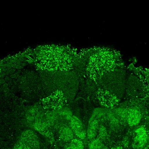
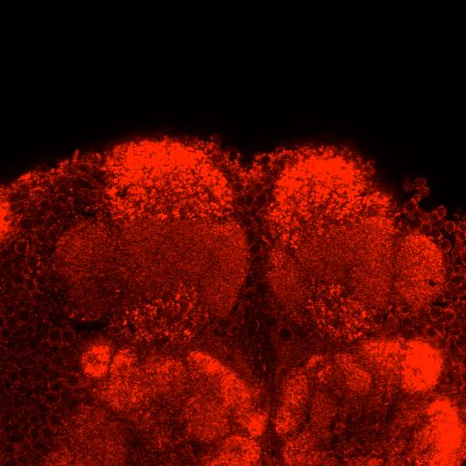
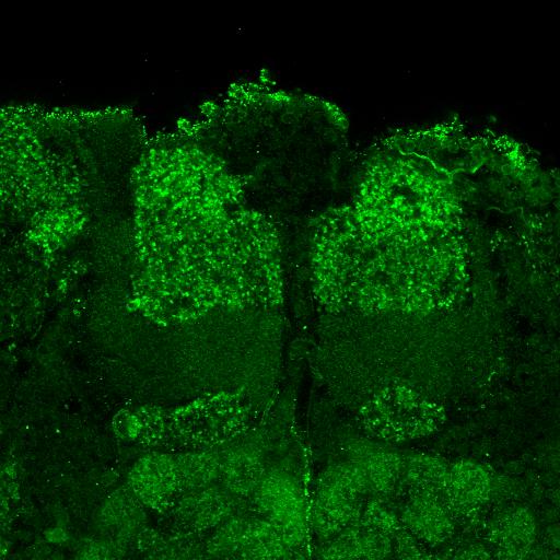
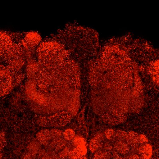

2 OK107-GAL4 + + ChATRNAi OK107-GAL4 ChATRNAi αchat SMP MB αvt 2 + VTRNAi OK107-GAL4 VTRNAi
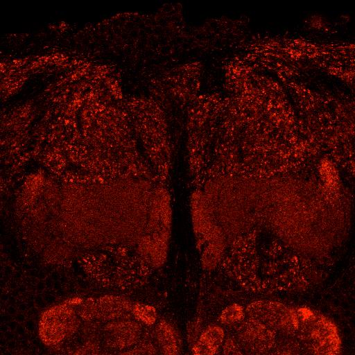
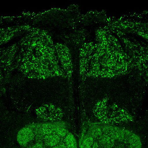
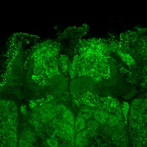
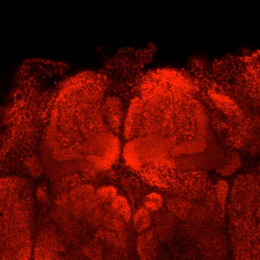
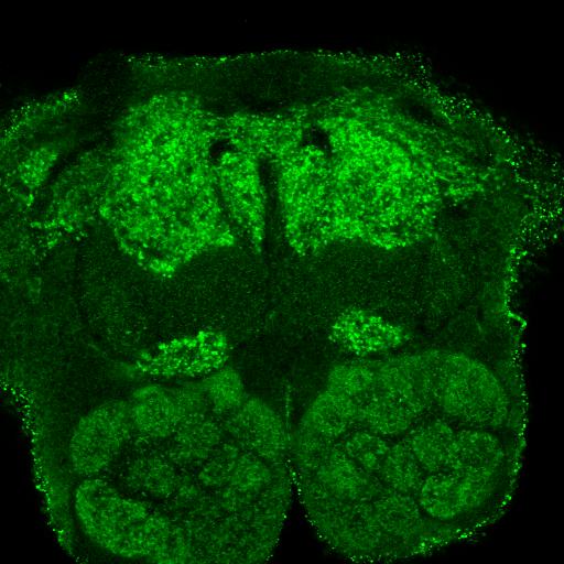
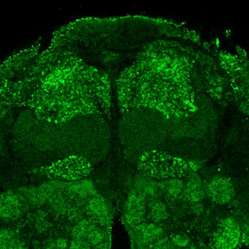
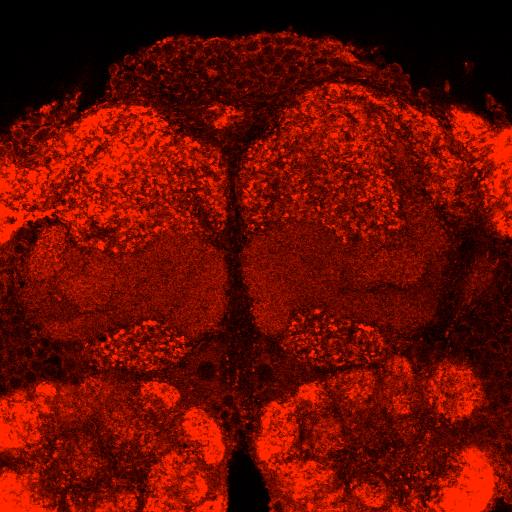
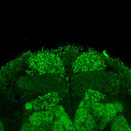
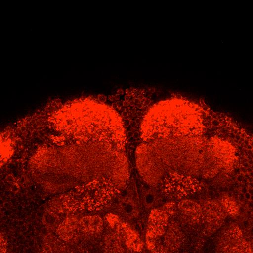
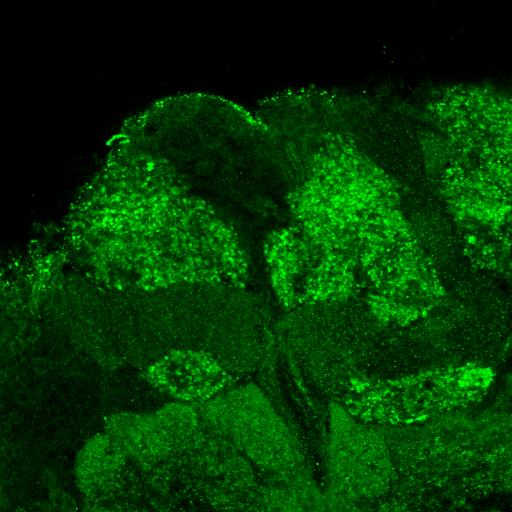
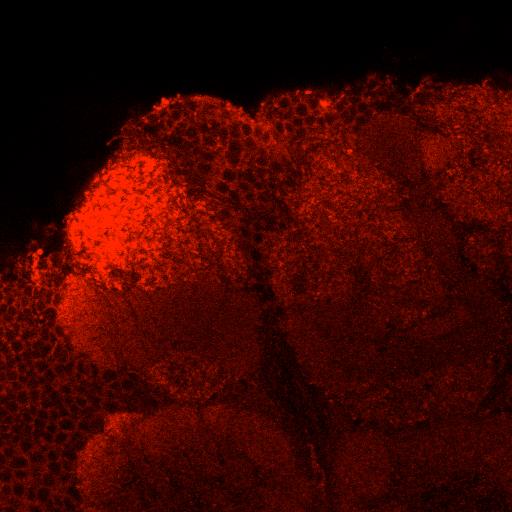
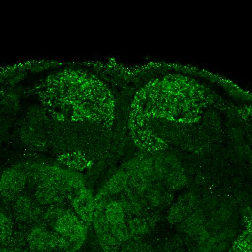
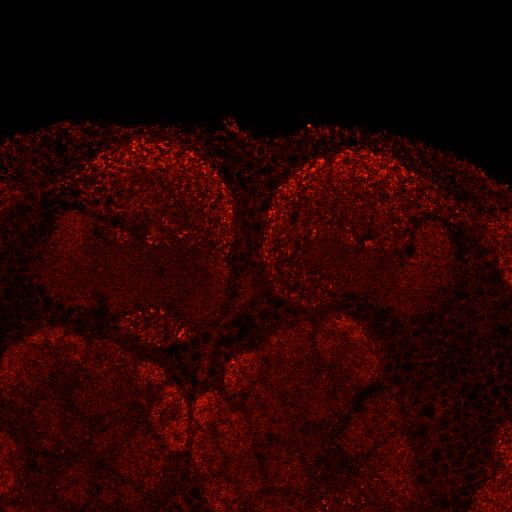
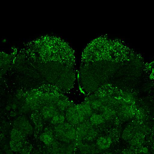
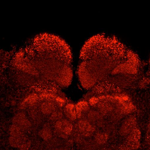
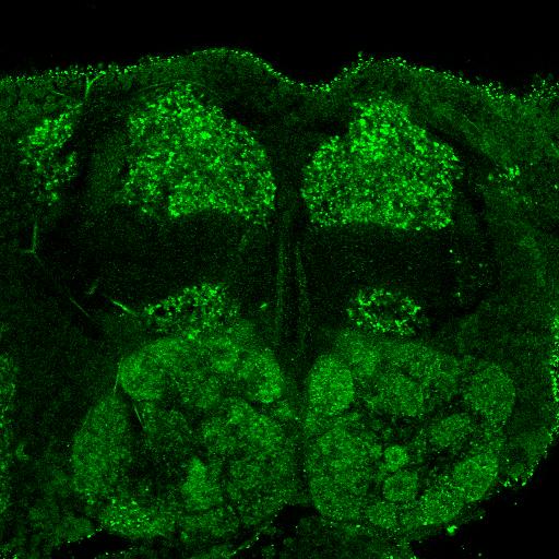
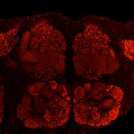
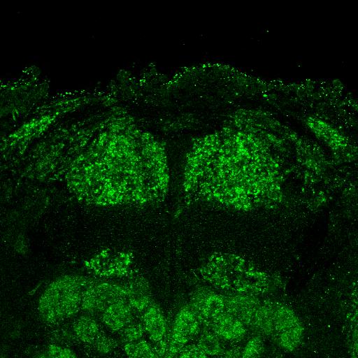
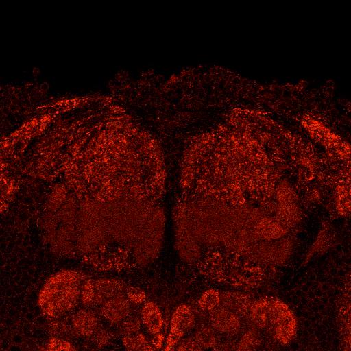
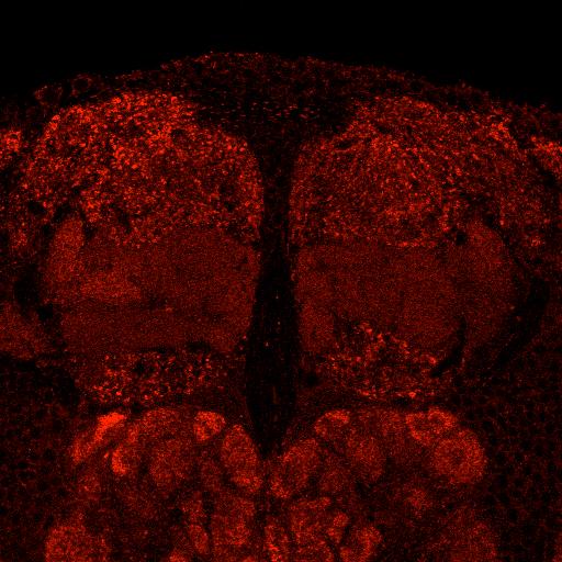
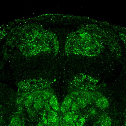
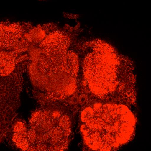
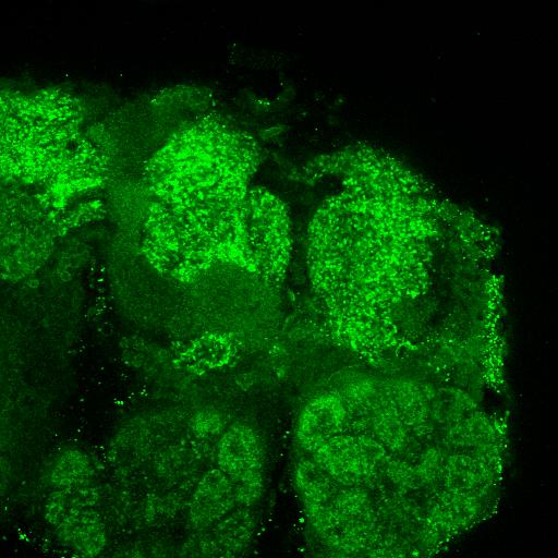
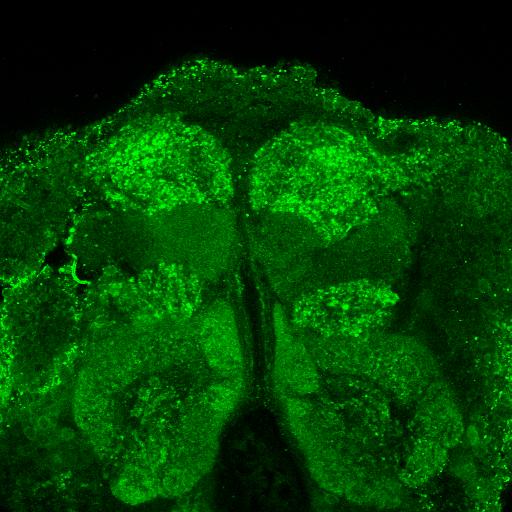
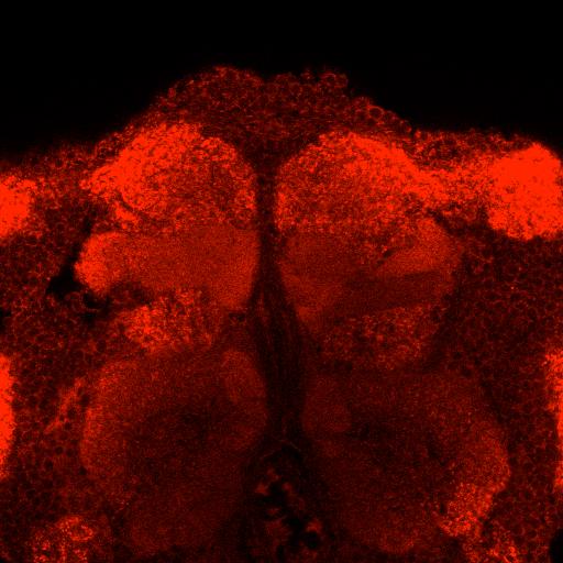
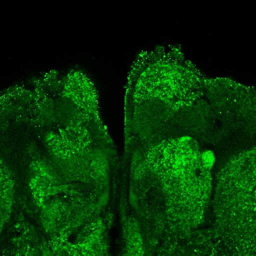
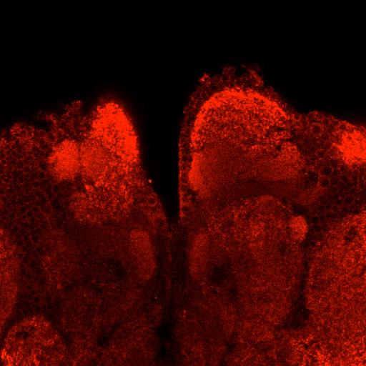
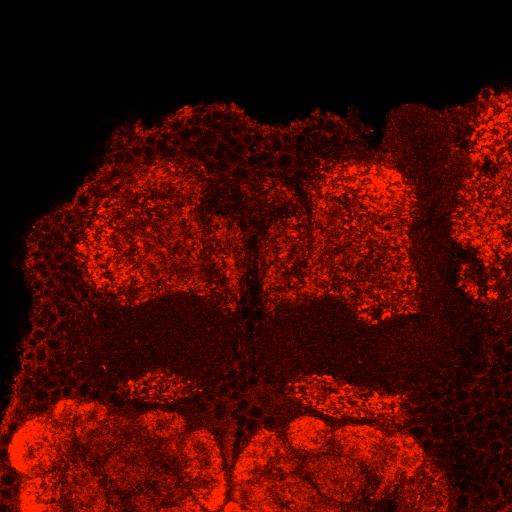
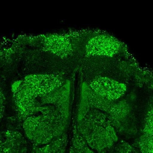
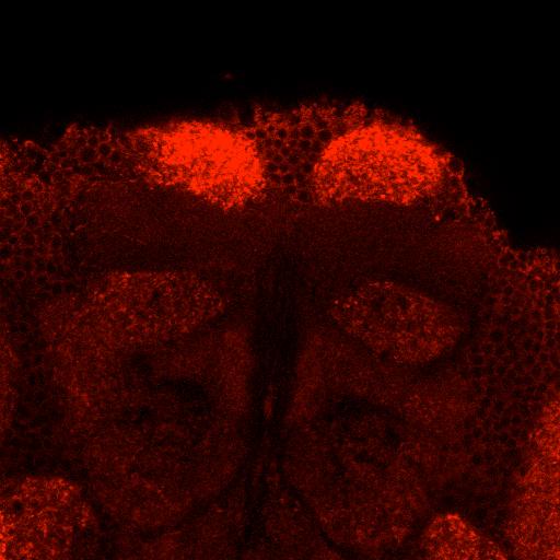
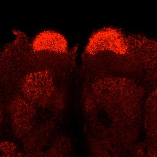
3 Figure S1, related to Figure 1A and B. ChAT and VT immunostaining labels Kenyon cells. Single confocal sections through resentative sample of brains of UAS- ChAT RNAi ; OK107-GAL4 and UAS-VT RNAi ; OK107-GAL4 flies and their genetic controls, shown at the level of the MB γ lobe neurons. αchat labeling is visibly lower in comparable images from brains of UAS-ChAT RNAi ; OK107-GAL4 flies demonstrating that the signal in control flies resents intrinsic labeling of KCs. Each lane sents both channels of single confocal sections at the level of the γ lobe from three to four resentative brains per genotype. All flies were raised and processed for immunostaining in parallel before being imaged using the same confocal settings. Scale bars 20 µm. White dashed rectangles denote areas taken for quantification. See Movie S1 for comparison of a whole stack. Similar levels of staining and RNAi-reduction were observed for the other MB lobes (not shown). 3
4 A Immediate Appetitive Memory 0.4 B Immediate Aversive Memory Performance Index Performance Index C 0 OK107 / + + / VT RNAi OK107 / VT RNAi!"#$%&'(%)%(%!%&+,- -0.8!"#$%&'(%)%(%!%&.,/ OK107 / + + / GAD RNAi OK107 / GAD RNAi + / VGAT RNAi OK107 / VGAT RNAi + / Vglut RNAi OK107 / Vglut RNAi "67!&8&3!&8&9 42:;3&<&2:27 42:1=&<&2:2>?@A4!"#$ BC@D!&8&3!&8&12 42:1;&<&2:2> 42:1E&<&2:2F?@A4%#!"$ BC@D!&8&9!&8&12 42:7=&<&2:29 42:1F&<&2:2>.0123&G&!"#$ &'#(!&8&3!&8&12 42:E7&<&2:29 42:EE&<&2:2F.0123&G&%#!"$ BC@D!&8&9!&8&12 42:7E&<&2:29 42:1=&<&2:2F Figure S2, related to Figure 1C. RNAi directed towards but not GABAergic and glutamatergic transmission compromises olfactory memory. (A) KC-driven UAS- VT RNAi impairs immediate appetitive memory (n = 10-12, asterisk denotes p < 5; oneway ANOVA, Tukey s HSD post-hoc test). (B) KC-driven UAS-GAD RNAi, UAS-VGAT RNAi (both required for GABAergic signaling), and UAS-VGlut RNAi (required for glutamatergic signaling) did not significantly impact immediate aversive memory performance, as compared to the relevant genetic control flies (n = 4-7, p > 5; one-way ANOVA, Tukey s HSD post-hoc test). Error bars in (A) and (B) resent SEM. (C) Naïve OCT and MCH avoidance performance of all fly strains tested for aversive behavioral memory in Figure 1C. Displayed are mean performance index values +/- SEM. No statistical differences were evident between the groups (n = 7-10, all p > 5, one-way ANOVA, Tukey s HSD post-hoc test) other than OK107; UAS-ChAT RNAi and +; UAS-ChAT RNAi flies which exhibit defective MCH avoidance compared to OK107; + (n = 7, p < 5, one-way ANOVA, Tukey s HSD post-hoc test). MCH concentration was 1:500. 4


5 5 ms A 7 ms 15 ms B 50 ms R21D02-GAL4 > UAS-GCaMP6f C 1% ΔF/F0 0. A Sad GCaMP6f ΔF x104 ΔF 1% ΔF/F0 20 msec D s 5% i 100 μm s 20% i s 200% i s i 100 μm onto M4/6 imaged from M4/6 R21D02-GAL4 > UAS-GCaMP6f 100 μm onto M4/6 imaged from M4/6 R21D02-GAL4 > UAS-GCaMP6f % ΔF/F sec 10 mm onto calyx imaged from M4/6 R21D02-GAL4 > UAS-GCaMP6f Mean Peak ΔF/F0 Mean Peak ΔF/F0 0.8 Mean Peak ΔF/F0 E 10 mm onto calyx imaged from calyx 247-LexA > lexaop-gcamp6f 0.4 normal Mg mm Mg2+ F 247::GCaMP5 1mM R21D02 > GCaMP5 1mM normal Mg2+ df/f0 normal Mg mm Mg mm Mg2+ 247::GCaMP5 Vehicle 247::GCaMP5 Nic 100 um R21D02 > GCaMP5 Nic 100 um 5 normal Mg mm Mg2+
6 Figure S3, related to Figure 2. Only local dendritic application of evokes calcium transients in M4/6 neurons. 100 µm was applied to the M4/6 dendritic region in R21D02-GAL4; UAS-GCaMP6f brains. (A) The breadth of the evoked calcium response in M4/6 dendrites depends on the sad of the applied. Upper panels follow the sad of Texas Red that was co-applied with 100 μm. The ejection dose was successively increased from a 5 ms to 50 ms application. Lower panels depict ΔF (mean of responses 1.5 s after application mean 1 s before application). Each pixel is color-coded. (B and C) elicits calcium responses in M4/6 neurons only when locally applied to the dendritic region. (B) 100 μm was ejected at different positions within the protocerebrum, while the sad of the solution was monitored with Texas Red. Sad outlines are marked with colored dashed ellipses. (C) Mean trace of three application trials, each trace relates to the corresponding application outline shown in (B). Only delivery close to M4/6 dendrites (the orange and bright blue traces) evoked calcium responses. A magnified view of the individual traces underlying the bright blue response reveals a ms response onset. The experiments in A-C contained 1 µm tetradotoxin (TTX). (D) The injection pipette was moved to successively more distant locations away from the dendrite, while calcium responses were recorded using widefield imaging. Each row depicts a different brain. Dashed white line resents the midline of the brain for orientation purposes; s - superior, i - inferior. Colored dashed ellipses resent sad of, monitored by Texas Red; colors of calcium traces correspond to the matching dashed colored ellipses. Scale bars in A, B and D 20 µm. (E) 100 μm still evokes M4/6 responses under conditions of high magnesium in AHLS. Addition of 10 mm Mg 2+ did not affect responses evoked in M4/6 neurons when 100 μm was applied to the M4/6 neuron dendrites (first panel; n = 6, p > 5, independent samples t-test). Calcium responses in M4/6 also show similar kinetics with and without additional 10 mm Mg 2+ (second panel). Mean traces are shown with SEM in shade. Calcium responses in M4/6 neurons were significantly decreased at 10 mm Mg 2+ when they were stimulated via 10 mm applied in the calyx (third panel; n = 3-4, p < 5, independent samples t-test). Additional 10 mm Mg 2+ did not decrease KC responses evoked by applying 10 mm to the MB calyx (fourth panel; n = 2, p > 5, independent samples t-test). Error bars resent SEM. (F) KCs do not respond to cholinergic stimulation in the MB lobes. Application of 1 mm or 100 μm nicotine to the MB lobes does not elicit calcium responses in KCs (upper row), while both agonists evoke strong calcium responses in M4/6 neurons when applied in the same location (bottom panels). Displayed are mean traces with the respective SEM as shade, each color resenting an individual brain. Note that the reduced signal observed in the upper panels likely results from tissue displacement caused by ssure-evoked release of or vehicle. 6
 MBON-β 2mp (M4β ) 20 μm D MBON-β 2mp (M4β ) MBON-β2β 2a (M4β) E MBON-α 3ap (V2α")


 MB002B labels MBON-βʹ2mp (M4β ).")
, MB210B labels MBON-γ5βʹ2a (M6) and MBON-βʹ2mp (M4β ).")
7 A MBON-β 2mp (M4β ) B MBON-γ5β 2a (M6) C MBON-β 2mp (M4β ) MBON-β 2mp_bi MBON-γ5β 2a (M6) MBON-β 2mp (M4β ) 20 μm D MBON-β 2mp (M4β ) MBON-β2β 2a (M4β) E MBON-α 3ap (V2α ) MBON-α 3m (V2α ) Figure S4, related to Figure 3. evokes responses in multiple classes of MBONs. Each panel shows the anatomy of the relevant MBON and below the corresponding GCaMP6f measured physiological responses elicited by 1 mm acetylcholine application. Data resent individual -evoked traces from one brain per genotype. (A) MB002B labels MBON-βʹ2mp (M4β ). (B), MB011B labels MBON-γ5βʹ2a (M6), MBON-βʹ2mp (M4β ) and MBON-βʹ2mp_bi.(C), MB210B labels MBON-γ5βʹ2a (M6) and MBON-βʹ2mp (M4β ). (D) R93F01-GAL4 labels MBON-βʹ2mp (M4β ) and MBON-γ5βʹ2a (M6). (E) MB027B labels MBON-αʹ3ap (V2α ) and MBON-αʹ3m (V2α ). Initial increase in traces of (D) is an artifact of the LED trigger. 7
8 A 100 μm Acetylcholine 250 μm Mecamylamine B post 10 sec post Mecamylamine Peak 0.1 post C 100 μm 100 μm D Acetylcholine Mecamylamine E G 100 μm Acetylcholine Vehicle 10 sec 10 sec post Mecamylamine post Vehicle Peak F Peak post 100 μm 100 μm Acetylcholine Hexamethonium post H Peak 0.4 I 100 μm Acetylcholine 10 μm MLA 20% 10 sec post Hexamethonium J post Peak % 10 sec post MLA K 100 μm Mecamylamine 30 min washout L ns Norm. Peak Δ F/F % 5 sec Meca wash M 250 μm Mecamylamine 30 min washout N ns Norm. Peak Δ F/F % df/f0 5 sec Meca wash 8
9 Figure S5, related to Figure 4. M4/6 neuron calcium responses can be reversibly blocked by nicotinic receptor antagonists. (A-J) Local evoked calcium responses in M4/6 dendrites are blocked by the non-selective nicotinic receptor antagonists mecamylamine, hexamethonium, and MLA. (A-B) responses vanish within 100 s of applying 250 µm mecamylamine to the recording chamber. (A) Sample trace and (B) quantification, n = 5 brains, average of four trials, asterisk denotes p < 5, paired samples t-test. (C-D) responses also decrease within 100 s of applying 100 µm mecamylamine to the recording chamber. (C) Sample trace and (D) quantification, n = 3 brains, average of four trials, asterisk denotes p < 5, paired samples t-test. (E-F) Application of a vehicle control has no effect on the -evoked responses. (E) Sample trace and (F) quantification, n = 5 brains, average of four trials, p > 5, paired samples t-test. (G-H) responses vanish within 100 s of applying 100 µm hexamethonium to the recording chamber. (G) Sample trace and (H) quantification, n = 3 brains, average of four trials, asterisk denotes p < 5, paired samples t-test. (I-J) responses vanish within 100 s of applying 10 µm MLA to the recording chamber. (I) Sample trace and (J) quantification, n = 3 brains, average of four trials, asterisk denotes p < 5, paired samples t-test. (K-N) Optogenetic stimulation of KCs triggers M4/6 neuron calcium responses that are significantly reduced by application of mecamylamine, and partially recovered after washout. (K) Sample trace and (L) quantification of applying 100 µm mecamylamine, n = 2 brains, average of four trials, asterisk denotes p < 5, paired samples t-test. Genotype: lexaop-cschrimson / UAS- GCaMP6f; R21D02-GAL4 / 247-LexA::VP16. (M) Sample trace and (N) quantification of applying 250 µm mecamylamine, n = 2 brains, average of four trials, asterisk denotes p < 5, paired samples t-test. Genotype: R15B01-LexA, lexaop-gcamp6f / UAS-CsChrimson; R13F02-GAL4 / +. was normalized to each experiment s average initial four responses. 9
10 Movie S1, related to Figure 1A. Kenyon cells are ChAT- and VT-immunopositive. ChAT and VT immunofluorescence in the mushroom body lobes are greatly reduced by OK107-GAL4 directed exssion of UAS-ChAT RNAi or UAS-VT RNAi respectively (middle panels). Movie shows a resentative sample stack at 0.5 µm/frame. All flies were raised and processed for immunostaining in parallel before being imaged using the same confocal settings. Scale bar 50 µm. Movie S2, related to Figure 4A and D. Mecamylamine abolishes Kenyon cell-driven M4/6 responses. Optogenetic KC activation via 247-LexA exssed lexaop-cschrimson and red light illumination leads to robust calcium responses in M4/6 axons. R21D02-GAL4; UAS-GCaMP6f explant brains monitored with two-photon imaging. Frame rate: 5.92 Hz. Two resentative samples before and after addition of the nicotinic receptor antagonist mecamylamine (250 µm) or vehicle, are shown. The data in these movies is also sented as static views in Figures 4 G-L. White horizontal stripes that appear at regular intervals are artifacts from LED stimulation. 10
Supplementary Figure 1. Flies form water-reward memory only in the thirsty state
 1 2 3 4 5 6 7 Supplementary Figure 1. Flies form water-reward memory only in the thirsty state Thirsty but not sated wild-type flies form robust 3 min memory. For the thirsty group, the flies were water-deprived
Supplementary Figure 1. Procedures to independently control fly hunger and thirst states.
 Supplementary Figure 1 Procedures to independently control fly hunger and thirst states. (a) Protocol to produce exclusively hungry or thirsty flies for 6 h water memory retrieval. (b) Consumption assays
Hormonal gain control of a medial preoptic area social reward circuit
 CORRECTION NOTICE Nat. Neurosci. 20, 449 458 (2017) Hormonal gain control of a medial preoptic area social reward circuit Jenna A McHenry, James M Otis, Mark A Rossi, J Elliott Robinson, Oksana Kosyk,
A Drosophila model for alcohol reward. Supplemental Material
 Kaun et al. Supplemental Material 1 A Drosophila model for alcohol reward Supplemental Material Kaun, K.R. *, Azanchi, R. *, Maung, Z. *, Hirsh, J. and Heberlein, U. * * Department of Anatomy, University
Nature Neuroscience: doi: /nn Supplementary Figure 1. Trial structure for go/no-go behavior
 Supplementary Figure 1 Trial structure for go/no-go behavior a, Overall timeline of experiments. Day 1: A1 mapping, injection of AAV1-SYN-GCAMP6s, cranial window and headpost implantation. Water restriction
Nature Neuroscience: doi: /nn.4335
 Supplementary Figure 1 Cholinergic neurons projecting to the VTA are concentrated in the caudal mesopontine region. (a) Schematic showing the sites of retrograde tracer injections in the VTA: cholera toxin
Nature Neuroscience: doi: /nn Supplementary Figure 1
 Supplementary Figure 1 Drd1a-Cre driven ChR2 expression in the SCN. (a) Low-magnification image of a representative Drd1a-ChR2 coronal brain section (n = 2) showing endogenous tdtomato fluorescence (magenta).
Neural circuits for long-term water-reward memory processing in thirsty Drosophila
 Received 2 Jun 216 Accepted 3 Mar 217 Published 15 May 217 DOI: 1.138/ncomms1523 OPEN Neural circuits for long-term water-reward processing in thirsty Drosophila Wei-Huan Shyu 1, Tai-Hsiang Chiu 1, Meng-Hsuan
Supplementary Figure 1. SDS-FRL localization of CB 1 in the distal CA3 area of the rat hippocampus. (a-d) Axon terminals (t) in stratum pyramidale
 Axon terminals (t) in stratum pyramidale") Supplementary Figure 1. SDS-FRL localization of CB 1 in the distal CA3 area of the rat hippocampus. (a-d) Axon terminals (t) in stratum pyramidale (b) show stronger immunolabeling for CB 1 than those in
Supplementary Figure 1. SDS-FRL localization of CB 1 in the distal CA3 area of the rat hippocampus. (a-d) Axon terminals (t) in stratum pyramidale (b) show stronger immunolabeling for CB 1 than those in
Supplementary Figure 1. SybII and Ceb are sorted to distinct vesicle populations in astrocytes. Nature Neuroscience: doi: /nn.
 Supplementary Figure 1 SybII and Ceb are sorted to distinct vesicle populations in astrocytes. (a) Exemplary images for cultured astrocytes co-immunolabeled with SybII and Ceb antibodies. SybII accumulates
A genetically targeted optical sensor to monitor calcium signals in astrocyte processes
 A genetically targeted optical sensor to monitor calcium signals in astrocyte processes 1 Eiji Shigetomi, 1 Sebastian Kracun, 2 Michael V. Sofroniew & 1,2 *Baljit S. Khakh Ψ 1 Departments of Physiology
Supplementary Figure 1. GABA depolarizes the majority of immature neurons in the
 Supplementary Figure 1. GABA depolarizes the majority of immature neurons in the upper cortical layers at P3 4 in vivo. (a b) Cell-attached current-clamp recordings illustrate responses to puff-applied
doi: /nature09554
 SUPPLEMENTARY INFORMATION doi:10.1038/nature09554 Supplementary Figure 1: Optical Tracing with New Photoactivatable GFP Variants Reveals Enhanced Labeling of Neuronal Processes We qualitatively compare
Nature Neuroscience: doi: /nn Supplementary Figure 1. Large-scale calcium imaging in vivo.
 Supplementary Figure 1 Large-scale calcium imaging in vivo. (a) Schematic illustration of the in vivo camera imaging set-up for large-scale calcium imaging. (b) High-magnification two-photon image from
STDP enhances synchrony in feedforward network
 1 1 of 10 STDP enhances synchrony in feedforward network STDP strengthens/weakens synapses driving late/early-spiking cells [Laurent07] Olfactory neurons' spikes phase-lock (~2ms) to a 20Hz rhythm. STDP
Supplementary figure 1: LII/III GIN-cells show morphological characteristics of MC
 1 2 1 3 Supplementary figure 1: LII/III GIN-cells show morphological characteristics of MC 4 5 6 7 (a) Reconstructions of LII/III GIN-cells with somato-dendritic compartments in orange and axonal arborizations
Unique functional properties of somatostatin-expressing GABAergic neurons in mouse barrel cortex
 Supplementary Information Unique functional properties of somatostatin-expressing GABAergic neurons in mouse barrel cortex Luc Gentet, Yves Kremer, Hiroki Taniguchi, Josh Huang, Jochen Staiger and Carl
Nature Neuroscience: doi: /nn Supplementary Figure 1
 Supplementary Figure 1 Bidirectional optogenetic modulation of the tonic activity of CEA PKCδ + neurons in vitro. a, Top, Cell-attached voltage recording illustrating the blue light-induced increase in
File name: Supplementary Information Description: Supplementary Figures, Supplementary Table and Supplementary References
 File name: Supplementary Information Description: Supplementary Figures, Supplementary Table and Supplementary References File name: Supplementary Data 1 Description: Summary datasheets showing the spatial
-51mV 30s 3mV. n=14 n=4 p=0.4. Depolarization (mv) 3
 3") Supplementary Figure 1 a optoβ 2 -AR b ChR2-51mV 30s 3mV -50mV 30s 3mV c 4 n=14 n=4 p=0.4 Depolarization (mv) 3 2 1 0 Both optogenetic actuators, optoβ 2 AR and ChR2, were effective in stimulating astrocytes
Supplementary Figure 1 a optoβ 2 -AR b ChR2-51mV 30s 3mV -50mV 30s 3mV c 4 n=14 n=4 p=0.4 Depolarization (mv) 3 2 1 0 Both optogenetic actuators, optoβ 2 AR and ChR2, were effective in stimulating astrocytes
PEER REVIEW FILE. Reviewers' Comments: Reviewer #1 (Remarks to the Author):
:") PEER REVIEW FILE Reviewers' Comments: Reviewer #1 (Remarks to the Author): In this manuscript entitled "Neural circuits for long-term water-reward memory processing in thirsty Drosophila", the authors
PEER REVIEW FILE Reviewers' Comments: Reviewer #1 (Remarks to the Author): In this manuscript entitled "Neural circuits for long-term water-reward memory processing in thirsty Drosophila", the authors
Supporting Online Material for
 www.sciencemag.org/cgi/content/full/312/5779/1533/dc1 Supporting Online Material for Long-Term Potentiation of Neuron-Glia Synapses Mediated by Ca 2+ - Permeable AMPA Receptors Woo-Ping Ge, Xiu-Juan Yang,
Structural basis for the role of inhibition in facilitating adult brain plasticity
 Structural basis for the role of inhibition in facilitating adult brain plasticity Jerry L. Chen, Walter C. Lin, Jae Won Cha, Peter T. So, Yoshiyuki Kubota & Elly Nedivi SUPPLEMENTARY FIGURES 1-6 a b M
Ube3a is required for experience-dependent maturation of the neocortex
 Ube3a is required for experience-dependent maturation of the neocortex Koji Yashiro, Thorfinn T. Riday, Kathryn H. Condon, Adam C. Roberts, Danilo R. Bernardo, Rohit Prakash, Richard J. Weinberg, Michael
Supplemental Information. Octopamine Neurons Mediate Flight-Induced Modulation of Visual Processing in Drosophila. Supplemental Inventory
 1 Current Biology, Volume 22 Supplemental Information Octopamine Neurons Mediate Flight-Induced Modulation of Visual Processing in Drosophila Marie P. Suver, Akira Mamiya, and Michael H. Dickinson Supplemental
Nature Neuroscience: doi: /nn Supplementary Figure 1
 Supplementary Figure 1 Atlas representations of the midcingulate (MCC) region targeted in this study compared against the anterior cingulate (ACC) region commonly reported. Coronal sections are shown on
Supplemental Information. A Visual-Cue-Dependent Memory Circuit. for Place Navigation
 Neuron, Volume 99 Supplemental Information A Visual-Cue-Dependent Memory Circuit for Place Navigation Han Qin, Ling Fu, Bo Hu, Xiang Liao, Jian Lu, Wenjing He, Shanshan Liang, Kuan Zhang, Ruijie Li, Jiwei
Supplementary Figure 1. Basic properties of compound EPSPs at
 Supplementary Figure 1. Basic properties of compound EPSPs at hippocampal CA3 CA3 cell synapses. (a) EPSPs were evoked by extracellular stimulation of the recurrent collaterals and pharmacologically isolated
SUPPLEMENTARY INFORMATION. Supplementary Figure 1
 SUPPLEMENTARY INFORMATION Supplementary Figure 1 The supralinear events evoked in CA3 pyramidal cells fulfill the criteria for NMDA spikes, exhibiting a threshold, sensitivity to NMDAR blockade, and all-or-none
SUPPLEMENTARY INFORMATION
 doi:1.138/nature1139 a d Whisker angle (deg) Whisking repeatability Control Muscimol.4.3.2.1 -.1 8 4-4 1 2 3 4 Performance (d') Pole 8 4-4 1 2 3 4 5 Time (s) b Mean protraction angle (deg) e Hit rate (p
SUPPLEMENTARY INFORMATION
 doi: 1.138/nature6416 Supplementary Notes Spine Ca 2+ signals produced by glutamate uncaging We imaged uncaging-evoked [Ca 2+ ] transients in neurons loaded with a green Ca 2+ - sensitive indicator (G;
Activity of Defined Mushroom Body Output Neurons Underlies Learned Olfactory Behavior in Drosophila
 Article Activity of Defined Mushroom Body Output Neurons Underlies Learned Olfactory Behavior in Drosophila Highlights d Glutamatergic mushroom body output neurons are required for memory expression Authors
Supplementary Figure 1
 Supplementary Figure 1 Supplementary Figure 1 SNARE Probes for FRET/2pFLIM Analysis Used in the Present Study. mturquoise (mtq) and Venus (Ven) are in blue and yellow, respectively. The soluble N-ethylmaleimide-sensitive
Supplementary Figure 1) GABAergic enhancement by leptin hyperpolarizes POMC neurons A) Representative recording samples showing the membrane
 GABAergic enhancement by leptin hyperpolarizes POMC neurons A) Representative recording samples showing the membrane") Supplementary Figure 1) GABAergic enhancement by leptin hyperpolarizes POMC neurons A) Representative recording samples showing the membrane potential recorded from POMC neurons following treatment with
Supplementary Figure 1) GABAergic enhancement by leptin hyperpolarizes POMC neurons A) Representative recording samples showing the membrane potential recorded from POMC neurons following treatment with
How Nicotinic Signaling Shapes Neural Networks
 How Nicotinic Signaling Shapes Neural Networks Darwin K. Berg Division of Biological Sciences University of California, San Diego Nicotinic Cholinergic Signaling Uses the transmitter ACh to activate cation-selective
Supporting Information
 ATP from synaptic terminals and astrocytes regulates NMDA receptors and synaptic plasticity through PSD- 95 multi- protein complex U.Lalo, O.Palygin, A.Verkhratsky, S.G.N. Grant and Y. Pankratov Supporting
Supplementary Figure 1 hlrrk2 promotes CAP dependent protein translation.
 ` Supplementary Figure 1 hlrrk2 promotes CAP dependent protein translation. (a) Overexpression of hlrrk2 in HeLa cells enhances total protein synthesis in [35S] methionine/cysteine incorporation assays.
Drosophila DPM Neurons Form a Delayed and Branch-Specific Memory Trace after Olfactory Classical Conditioning
 Drosophila DPM Neurons Form a Delayed and Branch-Specific Memory Trace after Olfactory Classical Conditioning Dinghui Yu, 1 Alex C. Keene, 4 Anjana Srivatsan, 2 Scott Waddell, 4 and Ronald L. Davis 1,3,
TRPA1 channels regulate astrocyte resting calcium. and inhibitory synapse efficacy through GAT-3
 TRPA1 channels regulate astrocyte resting calcium and inhibitory synapse efficacy through GAT-3 * 1 Eiji Shigetomi, * 1 Xiaoping Tong 3 Kelvin Y. Kwan, 3 David P. Corey & 1,2 Baljit S. Khakh Ψ 1 Departments
Supplementary Materials for VAMP4 directs synaptic vesicles to a pool that selectively maintains asynchronous neurotransmission
 Supplementary Materials for VAMP4 directs synaptic vesicles to a pool that selectively maintains asynchronous neurotransmission Jesica Raingo, Mikhail Khvotchev, Pei Liu, Frederic Darios, Ying C. Li, Denise
Nature Neuroscience: doi: /nn Supplementary Figure 1
 Supplementary Figure 1 Subcellular segregation of VGluT2-IR and TH-IR within the same VGluT2-TH axon (wild type rats). (a-e) Serial sections of a dual VGluT2-TH labeled axon. This axon (blue outline) has
Supplementary Figure 1: Validation of labeling specificity of immature OSNs and presynaptic terminals. (A) (B) (C) (D) (E)
 (B) (C) (D) (E)") Supplementary Figure 1: Validation of labeling specificity of immature OSNs and presynaptic terminals. (A) Confocal images of septal olfactory epithelium of an adult Gγ8-sypGFP-tdTom mouse showing colocalization
Supplementary Figure 1: Validation of labeling specificity of immature OSNs and presynaptic terminals. (A) Confocal images of septal olfactory epithelium of an adult Gγ8-sypGFP-tdTom mouse showing colocalization
SUPPLEMENTARY INFORMATION
 doi:10.1038/nature10776 Supplementary Information 1: Influence of inhibition among blns on STDP of KC-bLN synapses (simulations and schematics). Unconstrained STDP drives network activity to saturation
Zhu et al, page 1. Supplementary Figures
 Zhu et al, page 1 Supplementary Figures Supplementary Figure 1: Visual behavior and avoidance behavioral response in EPM trials. (a) Measures of visual behavior that performed the light avoidance behavior
Nature Neuroscience: doi: /nn Supplementary Figure 1
 Supplementary Figure 1 Relative expression of K IR2.1 transcript to enos was reduced 29-fold in capillaries from knockout animals. Relative expression of K IR2.1 transcript to enos was reduced 29-fold
McWilliams et al., http :// /cgi /content /full /jcb /DC1
 Supplemental material JCB McWilliams et al., http ://www.jcb.org /cgi /content /full /jcb.201603039 /DC1 THE JOURNAL OF CELL BIOLOGY Figure S1. In vitro characterization of mito-qc. (A and B) Analysis
Cellular Neurobiology / BIPN 140
 SECOND MIDTERM EXAMINATION Fall, 2015 GENERAL INSTRUCTIONS 1. Please write your name on ALL 6 pages. 2. Please answer each question IN THE SPACE ALLOTTED. 1) /10 pts 2) /10 pts 3) /15 pts 4) /15 pts 5)
An acetylcholine-activated microcircuit drives temporal dynamics of cortical activity
 An acetylcholine-activated microcircuit drives temporal dynamics of cortical activity Naiyan Chen, Hiroki Sugihara, & Mriganka Sur Nature America, nc. All rights reserved. Cholinergic modulation of cortex
Dep. Control Time (min)
") aa Control Dep. RP 1s 1 mv 2s 1 mv b % potentiation of IPSP 2 15 1 5 Dep. * 1 2 3 4 Time (min) Supplementary Figure 1. Rebound potentiation of IPSPs in PCs. a, IPSPs recorded with a K + gluconate pipette
aa Control Dep. RP 1s 1 mv 2s 1 mv b % potentiation of IPSP 2 15 1 5 Dep. * 1 2 3 4 Time (min) Supplementary Figure 1. Rebound potentiation of IPSPs in PCs. a, IPSPs recorded with a K + gluconate pipette
Nature Methods: doi: /nmeth Supplementary Figure 1. Activity in turtle dorsal cortex is sparse.
 Supplementary Figure 1 Activity in turtle dorsal cortex is sparse. a. Probability distribution of firing rates across the population (notice log scale) in our data. The range of firing rates is wide but
Supplemental Information. Dorsal Raphe Dual Serotonin-Glutamate Neurons. Drive Reward by Establishing Excitatory Synapses
 Cell Reports, Volume 26 Supplemental Information Dorsal Raphe Dual Serotonin-Glutamate Neurons Drive Reward by Establishing Excitatory Synapses on VTA Mesoaccumbens Dopamine Neurons Hui-Ling Wang, Shiliang
Supplementary Figure 1
 Supplementary Figure 1 Arcuate ChIEF-tdTomato neurons expressed TH These micrographs show that TH-Cre-ChIEF-tdTomato (magenta), expressed by AAV in a TH-Cre mouse, were immunostained with TH (green) in
Wenqin Hu, Cuiping Tian, Tun Li, Mingpo Yang, Han Hou & Yousheng Shu
 Distinct contributions of Na v 1.6 and Na v 1.2 in action potential initiation and backpropagation Wenqin Hu, Cuiping Tian, Tun Li, Mingpo Yang, Han Hou & Yousheng Shu Supplementary figure and legend Supplementary
Fig. S1. High K+ increases intracellular calcium level.
 Fig. S1. High K + increases intracellular calcium level. (A) Neuronal activation measured by calcium imaging using Fura-2. Intracellular calcium levels were continuously monitored by the fura-2 florescence
Nature Neuroscience: doi: /nn Supplementary Figure 1. Confirmation that optogenetic inhibition of dopaminergic neurons affects choice
 Supplementary Figure 1 Confirmation that optogenetic inhibition of dopaminergic neurons affects choice (a) Sample behavioral trace as in Figure 1d, but with NpHR stimulation trials depicted as green blocks
Tuning properties of individual circuit components and stimulus-specificity of experience-driven changes.
 Supplementary Figure 1 Tuning properties of individual circuit components and stimulus-specificity of experience-driven changes. (a) Left, circuit schematic with the imaged component (L2/3 excitatory neurons)
SUPPLEMENTARY INFORMATION
 Supplementary Figure 1. Behavioural effects of ketamine in non-stressed and stressed mice. Naive C57BL/6 adult male mice (n=10/group) were given a single dose of saline vehicle or ketamine (3.0 mg/kg,
Astrocyte signaling controls spike timing-dependent depression at neocortical synapses
 Supplementary Information Astrocyte signaling controls spike timing-dependent depression at neocortical synapses Rogier Min and Thomas Nevian Department of Physiology, University of Berne, Bern, Switzerland
marker. DAPI labels nuclei. Flies were 20 days old. Scale bar is 5 µm. Ctrl is
 Supplementary Figure 1. (a) Nos is detected in glial cells in both control and GFAP R79H transgenic flies (arrows), but not in deletion mutant Nos Δ15 animals. Repo is a glial cell marker. DAPI labels
Supplemental Data Figure S1 Effect of TS2/4 and R6.5 antibodies on the kinetics of CD16.NK-92-mediated specific lysis of SKBR-3 target cells.
 Supplemental Data Figure S1. Effect of TS2/4 and R6.5 antibodies on the kinetics of CD16.NK-92-mediated specific lysis of SKBR-3 target cells. (A) Specific lysis of IFN-γ-treated SKBR-3 cells in the absence
Nature Neuroscience: doi: /nn Supplementary Figure 1. Diverse anorexigenic signals induce c-fos expression in CEl PKC-δ + neurons
 Supplementary Figure 1 Diverse anorexigenic signals induce c-fos expression in CEl PKC-δ + neurons a-c. Quantification of CEl c-fos expression in mice intraperitoneal injected with anorexigenic drugs (a),
Social transmission and buffering of synaptic changes after stress
 SUPPLEMENTARY INFORMATION Articles https://doi.org/10.1038/s41593-017-0044-6 In the format provided by the authors and unedited. Social transmission and buffering of synaptic changes after stress Toni-Lee
Regulation of sleep plasticity by a thermo-sensitive circuit in Drosophila
 Regulation of sleep plasticity by a thermo-sensitive circuit in Drosophila Angelique Lamaze 1, Arzu Öztürk-Çolak 2, Robin Fischer 3, Nicolai Peschel 3, Kyunghee Koh 2 and James E.C. Jepson 1* 1 UCL Institute
Supplementary Figure 1 Madm is not required in GSCs and hub cells. (a,b) Act-Gal4-UAS-GFP (a), Act-Gal4-UAS- GFP.nls (b,c) is ubiquitously expressed
 Act-Gal4-UAS-GFP (a), Act-Gal4-UAS- GFP.nls (b,c) is ubiquitously expressed") Supplementary Figure 1 Madm is not required in GSCs and hub cells. (a,b) Act-Gal4-UAS-GFP (a), Act-Gal4-UAS- GFP.nls (b,c) is ubiquitously expressed in the testes. The testes were immunostained with GFP
Supplementary Figure 1 Madm is not required in GSCs and hub cells. (a,b) Act-Gal4-UAS-GFP (a), Act-Gal4-UAS- GFP.nls (b,c) is ubiquitously expressed in the testes. The testes were immunostained with GFP
Supplementary Table I Blood pressure and heart rate measurements pre- and post-stroke
 SUPPLEMENTARY INFORMATION doi:10.1038/nature09511 Supplementary Table I Blood pressure and heart rate measurements pre- and post-stroke Pre Post 7-days Systolic Diastolic BPM Systolic Diastolic BPM Systolic
Title: Smooth muscle cell-specific Tgfbr1 deficiency promotes aortic aneurysm formation by stimulating multiple signaling events
 Title: Smooth muscle cell-specific Tgfbr1 deficiency promotes aortic aneurysm formation by stimulating multiple signaling events Pu Yang 1, 3, radley M. Schmit 1, Chunhua Fu 1, Kenneth DeSart 1, S. Paul
Neuroscience 201A (2016) - Problems in Synaptic Physiology
 - Problems in Synaptic Physiology") Question 1: The record below in A shows an EPSC recorded from a cerebellar granule cell following stimulation (at the gap in the record) of a mossy fiber input. These responses are, then, evoked by stimulation.
Question 1: The record below in A shows an EPSC recorded from a cerebellar granule cell following stimulation (at the gap in the record) of a mossy fiber input. These responses are, then, evoked by stimulation.
Light-evoked hyperpolarization and silencing of neurons by conjugated polymers
 Light-evoked hyperpolarization and silencing of neurons by conjugated polymers Paul Feyen 1,, Elisabetta Colombo 1,2,, Duco Endeman 1, Mattia Nova 1, Lucia Laudato 2, Nicola Martino 2,3, Maria Rosa Antognazza
B220 CD4 CD8. Figure 1. Confocal Image of Sensitized HLN. Representative image of a sensitized HLN
 B220 CD4 CD8 Natarajan et al., unpublished data Figure 1. Confocal Image of Sensitized HLN. Representative image of a sensitized HLN showing B cell follicles and T cell areas. 20 µm thick. Image of magnification
SUPPLEMENTARY INFORMATION
 SUPPLEMENTARY INFORMATION doi:10.1038/nature11306 Supplementary Figures Supplementary Figure 1. Basic characterization of GFP+ RGLs in the dentate gyrus of adult nestin-gfp mice. a, Sample confocal images
Supplementary Figure 1. Nature Neuroscience: doi: /nn.4547
 Supplementary Figure 1 Characterization of the Microfetti mouse model. (a) Gating strategy for 8-color flow analysis of peripheral Ly-6C + monocytes from Microfetti mice 5-7 days after TAM treatment. Living
Shocking revelations and saccharin sweetness in the study of Drosophila olfactory memory
 University of Massachusetts Medical School escholarship@umms University of Massachusetts Medical School Faculty Publications 9-9-2013 Shocking revelations and saccharin sweetness in the study of Drosophila
Nature Neuroscience: doi: /nn Supplementary Figure 1
 Supplementary Figure 1 Reward rate affects the decision to begin work. (a) Latency distributions are bimodal, and depend on reward rate. Very short latencies (early peak) preferentially occur when a greater
Tyrodes solution in a custom-built imaging chamber as described previously. Images were acquired
 Supplemental Material Supplemental Methods Electrical stimulation of CX-G3-labeled hippocampal neurons Following 5 min incubation in 0.5 µm CX-G3 and washes, 18-20 DIV neurons were imaged in normal Tyrodes
SUPPLEMENTARY INFORMATION
 Supplementary Figure 1. Normal AMPAR-mediated fepsp input-output curve in CA3-Psen cdko mice. Input-output curves, which are plotted initial slopes of the evoked fepsp as function of the amplitude of the
Nature Neuroscience: doi: /nn.4581
 Supplementary Figure 1 Anatomy and functional connectivity suggest that TB neurons are part of an excitatory feedforward pathway from the AOTU to the BU composed of parallel channels. a, Different channels
Nature Neuroscience: doi: /nn.4642
 Supplementary Figure 1 Recording sites and example waveform clustering, as well as electrophysiological recordings of auditory CS and shock processing following overtraining. (a) Recording sites in LC
Schwarz et al. Activity-Dependent Ubiquitination of GluA1 Mediates a Distinct AMPAR Endocytosis
 Schwarz et al Activity-Dependent Ubiquitination of GluA1 Mediates a Distinct AMPAR Endocytosis and Sorting Pathway Supplemental Data Supplemental Fie 1: AMPARs undergo activity-mediated ubiquitination
50mM D-Glucose. Percentage PI. L-Glucose
 Monica Dus, Minrong Ai, Greg S.B Suh. Taste-independent nutrient selection is mediated by a brain-specific Na+/solute cotransporter in Drosophila. Control + Phlorizin mm D-Glucose 1 2mM 1 L-Glucose Gr5a;Gr64a
Brief presynaptic bursts evoke synapse-specific retrograde inhibition mediated by endogenous cannabinoids
 Brief presynaptic bursts evoke synapse-specific retrograde inhibition mediated by endogenous cannabinoids Solange P Brown 1 3,Stephan D Brenowitz 1,3 & Wade G Regehr 1 Many types of neurons can release
T H E J O U R N A L O F C E L L B I O L O G Y
 Supplemental material Chen et al., http://www.jcb.org/cgi/content/full/jcb.201210119/dc1 T H E J O U R N A L O F C E L L B I O L O G Y Figure S1. Lack of fast reversibility of UVR8 dissociation. (A) HEK293T
SUPPLEMENTARY INFORMATION. Rett Syndrome Mutation MeCP2 T158A Disrupts DNA Binding, Protein Stability and ERP Responses
 SUPPLEMENTARY INFORMATION Rett Syndrome Mutation T158A Disrupts DNA Binding, Protein Stability and ERP Responses Darren Goffin, Megan Allen, Le Zhang, Maria Amorim, I-Ting Judy Wang, Arith-Ruth S. Reyes,
SUPPLEMENTARY INFORMATION
 SUPPLEMENTARY INFORMATION Human cerebral cortex development from pluripotent stem cells to functional excitatory synapses Yichen Shi 1,2, Peter Kirwan 1,2, James Smith 1,2, Hugh P.C. Robinson 3 and Frederick
Supplementary Figure 1. Properties of various IZUMO1 monoclonal antibodies and behavior of SPACA6. (a) (b) (c) (d) (e) (f) (g) .
 (b) (c) (d) (e) (f) (g) .") Supplementary Figure 1. Properties of various IZUMO1 monoclonal antibodies and behavior of SPACA6. (a) The inhibitory effects of new antibodies (Mab17 and Mab18). They were investigated in in vitro fertilization
Supplementary Figure 1. Properties of various IZUMO1 monoclonal antibodies and behavior of SPACA6. (a) The inhibitory effects of new antibodies (Mab17 and Mab18). They were investigated in in vitro fertilization
GABAergic Projection Neurons Route Selective Olfactory Inputs to Specific Higher-Order Neurons
 Article GABAergic Projection s Route Selective Olfactory Inputs to Specific Higher-Order s Liang Liang, 1,2 Yulong Li, 3,5 Christopher J. Potter, 1,6 Ofer Yizhar, 4,7 Karl Deisseroth, 4 Richard W. Tsien,
Supplementary Figure 1 Information on transgenic mouse models and their recording and optogenetic equipment. (a) 108 (b-c) (d) (e) (f) (g)
 108 (b-c) (d) (e) (f) (g)") Supplementary Figure 1 Information on transgenic mouse models and their recording and optogenetic equipment. (a) In four mice, cre-dependent expression of the hyperpolarizing opsin Arch in pyramidal cells
Supplementary Figure 1 Information on transgenic mouse models and their recording and optogenetic equipment. (a) In four mice, cre-dependent expression of the hyperpolarizing opsin Arch in pyramidal cells
Layer-specific cholinergic control of human and mouse cortical synaptic plasticity
 Received 15 Oct 215 Accepted 4 Aug 216 Published 8 Sep 216 DOI: 1.138/ncomms12826 OPEN Layer-specific cholinergic control of human and mouse cortical synaptic plasticity Matthijs B. Verhoog 1, Joshua Obermayer
The Journal of Physiology
 J Physiol 589.24 (2011) pp 5941 5947 5941 TECHNIQUES FOR PHYSIOLOGY Fast calcium and voltage-sensitive dye imaging in enteric neurones reveal calcium peaks associated with single action potential discharge
Supplementary Information
 Supplementary Information Supplementary s Supplementary 1 All three types of foods suppress subsequent feeding in both sexes when the same food is used in the pre-feeding test feeding. (a) Adjusted pre-feeding
Supplementary Information
 Supplementary Information Title Degeneration and impaired regeneration of gray matter oligodendrocytes in amyotrophic lateral sclerosis Authors Shin H. Kang, Ying Li, Masahiro Fukaya, Ileana Lorenzini,
Supplementary Figure 1. Microglia do not show signs of classical immune activation following MD a-b. Images showing immunoreactivity for MHCII (a)
") 1 Supplementary Figure 1. Microglia do not show signs of classical immune activation following MD a-b. Images showing immunoreactivity for MHCII (a) and CD45 (b) in fixed sections of binocular visual cortex
1 Supplementary Figure 1. Microglia do not show signs of classical immune activation following MD a-b. Images showing immunoreactivity for MHCII (a) and CD45 (b) in fixed sections of binocular visual cortex
Supplementary Figure S1: TIPF reporter validation in the wing disc.
 Supplementary Figure S1: TIPF reporter validation in the wing disc. a,b, Test of put RNAi. a, In wildtype discs the Dpp target gene Sal (red) is expressed in a broad stripe in the centre of the ventral
Two distinct mechanisms for experiencedependent
 SUPPLEMENTARY INFORMATION Articles https://doi.org/10.1038/s41593-018-0150-0 In the format provided by the authors and unedited. Two distinct mechanisms for experiencedependent homeostasis Michelle C.
Macrophages form functional vascular mimicry channels in vivo. SI Figures and Legend
 Macrophages form functional vascular mimicry channels in vivo Authors: *Faith H. Barnett, *Mauricio Rosenfeld, Malcolm Wood, William Kiosses, Yoshihiko Usui, Valentina Marchetti, Edith Aguilar, and Martin
Supplementary Figure 1. Western blot of hippocampal lysates from WT and Adcy1 KO mice demonstrates the specificity of the ADCY1 antibody.
 ADCY1 13 kda β-actin 45 kda Supplementary Figure 1. Western blot of hippocampal lysates from and mice demonstrates the specificity of the ADCY1 antibody. a DHPG perk1/2 ERK1/2 Relative level min 1.6 *
GFP/Iba1/GFAP. Brain. Liver. Kidney. Lung. Hoechst/Iba1/TLR9!
 Supplementary information a +KA Relative expression d! Tlr9 5!! 5! NSC Neuron Astrocyte Microglia! 5! Tlr7!!!! NSC Neuron Astrocyte! GFP/Sβ/! Iba/Hoechst Microglia e Hoechst/Iba/TLR9! GFP/Iba/GFAP f Brain
Supplementary Figure 1
 8w Pia II/III IV V VI PV EYFP EYFP PV EYFP PV d PV EYFP Supplementary Figure a Spike probability x - PV-Cre d Spike probability x - RS RS b e Spike probability Spike probability.6......8..... FS FS c f
P/Q And N Channels Control Baseline and Spike-Triggered Calcium Levels in Neocortical Axons And Synaptic Boutons
 P/Q And N Channels Control Baseline and Spike-Triggered Calcium Levels in Neocortical Axons And Synaptic Boutons Yuguo Yu, Carlos Maureira, Xiuxin Liu and David McCormick Supplemental Figures 1-9 1 Figure
Nature Neuroscience: doi: /nn Supplementary Figure 1. Splenic atrophy and leucopenia caused by T3 SCI.
 Supplementary Figure 1 Splenic atrophy and leucopenia caused by T3 SCI. (a) Gross anatomy of representative spleens from control and T3 SCI mice at 28 days post-injury. (b and c) Hematoxylin and eosin