DIENCEPHALON. ..Central core of the forebrain..consists of three paired structures
|
|
|
- Judith Casey
- 5 years ago
- Views:
Transcription
1
2 DIENCEPHALON..Central core of the forebrain..consists of three paired structures thalamus, hypothalamus, epithalamus..encloses the third ventricle

3 Diencephalon between cerebral hemispheres and brainstem surrounds third ventricle thalamus hypothalamus optic tracts optic chiasm infundibulum posterior pituitary mammillary bodies pineal gland 11-32

4
5 Thalamus Paired, egg-shaped masses that form the superolateral walls of the third ventricle Connected at the midline by the intermediate mass Nuclei project and receive fibers from the cerebral cortex Final relay point for ascending sensory information Coordinates the activities of the cerebral cortex and basal nuclei
6 Grey matter nucleus of thalamus Subdivisions of the Thalamus Medial nuclear group, lateral nuclear group, anterior nuclear group Internal medullary lamina Intralaminar nuclei Midline thalamic nuclei Thalamic reticular nucleus

7 Relay Nuclei Most of thalamus consists of relay nuclei. Relay nuclei receive inputs from pathways, process the input and send it on to a particular region of cerebral cortex. All sensory modalities except for olfaction have a relay in the lateral thalamus that projects to a specific region of cerebral cortex. Ventral posterior lateral nucleus (VPL): somatosensory relay from body (C2 and below) Ventral posterior medail nucleus (VPM): somatosensory relay from cranial nerves Lateral geniculate nucleus: visual relay Medial geniculate nucleus: auditory relay Ventral lateral nucleus: motor relays from cerebellum and basal ganglia Anterior nuclear group: limbic connections to cingulate gyrus
8 Intralaminar Nuclei Unlike relay nuclei, their main inputs and outputs are from the basal ganglia. Caudal intralaminar nuclei: including large centromedian nucleus; involved mainly in basal ganglia circuitry Rostral intralaminar nuclei: reciprocal connections with basal ganglia; connections with ascending reticular activating system Reticular Nucleus Only nucleus of thalamus not projecting to cerebral cortex. Inputs from other thalamic nuclei and cortex and projects to other thalamic nuclei. Inhibitory control of other thalamic nuclei Also may function in alertness & attention
9 Hypothalamus Located below the thalamus, it caps the brainstem and forms the inferolateral walls of the third ventricle Mammillary bodies Small, paired nuclei bulging anteriorly from the hypothalamus Relay station for olfactory pathways Infundibulum stalk of the hypothalamus; connects to the pituitary gland Main visceral control center of the body
10

11 --optic part --tubersl part --mamillary part

12 Optic part: 1. Supraoptic nucleius 2. Paraventrical nucleus Tuberal part: 3. Ventromeddial nucleus 4.Dorsomedial nucleus 5.Tuberal nucleus Mamillary part 6.Posterior nucleius 7.Lateral nucleus
13 Functions 1. Endocrine control: regulate secretion of TSH, ACTH, STH, LH,FSH 2. Neurosecretion; supraoptic and paraventricular nuclei produce ADH and oxytocin 3. Controls autonomic function, Regulates blood pressure, rate and force of heartbeat, digestive tract motility, rate and depth of breathing, and many other visceral activities 4. Produces emotions and behavioral drives 5. Regulates feelings of hunger and satiety 6. Regulates sleep and the sleep cycle
14 Epithalamus Most dorsal portion of the diencephalon; forms roof of the third ventricle Pineal gland extends from the posterior border and secretes melatonin Melatonin a hormone involved with sleep regulation, sleep-wake cycles, and mood Choroid plexus a structure that secretes cerebral spinal fluid (CSF)
15
16
17
18 Cerebral Hemispheres Form the superior part of the brain and make up 83% of its mass Contain ridges (gyri) and shallow grooves (sulci) Contain deep grooves called fissures Are separated by the longitudinal fissure Have three basic regions: cortex, white matter, and basal nuclei
19
20 Deep sulci divide the hemispheres into five lobes: Frontal, parietal, temporal, occipital, and insula Three fissure: 1.Central sulcus( sulcus of Roando) separates the frontal and parietal lobes 2. Parieto-occipital sulcus separates the parietal and occipital lobes 3. Lateral sulcus separates the parietal and temporal lobes
21 FRONTAL LOBE --PRECENTRAL SULCUS : is in front of & parallel to the central sulcus --precentral gyrus: between precentral and c entral sulcus; -- Superior & inferior frontal sulci : divide remaining front lobe into SUPERIOR, MIDDLE & INFERIOR.
22 PARIETAL LOBE --POSTCENTRAL SULCUS : is behind & parallel to central sulcus, bound the POSTCENTRAL GYRUS. --Interparietal sulcus extend from middle of the postcentral sulcus, divide into SUPERIOR & INFERIOR PARIETAL lobules. --inferior parietal lobule have SUPRAMARGINAL GYRUS & ANGULAR GYRUS, respectively
23 TEMPORAL LOBE: --superior & inferior temporal sulci divide the laeral surface of temporal lobe into SUPERIOR, MIDDLE & INFERIOR TEMPORAL GYRI.

24

25 OCCIPITAL LOBE -Calcarine sulcus: continues for a short distance over the occipital pole & joins the parieto-occipital sulcus. The cortex on both sides of calcarine sulcus represents the striate cortex. CINGULATE gyrus: is seperated from the corpus callosum by the callosal sulcus.

26

27
28 The cortex superficial gray matter; accounts for 40% of the mass of the brain Each hemisphere acts contralaterally (controls the opposite side of the body) The three types of functional areas are: Motor areas control voluntary movement Sensory areas conscious awareness of sensation Association areas integrate diverse information
29 MOTOR AREA: Located in the precentral gyrus Composed of pyramidal cells whose axons make up the corticospinal tracts Allows conscious control of precise, skilled, voluntary movements BRODMANN S 4, giant pyramidal dell of Betz present here.

30
31 PREMOTOR AREA: Located anterior to the precentral gyrus Controls learned, repetitious, or patterned motor skills Brodmann;s area..6
32 SENSORY AREA: Located in the postcentral gyrus, this area: Receives information from the skin and skeletal muscles BRODMANN;S area..3,1.2 Associate sensory area Located posterior to the primary somatosensory cortex Integrates sensory information Forms comprehensive understanding of the stimulus Determines size, texture, and relationship of parts

33
34 VISUAL AREA: Primary visual (striate) cortex Seen on the extreme posterior tip of the occipital lobe Most of it is buried in the calcarine sulcus Receives visual information from the retinas BROADMANN S AREA 17 Visual association area Surrounds the primary visual cortex Interprets visual stimuli (e.g., color, form, and movement)
35 ACOUSTIC AREA: Primary auditory cortex Located at the superior margin of the temporal lobe Receives information related to pitch, rhythm, and loudness BRODMANN AREA: 41, 42 Auditory association area Located posterior to the primary auditory cortex Stores memories of sounds and permits perception of sounds Wernicke s area: which is nearby Broca's area but in the temporal lobe, right next to the auditory cortex. This is were we understand the meaning of speech, and damage to this area will leave you with receptive aphasia, meaning that you will be unable to understand what is being said to you
36 Broca s area Located anterior to the inferior region of the premotor area Present in one hemisphere (usually the left) A motor speech area that directs muscles of the tongue Is active as one prepares to speak


37


38 Fron tal Lobe Parie Parie Primary tal MotorPrimary Lobe Area Sensory Area Premotor leg Area trunk Sensory arm Association Higher Area Intellectual hand Functions Visual face Association tongue Area Speech Primary Language Motor Visual Comprehension Area Primary Area & Formation Auditory Area Memory The Cerebr al Cortex Temp oral Lobe Occipi tal Lobe
 Neostriatum: caudate + putamen Paleostriatum: globus")
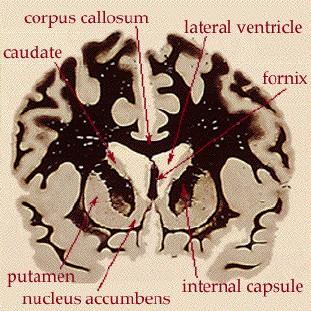
39 INTERNAL STRUCTURE BASAL GANGLIA : consists Corpus striatum Lentiform nucleus( putamen & globus pallidus) Caudate nucleus Claustrum Amygdaloid body Function Regulate voluntary movement Integrative or just a relay station? Pathology Movement disorders (e.g. Parkinson s) Neostriatum: caudate + putamen Paleostriatum: globus
40
41 The limbic system has three functions: It mediates the sense of smell It mediates emotional behaviour It plays an important role in memory. Structures on medial and basal surfaces of cerebral hemispheres Cingulate gyrus + parahippocampal gyrus + hippocampal formation + fornix + amygdala + septum + mammillary bodies Limbic system include the amygdala, the hippocampus and the fornix Recall that the amygdala is for emotion regulation,and perception of odor Hippocampus and fornix are important for learning and memory

42 Cingulate gyrus Anterior nucleus of thalamus Thalamus Para-olfactory area Fornix Mamillary bodies of hypothalamus Hypothalamus Hippocampus Uncus Amygdala Para-hippocampal gyrus


43

 -Form partitions")
44 Three connective tissue membranes external to the CNS dura mater, arachnoid mater, and pia mater Functions: -Cover and protect the CNS -Protect blood vessels and enclose venous sinuses -Contain cerebrospinal fluid (CSF) -Form partitions within the skull


45 BLOOD SUPPLY OF BRAIN & SPINAL CORD Brain recive the blood from vertebral artery and internal carotid artery Vertebral Deep cervical Ascending cervical Transverse cervical Inf. thyroid Thyrocervical Suprascapular Subclavian

46 Blood supply to brain Brain is supplied by: Internal carotid Aa. Basilar A. (formed by fusion of vertebral Aa.) Together they contribute to the Circle of Willis that sends branches to supply cerebral hemispheres
47

48 Venous Drainage Internal Jugular Brain External Jugular Scalp, face Vertebral cervical vertebrae, spinal cord, neck muscles Dural sinuses Veins in brain to Internal Jugular
49 Ventricles of the Brain Arise from expansion of the lumen of the neural tube The ventricles are: The paired C-shaped lateral ventricles The third ventricle found in the diencephalon The fourth ventricle found in the hindbrain dorsal to the pons

50 Ventricles of the Brain Figure 12.5

51

52 Flow of CSF Choroid plexus Ventricles Subarachnoid space through lateral and median apertures of 4th ventricle Blood of dural sinuses via arachnoid villi
53 There are four cavities in the brain, called ventricles. The ventricles are filled with cerebrospinal fluid (CSF), which provides the following functions: Absorbs physical shocks to the brain Distributes nutritive materials to and removes wastes from nervous tissue Provides a chemically stable environment There are four ventricles: Each of two lateral ventricles occupies a cerebral hemisphere The third ventricle is connected by a passage (interventricular foramen) to each of the two lateral ventricles The fourth ventricle connects to the third ventricle (via the cerebral aqueduct) and to the central canal of the spinal cord (a narrow, central tube extending the length of the spinal cord). Additional openings in the fourth ventricle also allow CSF to flow into the subarachnoid space.

54 Blood-Brain Barrier (BBB) Selective Permeability: Substances that can pass include oxygen, glucose, carbon dioxide, alcohol, anesthetics & water Substances that can not pass include medications such as many antibiotics & systemic chemotherapy agents. Brain capillary endothelium less permeable, tightly joined Toxins cannot penetrate O2 & glucose pass through Lipid soluble substances pass

55 Major Functions 1. Sensory perception 2. Voluntary control of movement 3. Language 4. Personality traits 5. Sophisticated mental events, such as thinking memory, decision making, creativity, and self-consciousness 1. Inhibition of muscle tone 2. Coordination of slow, sustained movements 3. Suppression of useless patterns of movements 1. Relay station for all synaptic input 2. Crude awareness of sensation 3. Some degree of consciousness 4. Role in motor control 1. Regulation of many homeostatic functions, such as temperature control, thirst, urine output, and food intake 2. Important link between nervous and endocrine systems 3. Extensive involvement with emotion and basic behavioral patterns 1. Maintenance of balance 2. Enhancement of muscle tone 3. Coordination and planning of skilled voluntary muscle activity 1. Origin of majority of peripheral cranial nerves 2. Cardiovascular, repiratory, and digestive control centers 3. Regulation of muscle reflexes involved with equilibrium and posture 4. Reception and intergration of all synaptic input from spinal cord; arousal and activation of cerebral cortex 5. Role in sleep-wake cycle Brain component Cerebral cortex Basal nuclei Thalamus Hypothalamus Cerebellum Brain stem (midbrain, pons, and medulla)

56
Anatomy and Physiology (Bio 220) The Brain Chapter 14 and select portions of Chapter 16
 The Brain Chapter 14 and select portions of Chapter 16") Anatomy and Physiology (Bio 220) The Brain Chapter 14 and select portions of Chapter 16 I. Introduction A. Appearance 1. physical 2. weight 3. relative weight B. Major parts of the brain 1. cerebrum 2.
Anatomy and Physiology (Bio 220) The Brain Chapter 14 and select portions of Chapter 16 I. Introduction A. Appearance 1. physical 2. weight 3. relative weight B. Major parts of the brain 1. cerebrum 2.
Chapter 18: The Brain & Cranial Nerves. Origin of the Brain
 Chapter 18: The Brain & Cranial Nerves BIO 218 Fall 2015 Origin of the Brain The brain originates from a structure called the neural tube, which arises during a developmental stage called neurulation.
BRAIN PART I (A & B): VENTRICLES & MENINGES
: VENTRICLES & MENINGES") BRAIN PART I (A & B): VENTRICLES & MENINGES Cranial Meninges Cranial meninges are continuous with spinal meninges Dura mater: inner layer (meningeal layer) outer layer (endosteal layer) fused to periosteum
BRAIN PART I (A & B): VENTRICLES & MENINGES Cranial Meninges Cranial meninges are continuous with spinal meninges Dura mater: inner layer (meningeal layer) outer layer (endosteal layer) fused to periosteum
CEREBRUM Dr. Jamila Elmedany Dr. Essam Eldin Salama
 CEREBRUM Dr. Jamila Elmedany Dr. Essam Eldin Salama Objectives At the end of the lecture, the student should be able to: List the parts of the cerebral hemisphere (cortex, medulla, basal nuclei, lateral
b. The groove between the two crests is called 2. The neural folds move toward each other & the fuse to create a
 Chapter 13: Brain and Cranial Nerves I. Development of the CNS A. The CNS begins as a flat plate called the B. The process proceeds as: 1. The lateral sides of the become elevated as waves called a. The
Biological Bases of Behavior. 3: Structure of the Nervous System
 Biological Bases of Behavior 3: Structure of the Nervous System Neuroanatomy Terms The neuraxis is an imaginary line drawn through the spinal cord up to the front of the brain Anatomical directions are
Chapter 3. Structure and Function of the Nervous System. Copyright (c) Allyn and Bacon 2004
 Allyn and Bacon 2004") Chapter 3 Structure and Function of the Nervous System 1 Basic Features of the Nervous System Neuraxis: An imaginary line drawn through the center of the length of the central nervous system, from the
Chapter 3 Structure and Function of the Nervous System 1 Basic Features of the Nervous System Neuraxis: An imaginary line drawn through the center of the length of the central nervous system, from the
M555 Medical Neuroscience Lab 1: Gross Anatomy of Brain, Crainal Nerves and Cerebral Blood Vessels
 M555 Medical Neuroscience Lab 1: Gross Anatomy of Brain, Crainal Nerves and Cerebral Blood Vessels Anatomical Directions Terms like dorsal, ventral, and posterior provide a means of locating structures
Telencephalon (Cerebral Hemisphere)
") Telencephalon (Cerebral Hemisphere) OUTLINE The Cortex - Lobes, Sulci & Gyri - Functional Subdivisions - Limbic Lobe & Limbic System The Subcortex - Basal Ganglia - White Matter (Internal Capsule) - Relations
Telencephalon (Cerebral Hemisphere) OUTLINE The Cortex - Lobes, Sulci & Gyri - Functional Subdivisions - Limbic Lobe & Limbic System The Subcortex - Basal Ganglia - White Matter (Internal Capsule) - Relations
Homework Week 2. PreLab 2 HW #2 Synapses (Page 1 in the HW Section)
") Homework Week 2 Due in Lab PreLab 2 HW #2 Synapses (Page 1 in the HW Section) Reminders No class next Monday Quiz 1 is @ 5:30pm on Tuesday, 1/22/13 Study guide posted under Study Aids section of website
Homework Week 2 Due in Lab PreLab 2 HW #2 Synapses (Page 1 in the HW Section) Reminders No class next Monday Quiz 1 is @ 5:30pm on Tuesday, 1/22/13 Study guide posted under Study Aids section of website
CEREBRUM. Dr. Jamila EL Medany
 CEREBRUM Dr. Jamila EL Medany Objectives At the end of the lecture, the student should be able to: List the parts of the cerebral hemisphere (cortex, medulla, basal nuclei, lateral ventricle). Describe
Organization of The Nervous System PROF. SAEED ABUEL MAKAREM
 Organization of The Nervous System PROF. SAEED ABUEL MAKAREM Objectives By the end of the lecture, you should be able to: List the parts of the nervous system. List the function of the nervous system.
The Brain. Brain. Spinal Cord. Cauda Equina
 The Brain Brain Spinal Cord Cauda Equina The Brain Ventricles- cavities in the brain filled with cerebrospinal fluid connected to the subarachnoid space- fluid filled space surrounding the brain Brain
The Nervous System PART B
 7 The Nervous System PART B PowerPoint Lecture Slide Presentation by Jerry L. Cook, Sam Houston University ESSENTIALS OF HUMAN ANATOMY & PHYSIOLOGY EIGHTH EDITION ELAINE N. MARIEB The Reflex Arc Reflex
Principles of Anatomy and Physiology
 Principles of Anatomy and Physiology 14 th Edition CHAPTER 14 The Brain and Cranial Nerves Introduction The purpose of the chapter is to: 1. Understand how the brain is organized, protected, and supplied
Blood supply to the brain Blood brain barrier isolates neural tissue from general circulation
 The Brain and Cranial Nerves Objectives Name the major regions of the brain and describe their functions. Discuss the formation, circulation, and functions of the CSF. List the main components of the medulla
Central Nervous System (CNS) -> brain and spinal cord. Major Divisions of the nervous system:
 -> brain and spinal cord. Major Divisions of the nervous system:") Central Nervous System (CNS) -> brain and spinal cord Major Divisions of the nervous system: Afferent (sensory input) -> cell bodies outside of the central nervous system (CNS), carry info into the CNS
Central Nervous System (CNS) -> brain and spinal cord Major Divisions of the nervous system: Afferent (sensory input) -> cell bodies outside of the central nervous system (CNS), carry info into the CNS
Psyc 311A, fall 2008 Conference week 3 TA: Jürgen Germann
 Psyc 311A, fall 2008 Conference week 3 TA: Jürgen Germann e-mail: jurgen.germann@mcgill.ca Overview: 1. Meninges 2. Cerebral cortex-cytoarchitecture 3. Diencephalon (thalamus/hypothalamus) (this replaces
Unit 12a: The Nervous System The Brain. MDL231 Principle of Anatomy
 Unit 12a: The Nervous System The Brain MDL231 Principle of Anatomy The Brain - Overview Cerebrum T PP H midbrain Cerebellum pons m.o. Brain stem medulla oblongata (M.O.) pons midbrain (mesencephalon) Diencephalon
Lecture - Chapter 13: Central Nervous System
 Lecture - Chapter 13: Central Nervous System 1. Describe the following structures of the brain, what is the general function of each: a. Cerebrum b. Diencephalon c. Brain Stem d. Cerebellum 2. What structures
meninges Outermost layer of the meninge dura mater arachnoid mater pia mater membranes located between bone and soft tissue of the nervous system
 membranes located between bone and soft tissue of the nervous system meninges Outermost layer of the meninge dura mater middle layer of the meninges, contains no blood vessels arachnoid mater Innermost
PROPERTY OF ELSEVIER SAMPLE CONTENT - NOT FINAL. Gross Anatomy and General Organization of the Central Nervous System
 3 Gross Anatomy and General Organization of the Central Nervous System C h a p t e r O u t l i n e The Long Axis of the CNS Bends at the Cephalic Flexure Hemisecting a Brain Reveals Parts of the Diencephalon,
The Central Nervous System I. Chapter 12
 The Central Nervous System I Chapter 12 The Central Nervous System The Brain and Spinal Cord Contained within the Axial Skeleton Brain Regions and Organization Medical Scheme (4 regions) 1. Cerebral Hemispheres
Neurology study of the nervous system. nervous & endocrine systems work together to maintain homeostasis
 Nervous System Neurology study of the nervous system nervous & endocrine systems work together to maintain homeostasis Nervous System works very fast Uses electrical signals called nerve impulses Short-lived
If I Only Had a Brain
 If I Only Had a Brain A Heart. (The Nerve!) Regions of the Brain Cerebral hemisphere Diencephalon Cerebellum (b) Adult brain Brain stem Regions of the Brain: Cerebrum Precentral gyrus Frontal lobe Central
Human Anatomy. Brain and Cranial Nerves
 Human Anatomy Brain and Cranial Nerves 1 Brain and Cranial Nerves An adult brain weighs between 1.35 and 1.4 kilograms (kg) (around 3 pounds) and has a volume of about 1200 cubic centimeters (cc). Brain
CNS consists of brain and spinal cord Cephalization Evolutionary development of rostral (anterior) portion of CNS Increased number of neurons in head
 portion of CNS Increased number of neurons in head") CNS consists of brain and spinal cord Cephalization Evolutionary development of rostral (anterior) portion of CNS Increased number of neurons in head Highest level reached in human brain 1 Mostly to orient
CNS consists of brain and spinal cord Cephalization Evolutionary development of rostral (anterior) portion of CNS Increased number of neurons in head Highest level reached in human brain 1 Mostly to orient
Anatomy Lab (1) Theoretical Part. Page (2 A) Page (2B)
 Theoretical Part. Page (2 A) Page (2B)") Anatomy Lab (1) This sheet only includes the extra notes for the lab handout regarding the theoretical part, as for the practical part it includes everything the doctor mentioned. Theoretical Part Page
Anatomy Lab (1) This sheet only includes the extra notes for the lab handout regarding the theoretical part, as for the practical part it includes everything the doctor mentioned. Theoretical Part Page
The neurvous system senses, interprets, and responds to changes in the environment. Two types of cells makes this possible:
 NERVOUS SYSTEM The neurvous system senses, interprets, and responds to changes in the environment. Two types of cells makes this possible: the neuron and the supporting cells ("glial cells"). Neuron Neurons
Organization of The Nervous System PROF. MOUSAED ALFAYEZ & DR. SANAA ALSHAARAWY
 Organization of The Nervous System PROF. MOUSAED ALFAYEZ & DR. SANAA ALSHAARAWY Objectives At the end of the lecture, the students should be able to: List the parts of the nervous system. List the function
Chapter 13 Brain and Cranial Nerves
 Chapter 13 Brain and Cranial Nerves 13-1 Brain and Cranial Nerves Brain Part of CNS contained in cranial cavity Control center for many of body s functions Much like a complex computer but more Parts of
Ch 13: Central Nervous System Part 1: The Brain p 374
 Ch 13: Central Nervous System Part 1: The Brain p 374 Discuss the organization of the brain, including the major structures and how they relate to one another! Review the meninges of the spinal cord and
Blood Supply of the CNS
 Blood Supply of the CNS Lecture Objectives Describe the four arteries supplying the CNS. Follow up each artery to its destination. Describe the circle of Willis and its branches. Discuss the principle
The Nervous system is divided into 2 major divisions: 1) Central Nervous System (CNS): found within bones & consists of:
 Central Nervous System (CNS): found within bones & consists of:") The Nervous system is divided into 2 major divisions: 1) Central Nervous System (CNS): found within bones & consists of: - The Brain: within the skull, composed of cerebrum, cerebellum and brain stem.
The Nervous system is divided into 2 major divisions: 1) Central Nervous System (CNS): found within bones & consists of: - The Brain: within the skull, composed of cerebrum, cerebellum and brain stem.
Unit Three. The brain includes: cerebrum, diencephalon, brain stem, & cerebellum. The brain lies within the cranial cavity of the skull.
 Human Anatomy & Physiology 11 Divisions of the Nervous System Karen W. Smith, Instructor Unit Three BRAIN & SPINAL CORD Refer to the following URLs. Be sure to study these along with your book. http://www.sirinet.net/~jgjohnso/nervous.html
stored information, making decisions, and taking action. 1. It is also the center for intellect, emotions, behavior, and memory.
 Chapter 14 - Outline I. INTRODUCTION A. The brain is the center for registering sensations, correlating them with one another and with stored information, making decisions, and taking action. 1. It is
The Brain and Cranial Nerves Pg Three Main Regions of the Brain. Forebrain
 The Brain and Cranial Nerves Pg. 129 Three Main Regions of the Brain Forebrain Cerbral hemispheres Diencephalon Midbrain Brain stem Hindbrain Pons Cerebellum Medulla oblongata Interprets sensory inputs
Brain ميهاربا لض اف دمح ا د The Meninges 1- Dura Mater of the Brain endosteal layer does not extend meningeal layer falx cerebri tentorium cerebelli
 .احمد د فاضل ابراهيم Lecture 15 Brain The Meninges Three protective membranes or meninges surround the brain in the skull: the dura mater, the arachnoid mater, and the pia mater 1- Dura Mater of the Brain
a) Central sulcus- shallow groove that runs across brain sagitally
 Central sulcus- shallow groove that runs across brain sagitally") KEY BRAIN Brain Gross Anatomy Terms 1) Explain each of the following in terms of structure of the brain a) Central sulcus- shallow groove that runs across brain sagitally b) Lateral fissure- deep groove
KEY BRAIN Brain Gross Anatomy Terms 1) Explain each of the following in terms of structure of the brain a) Central sulcus- shallow groove that runs across brain sagitally b) Lateral fissure- deep groove
The Brain and Cranial Nerves Pg. 129
 The Brain and Cranial Nerves Pg. 129 Three Main Regions of the Brain Forebrain Cerbral hemispheres Diencephalon Midbrain Brain stem Hindbrain Pons Cerebellum Medulla oblongata Forebrain Interprets sensory
Neuroanatomy lecture (1)
") Neuroanatomy lecture (1) Introduction: Neuroanatomy has two parts: the central and peripheral nervous system. The central nervous system is composed of brain and spinal cord. The brain has the following
Neuroanatomy lecture (1) Introduction: Neuroanatomy has two parts: the central and peripheral nervous system. The central nervous system is composed of brain and spinal cord. The brain has the following
The Nervous System. Divisions of the Nervous System. Branches of the Autonomic Nervous System. Central versus Peripheral
 The Nervous System Divisions of the Nervous System Central versus Peripheral Central Brain and spinal cord Peripheral Everything else Somatic versus Autonomic Somatic Nerves serving conscious sensations
I. Anatomy of the Brain A. Cranial Meninges and Ventricles of the Brain 1. Meninges a. Dura mater 1) Endosteal/Periosteal Layer - Outer 2) Meningeal
 Endosteal/Periosteal Layer - Outer 2) Meningeal") I. Anatomy of the Brain A. Cranial Meninges and Ventricles of the Brain 1. Meninges a. Dura mater 1) Endosteal/Periosteal Layer - Outer 2) Meningeal Layer - Inner 3) Falx cerebri a) Superior sagittal sinus
I. Anatomy of the Brain A. Cranial Meninges and Ventricles of the Brain 1. Meninges a. Dura mater 1) Endosteal/Periosteal Layer - Outer 2) Meningeal Layer - Inner 3) Falx cerebri a) Superior sagittal sinus
The Nervous System PART B
 7 The Nervous System PART B PowerPoint Lecture Slide Presentation by Jerry L. Cook, Sam Houston University ESSENTIALS OF HUMAN ANATOMY & PHYSIOLOGY EIGHTH EDITION ELAINE N. MARIEB Central Nervous System
Medical Neuroscience Tutorial Notes
 Medical Neuroscience Tutorial Notes Blood Supply to the Brain MAP TO NEUROSCIENCE CORE CONCEPTS 1 NCC1. The brain is the body's most complex organ. LEARNING OBJECTIVES After study of the assigned learning
Chapter 14: The Brain and Cranial Nerves. Copyright 2009, John Wiley & Sons, Inc.
 Chapter 14: The Brain and Cranial Nerves Development of the Brain Three to four-week embryo: prosencephalon, mesencephalon and rhombencephalon. Five-week embryo: telencephalon (cerebrum), diencephalon
14 - Central Nervous System. The Brain Taft College Human Physiology
 14 - Central Nervous System The Brain Taft College Human Physiology Development of the Brain The brain begins as a simple tube, a neural tube. The tube or chamber (ventricle) is filled with cerebrospinal
Embryonic Brain Development
 Chapter 14 The Brain and Cranial Nerves Largest organ in the body? Brain functions in sensations, memory, emotions, decision making, behavior 19-1 19-2 Embryonic Brain Development Principal Parts of the
The Nervous System 7PART B. PowerPoint Lecture Slide Presentation by Patty Bostwick-Taylor, Florence-Darlington Technical College
 PowerPoint Lecture Slide Presentation by Patty Bostwick-Taylor, Florence-Darlington Technical College The Nervous System 7PART B What is a reflex? What is a reflex? What is meant by the statement that
PSY 302: CHAPTER 3 NOTES THE BRAIN (PART II) - 9/5/17. By: Joseline
 - 9/5/17. By: Joseline") PSY 302: CHAPTER 3 NOTES THE BRAIN (PART II) - 9/5/17 By: Joseline Left 3 MAJOR FISSURES : 2HEMISPHERES Right Lateral Ventricle Central Fissure Third Ventricle Sulcus Lateral Fissure Gyros Fissure- Fissures
PSY 302: CHAPTER 3 NOTES THE BRAIN (PART II) - 9/5/17 By: Joseline Left 3 MAJOR FISSURES : 2HEMISPHERES Right Lateral Ventricle Central Fissure Third Ventricle Sulcus Lateral Fissure Gyros Fissure- Fissures
Announcement. Danny to schedule a time if you are interested.
 Announcement If you need more experiments to participate in, contact Danny Sanchez (dsanchez@ucsd.edu) make sure to tell him that you are from LIGN171, so he will let me know about your credit (1 point).
THE CENTRAL NERVOUS SYSTEM. The Brain & Spinal Cord
 THE CENTRAL NERVOUS SYSTEM The Brain & Spinal Cord Review: Nervous System Parallel Distributed Processing Composition of the CNS Nuclei: Clusters of neurons in the CNS ( neighborhoods ) Fiber Tracts/Pathways:
Sheep Brain Dissection
 Sheep Brain Dissection Mammalian brains have many features in common. Human brains may not be available, so sheep brains often are dissected as an aid to understanding the mammalian brain since he general
CEREBRUM & CEREBRAL CORTEX
 CEREBRUM & CEREBRAL CORTEX Seonghan Kim Dept. of Anatomy Inje University, College of Medicine THE BRAIN ANATOMICAL REGIONS A. Cerebrum B. Diencephalon Thalamus Hypothalamus C. Brain Stem Midbrain Pons
Chapter 13 Lecture Outline *
 Anatomy and Physiology, Seventh Edition Rod R. Seeley Idaho State University Trent D. Stephens Idaho State University Philip Tate Phoenix College Chapter 13 Lecture Outline * *See PowerPoint Image Slides
Introduction to the Central Nervous System: Internal Structure
 Introduction to the Central Nervous System: Internal Structure Objective To understand, in general terms, the internal organization of the brain and spinal cord. To understand the 3-dimensional organization
Instructor s Review for Final Exams. The Nervous System
 Instructor s Review for Final Exams The Nervous System Divisions of the Central Nervous System? Brain and spinal cord. Key word, central. Divisions of the nervous system Central and Peripheral Coverings
Department of Cognitive Science UCSD
 Department of Cognitive Science UCSD Verse 1: Neocortex, frontal lobe, Brain stem, brain stem, Hippocampus, neural node, Right hemisphere, Pons and cortex visual, Brain stem, brain stem, Sylvian fissure,
Somatic Nervous Systems. III. Autonomic Nervous System. Parasympathetic Nervous System. Sympathetic Nervous Systems
 7/21/2014 Outline Nervous System - PNS and CNS I. II. Two Parts of the Nervous System Central Nervous System vs Peripheral Nervous System Peripheral Nervous System A. B. Brain and Spinal Cord III. Autonomic
-Zeina Assaf. -Omar Odeh. - Maha Beltagy
 -3 -Zeina Assaf -Omar Odeh - Maha Beltagy 1 P a g e The Inferior Surface Of The Brain The inferior surface of the brain is divide by the stem of the lateral fissure into 2 parts : The orbital surface and
Prof. Saeed Abuel Makarem & Dr.Sanaa Alshaarawy
 Prof. Saeed Abuel Makarem & Dr.Sanaa Alshaarawy 1 Objectives By the end of the lecture, you should be able to: Describe the anatomy and main functions of the thalamus. Name and identify different nuclei
Gross Morphology of the Brain
 Gross Morphology of the Brain Done by : Marah Marahleh & Razan Krishan *slides in bold Principal Parts of the Brain Cerebrum : largest part of the brain Diencephalon Thalamus & hypothalamus Cerebellum
Developmental sequence of brain
 Cerebellum Developmental sequence of brain Fourth week Fifth week Location of cerebellum Lies above and behind the medullar and pons and occupies posterior cranial fossa Location of cerebellum External
Parts of the Brain. Hindbrain. Controls autonomic functions Breathing, Heartbeat, Blood pressure, Swallowing, Vomiting, etc. Upper part of hindbrain
 Parts of the Brain The human brain is made up of three main parts: 1) Hindbrain (or brainstem) Which is made up of: Myelencephalon Metencephalon 2) Midbrain Which is made up of: Mesencephalon 3) Forebrain
Chapter 12 The Central Nervous System Chapter Outline
 Chapter 12 The Central Nervous System Chapter Outline Module 12.1 Overview of the Central Nervous System (Figures 12.1, 12.2, 12.3) A. The central nervous system (CNS) includes the and, and is involved
Nervous System - PNS and CNS. Bio 105
 Nervous System - PNS and CNS Bio 105 Outline I. Central Nervous System vs Peripheral Nervous System II. Peripheral Nervous System A. Autonomic Nervous Systems B. Somatic Nervous Systems III. Autonomic
3/15/17. Outline. Nervous System - PNS and CNS. Two Parts of the Nervous System
 Nervous System - PNS and CNS Bio 105 Outline I. Central Nervous System vs Peripheral Nervous System II. Peripheral Nervous System A. Autonomic Nervous Systems B. Somatic Nervous Systems III. Autonomic
Central nervous system (CNS): brain and spinal cord Collections of cell body and dendrites (grey matter) are called nuclei/nucleus Nucleus can also
: brain and spinal cord Collections of cell body and dendrites (grey matter) are called nuclei/nucleus Nucleus can also") Chapter 3 Part 1 Orientation Directions in the nervous system are described relatively to the neuraxis An imaginary line drawn through the center of the length of the central nervous system, from the bottom
Chapter 3 Part 1 Orientation Directions in the nervous system are described relatively to the neuraxis An imaginary line drawn through the center of the length of the central nervous system, from the bottom
BIO 210 CHAPTER 13. The Central Nervous System SUPPLEMENT 2. PowerPoint by John McGill Supplemental Notes by Beth Wyatt CEREBELLUM
 BIO 210 CHAPTER 13 The Central Nervous System SUPPLEMENT 2 PowerPoint by John McGill Supplemental Notes by Beth Wyatt CEREBELLUM Second Largest Division of the Brain Lies Below the Posterior Portion of
ACTIVITY 7: NERVOUS SYSTEM HISTOLOGY, BRAIN, CRANIAL NERVES
 ACTIVITY 7: NERVOUS SYSTEM HISTOLOGY, BRAIN, CRANIAL NERVES LABORATORY OBJECTIVES: 1. Histology: Identify structures indicated on three different slides or images of nervous system tissue. These images
Cerebral hemisphere. Parietal Frontal Occipital Temporal
 Cerebral hemisphere Sulcus / Fissure Central Precental gyrus Postcentral gyrus Lateral (cerebral) Parieto-occipital Cerebral cortex Frontal lobe Parietal lobe Temporal lobe Insula Amygdala Hippocampus
NOTES CHAPTER 9 (Brief) The Nervous System LECTURE NOTES
 The Nervous System LECTURE NOTES") NOTES CHAPTER 9 (Brief) The Nervous System LECTURE NOTES I. Divisions of the Nervous System two major divisions A. Central Nervous System (CNS) 1. brain 2. spinal cord B. Peripheral Nervous System (PNS)
NOTES CHAPTER 9 (Brief) The Nervous System LECTURE NOTES I. Divisions of the Nervous System two major divisions A. Central Nervous System (CNS) 1. brain 2. spinal cord B. Peripheral Nervous System (PNS)
8.3 The Central Nervous System. SBI4U Ms. Ho-Lau
 8.3 The Central Nervous System SBI4U Ms. Ho-Lau The Central Nervous System the structural and functional centre for the entire nervous system the site of neural integration and processing The Central
MENTAL HOSPITAL PHONE MENU
 If you have low self-esteem, please hang up. Our operators are too busy to talk with you. MENTAL HOSPITAL PHONE MENU Hello and thank you for calling The State Mental Hospital. Please select from the following
Anatomy of the Human Brain
 Anatomy of the Human Brain Overview Lobes of the brain (Forebrain) Midbrain/Hindbrain Protection and Blood supply Structure and Function of a neuron Synaptic Transmission Neurotransmitters The brain Most
Divisions of the Nervous System
 Marieb s Human Anatomy and Physiology Marieb Hoehn Chapter 12 The Central Nervous System Lecture 19 1 Divisions of the Nervous System You are here CNS PNS 3 Brain Embryology & Overview Table & Figure From:
The Brain and Cranial Nerves Student Objectives
 The Brain and Cranial Nerves Student Objectives Chapter 14 Textbook and Laboratory Manual Name the major regions of the brain and describe their functions Name the ventricles of the brain and describe
Model 3-50B or 3-88 III VIII. Olfactory Nerve. Optic Nerve. Oculomotor Nerve. Trochlear Nerve. Trigeminal Nerve. Abducens Nerve.
 Model 3-50B or 3-88 I Olfactory Nerve II Optic Nerve Oculomotor Nerve III IV Trochlear Nerve Trigeminal Nerve V VI Abducens Nerve Glossopharyngeal Nerve IX VII Facial Nerve VIII Vestibocochlear Nerve or
Organization and Overview of the Central Nervous System CNS 424. By Prof. Hisham Al-Matubsi
 Organization and Overview of the Central Nervous System CNS 424 By Prof. Hisham Al-Matubsi Components Brain, spinal cord, nerves, sensory receptors Responsible for Sensory perceptions, mental activities,
Nervous System C H A P T E R 2
 Nervous System C H A P T E R 2 Input Output Neuron 3 Nerve cell Allows information to travel throughout the body to various destinations Receptive Segment Cell Body Dendrites: receive message Myelin sheath
Good Morning! Take out your notes and vocab 1-10! Copyright 2003 Pearson Education, Inc. publishing as Benjamin Cummings
 Good Morning! Take out your notes and vocab 1-10! Functions of the Nervous System 1. Sensory input gathering information To monitor changes occurring inside and outside the body (changes = stimuli) 2.
2/22/2012. Cerebrum CNS
 Chapter 8 outline CNS: Consists of???? Structural organization of the brain Cerebrum Diencephalon Midbrain and hindbrain Spinal cord tracts Cranial and spinal nerves Receives input from???? neurons Directs
Systems Neuroscience Dan Kiper. Today: Wolfger von der Behrens
 Systems Neuroscience Dan Kiper Today: Wolfger von der Behrens wolfger@ini.ethz.ch 18.9.2018 Neurons Pyramidal neuron by Santiago Ramón y Cajal (1852-1934, Nobel prize with Camillo Golgi in 1906) Neurons
Principles Arteries & Veins of the CNS LO14
 Principles Arteries & Veins of the CNS LO14 14. Identify (on cadaver specimens, models and diagrams) and name the principal arteries and veins of the CNS: Why is it important to understand blood supply
The Human Brain. I Think Therefore I am
 The Human Brain I Think Therefore I am The Beginning The simplest creatures have very simple nervous systems made up of nothing but a bunch of nerve cells They have neural nets, individual neurons linked
Bellringer: The central nervous system is comprised of: What is the name of the outermost layer of the brain? a. Brain. b.
 Bellringer: The central is comprised of: a. Brain b. Spinal cord c. Sensory receptors d. Both a and b What is the name of the outermost layer of the brain? a. Pia mater b. Dura mater c. Arachnoid d. Pons
Brain and Cranial Nerves (Ch. 15) Human Anatomy lecture. caudal = toward the spinal cord)
 Human Anatomy lecture. caudal = toward the spinal cord)") Insight: Some cranial nerve disorders Brain and Cranial Nerves (Ch. 15) Human Anatomy lecture I. Overview (Directional terms: rostral = toward the forehead caudal = toward the spinal cord) A. 3 Major parts
Insight: Some cranial nerve disorders Brain and Cranial Nerves (Ch. 15) Human Anatomy lecture I. Overview (Directional terms: rostral = toward the forehead caudal = toward the spinal cord) A. 3 Major parts
Protection of the Brain. Overview of the Brain. Visual Anatomy & Physiology First Edition. Martini & Ober. Chapter 13. Lecture 20
 Visual Anatomy & Physiology First Edition Martini & Ober Chapter 13 Brain and Cranial Nerves Lecture 20 1 Overview of the Brain Functions Major Parts regulates visceral activities cerebrum (two hemispheres)
Note to Self. The Brain and Cranial Nerves. Organization of the Brain
 Note to Self Despite what you think you can get through this chapter in 1 class period, or one with only a few slides left over The Brain and Cranial Nerves Chapter 14 Organization of the Brain The brain
Overview of Brain Structures
 First Overview of Brain Structures Psychology 470 Introduction to Chemical Additions Steven E. Meier, Ph.D. All parts are interrelated. You need all parts to function normally. Neurons = Nerve cells Listen
The Central Nervous System
 The Central Nervous System 1 The Central Nervous System Brain Spinal Cord Components 2 Protection of the Brain The brain is protected from injury by The skull enough said! You already know this. What else
Functional Organization of the Central Nervous System
 Functional Organization of the Central Nervous System Hierarchical orgnization CNS consists of the brain and the spinal cord The brain analyzes and interprets the information Response messages are
A recap of the Brain- Bio 230
 A recap of the Brain- Bio 230 This recap of the brain is to help you make sense of that 3 pound tofu blob that you carry around everyday. My hope is that if you get these basics, you can build and add
3/20/13. :: Slide 1 :: :: Slide 39 :: How Is the Nervous System Organized? Central Nervous System Peripheral Nervous System and Endocrine System
 :: Slide 1 :: :: Slide 39 :: How Is the Nervous System Organized? Central Nervous System Peripheral Nervous System and Endocrine System The nervous system is organized into several major branches, each
SHORT ANSWER. Write the word or phrase that best completes each statement or answers the question.
 Exam Name SHORT ANSWER. Write the word or phrase that best completes each statement or answers the question. Figure 12.3 Using Figure 12.3, match the following: 1) Site of efferent soma. 2) Site of axons
Nervous System: Part IV The Central Nervous System The Brain
 Nervous System: Part IV The Central Nervous System The Brain Can you survive when part of your brain is destroyed? 2 Essential Knowledge 3.D.2 2. Cells communicate with each other through direct contact
Outline of the next three lectures
 Outline of the next three lectures Lecture 35 Anatomy of the human cerebral cortex gross and microscopic cell types connections Vascular supply of the cerebral cortex Disorders involving the cerebral cortex
Student Lab #: Date. Lab: Gross Anatomy of Brain Sheep Brain Dissection Organ System: Nervous Subdivision: CNS (Central Nervous System)
") Lab: Gross Anatomy of Brain Sheep Brain Dissection Organ System: Nervous Subdivision: CNS (Central Nervous System) Student Lab #: Date 1 Objectives: 1. Learn the main components making up a motor neuron.
Lab: Gross Anatomy of Brain Sheep Brain Dissection Organ System: Nervous Subdivision: CNS (Central Nervous System) Student Lab #: Date 1 Objectives: 1. Learn the main components making up a motor neuron.
DISSECTION OF THE SHEEP'S BRAIN
 Sheep Brain Dissection Guide Page 1 DISSECTION OF THE SHEEP'S BRAIN Introduction The purpose of the sheep brain dissection is to familiarize you with the threedimensional structure of the brain and teach
CHAPTER 13&14: The Central Nervous System. Anatomy of the CNS
 CHAPTER 13&14: The Central Nervous System Anatomy of the CNS in human consists of brain and spinal cord as stated earlier neurons have little support from their extracellular matrix and depend on glial