Fundamentals of Nuclear Medicine Brain Imaging
|
|
|
- Hester York
- 6 years ago
- Views:
Transcription


1 Fundamentals of Nuclear Medicine Brain Imaging Nick Gulliver Chief Clinical Technologist, Department of Nuclear Medicine & PET-CT, King s College Hospital NHS Foundation Trust, London, UK EANM Technologist Committee (EANM-TC)


2
3 Overview Anatomy & Physiology Common Pathologies Caring for Patients in Imaging departments Positioning & Immobilisation Brain perfusion SPECT PET in neurological diseases PET in brain oncology imaging SPECT & PET in movement disorders PET/MR

4 Anatomy & Physiology Speech Reading Somatosensation Proprioception Olfactory Motor cortex Attention Speech Decisionmaking Vision memory Processing & interpreting sounds Balance Coordination Learning motor tasks Relays signals between brain and spinal cord
 Source: Gray s anatomy")
5 Cerebral blood supply Internal carotid & vertebral arteries provide oxygenated blood to brain Superficial & deep veins carry deoxygenated blood back to heart Complex arrangement of arteries to ensure continuous supply, Circle of Willis Occlusion leads to stroke (cerebrovascular accident CVA) Source: Gray s anatomy
 Grey")
 White matter")

6 Cerebral cortex Hills (gyrus) & valleys (sulcus) Grey matter mainly cell bodies (neurons, glia) White matter mainly axons, form major fibre bundles


7 Neuronal Communication Synaptic cleft Neurons communicate by action potential or release of neurochemicals which bind to receptors on next neuron lock and key system By Nrets (
8 Commonly seen pathologies in Nuclear Medicine Dementia e.g. Alzheimer s Movement disorders e.g. Parkinson s Epilepsy Brain infections & inflammation (encephalopathy) Brain tumours (primary & metastatic)

9 Dementia Graphic courtesy of Alzheimer s Disease International
10 Alzheimer s Disease Most common form of dementia Histopathologic analysis at autopsy is the standard of reference for the diagnosis of AD Neuronal loss due to aggregation of insoluble proteins: Amyloid β plaques Intracellular neurofibrillary tangles (consist of tau proteins) Graphic courtesy of IAEA
11 Other Dementia Types Dementia with Lewy Bodies (DLB) intracellular aggregations of a-synuclein related to Parkinson s disease Vascular Dementia (VD) Occurs because of brain injuries such as microscopic bleeding and blood vessel blockage Frontotemporal Dementia (FTD) Affects frontal and temporal lobes


12 Proteinopathies Neurofibrillary tangles Tau Protein Progressive Supranuclear Palsy Frontotemporal dementia Amyloid plaques b-amyloid Alzheimers Disease Lewy Bodies Dementia with Lewy Bodies a-synuclein Parkinsons Disease

13 Appropriate treatment depends on specific diagnosis use of expensive & ineffective medications avoided


14 Biomarker abnormality Neurodegenerative Process Time Jack et.al. Lancet Vol12, No. 2, Protein pathology process begins at least a decade before symptoms
15 Dementia Symptoms AD memory loss, depression, impaired communication, poor judgment, disorientation, behaviour changes and difficulty speaking and walking DLB - memory loss and thinking problems (like Alzheimer's) sleep disturbances, visual hallucinations, muscle rigidity or other parkinsonian movement features FTD - Typical symptoms include changes in personality and behaviour and difficulty with language. Patients often present with social impairment & disinhibited and impulsive behaviour


16 MMSE Score Dementia Common Symptoms of AD Any two people with dementia are unlikely to experience the condition in exactly the same way. Years from diagnosis DISEASE PROGRESSION
17 Patients with Parkinson s Disease Condition may vary hour to hour. Patient may appear fine at injection but not at scanning Common symptoms: Physical/motor symptoms - stiff rigid muscles, slow movements (bradykinesia), tremor, incontinence Cognitive symptoms - memory problems, hallucinations, difficulty completing day-to-day tasks, problems with reasoning Behavioural symptoms - behavioural inhibition, aggression, mood changes, problems with communicating
18 Patients with Parkinson s Disease Speak to patient and carer about how best to manage their condition within imaging department As Parkinson's disease progresses, it often results in a progressive dementia similar to Dementia with Lewy bodies or Alzheimer's All patients with dementia should be treated with dignity and respect whilst in hospital.


19 Preventing Motion Keep patients comfortable Blankets, straps (feelings of security) Patients with cognitive impairment, special support may be needed Reassure frequently Repeat instructions Get help from family/carer Sudden waking may cause more motion
 Use head holder to")

20 Standard brain positioning Patient supine, neck relaxed Brain in centre of field of view (FOV) Use head holder to reduce motion and improve comfort Use chin and forehead strap and wedges if required Laser position system if available
21 Brain imaging tracers For a tracer to be able to enter the brain it must cross blood brain barrier (BBB) Tracers divided into several groups: Regional cerebral blood flow (rcbf) tracers Metabolic tracers Tracers targeting neurotransmission and receptors Tracers targeting amyloid Tracers for brain tumour imaging
22 rcbf tracers rcbf tracers are used for measurement of perfusion of blood to brain Must be retained sufficiently in brain in their initial distribution sufficiently long to enable diagnostic imaging Two main tracers in EU: 99m Tc HMPAO 99m Tc-ECD Evaluation of cerebro-vascular disease, acute stroke, localisation of epileptogenic foci, traumatic brain injury, infection & inflammation, brain death
23 rcbf tracers 99m Tc-HMPAO hexamethyl propylene amine oxime, exametazime, Ceretec 99m Tc-ECD ethyl cysteine dimer, bicisate, Neurolite Brain images usually obtained 30-60min post-injection Diagnostic reference level = 750MBq, effective dose 6mSv EANM procedure guideline for brain perfusion SPECT using 99mTc-labelled radiopharmaceuticals (2009)

24 Brain HMPAO SPECT for AD Initial stage Hypoperfusion: parietal and/or posterior temporal cortex Unilateral or bilateral Intermediate stage Hypoperfusion: extensive, parietal, temporal, bilateral Advanced stage Diffuse cortical hypoperfusion Less/not affected: motor areas, occipital, basal ganglia, cerebellum

25 Brain Death HMPAO to determine complete absence of brain function Clinically diagnosed with legal standards Can also be used to determine the viability of internal organs for transplantation Hot nose sign absence of internal carotid artery flow increases external carotid artery flow, increases perfusion to nasal region Creative commons license: Case courtesy of Dr Andrew Dixon, Radiopaedia.org, rid: 19410
 Epilepsy (preoperative evaluation of foci) Movement disorders: differentiation between Parkinson s disease and other syndromes Also used in Neuro-oncology P neutrino + - positron")
26 18 F-FDG brain PET Alternative to HMPAO SPECT for brain perfusion imaging FDG PET superior to SPECT imaging for the early and differential diagnosis of Alzheimer s and frontotemporal dementia (FTD or Pick s disease) Epilepsy (preoperative evaluation of foci) Movement disorders: differentiation between Parkinson s disease and other syndromes Also used in Neuro-oncology P neutrino + - positron electron

27 18 F-FDG BLOOD TISSUE Glucose FDG Capillary Membrane FREE SPACE Glucose FDG CELLS Glucose-6- phosphate FDG-6- phosphate glycolysis FDG enters cell via GLUT proteins competing with glucose for hexokinase process, undergoes phosphorylation and trapped intraceullarly Tumours increase GLUT & hexokinase activity

28 18 F-FDG brain PET Healthy subjects Cerebral cortex Basal ganglia Thalamus Cerebellum Subcortical putamen, caudate nucleus, thalamus High uptake in the cortical grey matter Lower uptake in white matter ACG = anterior cingulate gyrus, CN = caudate nucleus, F = frontal lobe, O = occipital lobe, P = parietal lobe, PCG = posterior cingulate gyrus, PSMS = primary sensorimotor strip, Pu = putamen, T = temporal lobe, Th = thalamus. Brown et. al. RadioGraphics 2014; 34:
29 18 F-FDG brain PET Fast for at least 4h Patient should rest quietly with lights dimmed for 10 minutes prior to injection Check medication - psychotropic pharmaceuticals can influence regional metabolic rate Uptake in quiet dark room for minutes During uptake, the patient should remain silent 1 bed position - top of skull to base of brain (10 minute acquisition) Diagnostic reference level MBq (eff dose 5mSv) EANM procedure guidelines for PET brain imaging using FDG v2 (2009)
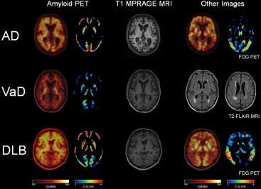
30 FDG in Neurology Clinical diagnosis for specific types of dementia (FTD, AD, DLB) with characteristic metabolic signatures Classic pattern of impaired metabolism - involvement of the posterior cingulate gyri, precuneus, and posterior temporal and parietal lobes Determine presence or absence of AD (vascular dementia has similar pattern) Differentiate DLB from AD


31 FDG in Neurology FDG PET is useful for imaging regional glucose consumption in the brain, where a pathologic change in neuronal activity is reflected by a corresponding decrease in glucose metabolism. Normal FDG PET findings in a patient undergoing evaluation for possible AD Broski et al RadioGraphics 2014; 34: Abnormal findings in a patient with known AD and progressive verbal difficulties (marked bilateral temporal hypometabolism)
 and interictal")
32 Epilepsy imaging HMPAO, ECD or FDG currently used clinically Dual role: Identify focal abnormalities in view of epilepsy surgery Explore mechanism of seizure onset Continuous EEG recording is required by telemetryexperienced staff Prepared syringes available to ensure quick injection time Ictal (immediately after seizure onset) and interictal scans
. Kumar et.al. J Nucl Med 2013;54:1775-1781")
33 Epilepsy imaging Interictal 99m Tc-ECD SPECT (left image) showing the usual hypoperfusion in presumed epileptogenic focus (arrow) in left frontal cortex region, which becomes hyperperfused during ictal SPECT (right image). Kumar et.al. J Nucl Med 2013;54:

34 Control AD b-amyloid imaging First amyloid tracer (2004): Carbon-11 labelled Pittsburgh compound B ( PiB ) short t½ (20min), widespread clinical use limited Selectively binds to amyloid plaque and cerebrovascular amyloid Significant retention seen in: >90% AD patients patients with mild cognitive impairment some normal elderly T1W-MRI PIB- PET Mathis et.al. J. Med. Chem., 2003, 46 (13), pp

 Flutemetamol (Vizamyl")


35 b-amyloid imaging Recognised need for 18 F amyloid tracer Development stages until st successful 18 F imaging in humans* Florbetapir (Amyvid ) Flutemetamol (Vizamyl ) Florbetaben (NeuraCeq )* All three 18 F current tracers approved by US FDA & EMA, all derived from 11 C-PiB for Mild Cognitive Impairment *Rowe et.al. Lancet Neurol. 2008;7:
 & grey matter (GM) Positive scan = GM uptake intense or >WM Rowe et.al.")
36 b-amyloid imaging Interpretation is independent of patient s clinical features and relies upon recognising unique image features Neg Pos Contrast between white matter (WM) & grey matter (GM) Positive scan = GM uptake intense or >WM Rowe et.al. J Nucl Med 2011;52:

37 b-amyloid imaging normal abnormal Courtesy of GE Healthcare
38 Neuro-Oncology imaging SPECT 99m Tc-HMPAO, 99m Tc-ECD 99m Tc-Sestamibi 201 Thallium chloride Amino acids (e.g. 123 I-methyl-tyrosine) PET Metabolic: 18 F-FDG, 15 O-water Amino acids: 11 C/ 18 F-Methionine, 18 F-DOPA 18 F-fluorotyrosine (FET) Cell proliferation: 18 F-fluorothymidine (FLT) Phospholipids: 11 C/ 18 F-Choline Tumour hypoxia: 18 F-MISO Somatastatin receptors: 68 Ga tracers
39 Metabolic PET brain imaging Main metabolic substrates in the brain are oxygen and glucose: 18 F-FDG (fluorodexyglucose) & 15 O (mainly used in research) High background uptake of FDG in normal brain limits its use in primary brain tumour imaging Paraneoplastic syndromes Common applications - tumour diagnosis, metabolic grading and prognosis, volume estimation and follow-up Not useful for assessing treatment response EANM FDG PET/CT: EANM procedure guidelines for tumour imaging: version 2.0 (2015); EANM procedure guidelines for PET brain imaging using [18F]FDG, version 2 (2009)


40 Metabolic PET brain imaging Standard brain positioning and technique as for brain perfusion FDG PET Suggested later imaging time for neurooncology (better tumour to normal tissue contrast) from 60min to several hours post injection Spence et.al. J Nucl Med. 2004;45: ) T1 MRI
41 PET Amino acid analogues 11 C- methyl-l-methionine 11 C-MET Drawback is 20min half-life of 11 C» requires on-site cyclotron Cellular amino acid uptake via overexpressed neurochemical transporters & increased metabolism 18 F-FET (fluorotyrosine) and 18 F-DOPA (fluorodopa) Can assess protein synthesis within malignant lesions Superior contrast to FDG Low uptake in normal brain More tumour specific not influenced by inflammation EANM Procedure Guidelines for Brain Tumour Imaging using Labelled Amino Acid Analogues (2006)
 37:1521 1528 Early imaging (i.e. 20 minutes post injection) for brain tumours (tumour uptake near maximum and early enough to avoid peak uptake in striatum)")
42 PET Amino acid analogues Detection of tumour tissue Tumour delineation & therapy planning Response assessment Selection of biopsy site FDG better for tumour grading Glioblastoma with markedly accumulating 18 F-FET in left temporal lobe Pichler et.al. Eur J Nucl Med Mol Imaging (2010) 37: Early imaging (i.e. 20 minutes post injection) for brain tumours (tumour uptake near maximum and early enough to avoid peak uptake in striatum)
 30:1561 1567 18 F-DOPA (A), shows an increased uptake in malignant glioblastoma, shown to be comparable")
43 18 F-DOPA in oncology Becherer A. et.al. Eur J Nucl Med Mol Imaging (2003) 30: F-DOPA (A), shows an increased uptake in malignant glioblastoma, shown to be comparable to 11 C-MET (B) lesions of matching size in temperoparietal, posterior basal ganglia and thalamus.

 11 C-choline or")

44 18 F-Choline Originally developed as 11 C-choline (a phospholipid component of cell membranes increased cellular metabolism choline uptake) 11 C-choline or 18 F- fluoro-methyl/ethyl choline 18 FDG 18 F Choline Courtesy of Charing Cross Hospital, London, UK


45 18 F-Choline Low concentration in normal cortex = excellent delineation of the tumour from normal brain Shows peri-tumoural uptake in high-grade gliomas Differentiate benign lesions from highgrade tumours and metastases T1 MR 18 F Choline increased uptake corresponding to the region of ring enhancement on T1 MRI, with increased peritumoral uptake extending anteriorly and contralaterally Kwee et.al. Radiology 2007; 244: 2
 Uptake in tumors is rapid, peaking at 5 10 min after injection and remaining")

46 18 F-FLT 39-deoxy-fluorothymidine ( 18 F-FLT) A marker of cell proliferation Assessment of grade and proliferation in gliomas Treatment response shown to correlate with histology markers (Ki-67) Uptake in tumors is rapid, peaking at 5 10 min after injection and remaining stable up to 75 min. Uptake of 11 C-MET and of 18 F-FLT in gliomas of low grade (top row) and high grade (bottom row). Uptake of 18 F-FLT is especially high in malignant glioma and also demonstrates infiltration into surrounding tissue Wolf-Dieter Heiss et al. JNM 2011;52:
 MRI (contrast-enhanced T1-weighted image) shows large area of contrast enhancement in right frontal lobe.")
47 18 F-FLT Wei Chen et al. J Nucl Med 2005;46: Newly diagnosed glioblastoma. (A) MRI (contrast-enhanced T1-weighted image) shows large area of contrast enhancement in right frontal lobe. Both 18 F-FDG PET (B) and 18 F-FLT PET (C) show increased uptake in same area.
48 18 F-MISO 18 F-fluoromisonidazole ( 18 F-FMISO) Evaluation of tissue hypoxia = relative resistance to radiation therapy Hypoxia predicts poor treatment response of malignant tumours Oxygen consumption is lowered in most brain tumours Greater 18 F-FMISO uptake is generally observed in high grade gliomas compared to low grade lesions 18 F-FMISO uptake associated with a decreased response to therapy and a worse prognosis
 Corresponding late 18 F-FMISO PET images show tumour hypoxia in low perfusion, in intermediate perfusion with an inverse pattern compared with")
49 18 F-MISO Bruehlmeier et.al. J Nucl Med 2004; 45: (A C) 15 O-H 2 O PET perfusion images in 3 patients with glioblastoma. (D F) Corresponding late 18 F-FMISO PET images show tumour hypoxia in low perfusion, in intermediate perfusion with an inverse pattern compared with hypoxia, and in high perfusion.
 68 Ga T½ = 68 minutes Requires 68 Ga generator onsite or near-site Medial temporal/sphenoidal meningioma (arrow) beneath pituitary gland (arrowhead) Henze et.")
50 Gallium-68 tracers Somatostatin receptor peptide imaging Some intracranial tumours express SSTRs 68 Ga-DOTATATE shows high binding affinity for receptor type 2 (SSTR2) DOTATOC/DOTANOC also available (different SSTRs) 68 Ga T½ = 68 minutes Requires 68 Ga generator onsite or near-site Medial temporal/sphenoidal meningioma (arrow) beneath pituitary gland (arrowhead) Henze et.al J Nucl Med 2001;42:
![Dopaminergic system Most common SPECT/PET tracers for mapping dopamine neurons: [ 123 I]FP-β-CIT [ 18 F]-DOPA Zhu et.al. Chem.Soc.](/docs-images/74/69743428/images/51-1.jpg "Rev (2014) 43: 6683-6691 Dopamine receptors divided into D1 & D2.")
51 Dopaminergic system Most common SPECT/PET tracers for mapping dopamine neurons: [ 123 I]FP-β-CIT [ 18 F]-DOPA Zhu et.al. Chem.Soc.Rev (2014) 43: Dopamine receptors divided into D1 & D2. Majority D2 located post-synaptically, most commonly 123I-IBZM SPECT and 18 F-fallypride PET
 from Alzheimer s disease Normal scan: crescent or")
52 123 I-DaTSCAN 123 I-Ioflupane manufactured by GE High binding affinity for presynaptic dopamine transporters in the striatal region of the brain Used to assess pre-synaptic striatal uptake in basal ganglia of brain Can differentiate Parkinsonian syndromes from essential tremor and Dementia with Lewy Bodies (DLB) from Alzheimer s disease Normal scan: crescent or comma

53 123 I-DaTSCAN Thyroid blocking required Potassium iodide or iodate tablets 100mg >1hr before (or Lugol s solution) DRL 185MBq Effective dose 4mSv Comes in referenced vial Imaging at 3-6 h p.i Imaging takes ~40 minutes EANM procedure guidelines for brain neurotransmission SPECT using 123I-labelled dopamine transporter ligands, version 2 (2009) Image Courtesy of GE

54 123 I-DaTSCAN Head motion Lateral shift has significant effect Striatum should appear bilaterally on same slice
 through caudate head that do not include putamen Correct")
 Covington et.al.")
55 123 I-DaTSCAN Mild sagittal tilt has little effect Severe sagittal motion can cause semicolon appearance Forward head tilt results in slices (red) through caudate head that do not include putamen Correct positioning - all slices through caudate head also include putamen Separate axial images of caudate heads and putamen (superimposed to illustrate semicolon appearance) Covington et.al. J Nucl Med Technol 2013; 41:

56 123 I-DaTSCAN Display raw data as cine loop to check for movement during acquisition Check for alignment and motion before patient leaves, repeat acquisition may be necessary Look out for kissing caudates Contingency measures required for repeat scans Images courtesy of Birmingham City Hospital
![18 F-DOPA 6-[ 18 F]-Fluorlevodopa: amino acid analogue which measures dopamine synthesis & neuron density Marker of presynaptic dopaminergic system Healthy control (left).](/docs-images/74/69743428/images/57-1.jpg "Patient with PD (right) - reduced uptake in right putamen and in posterior left putamen, uptake asymmetry between the heads of the two caudate nuclei Picco et.al.")
57 18 F-DOPA 6-[ 18 F]-Fluorlevodopa: amino acid analogue which measures dopamine synthesis & neuron density Marker of presynaptic dopaminergic system Healthy control (left). Patient with PD (right) - reduced uptake in right putamen and in posterior left putamen, uptake asymmetry between the heads of the two caudate nuclei Picco et.al. Eur J Nucl Med Mol Imaging (2015) 42: Useful for studies requiring repeated measures such as examinations of the course of a disease and the effect of treatment 4 hr fasting, imaging at 60-90min post injection

58 PET/MR in brain imaging
59 PET/MR challenges Different specs than conventional PET-CT Technical difficulties (e.g. attenuation correction) Staffing requires comprehensive understanding of both PET & MR Resource intensive, limited throughput ~70 scanners worldwide, very expensive Optimal applications & diagnostic medical benefits still being identified
60 Conclusions Vast number of tracers used in brain imaging Tracers are being used for diagnosis, tracking disease and measuring therapeutic effect in oncology Increasing used of PET tracers but there remains a strong clinical use for SPECT tracers Amyloid PET allows diagnosing AD before dementia (in future - tau PET?) PET/MR opening a whole new field

61 Thank you for your attention

62 All the staff in the Nuclear Medicine & PET-CT Department at King s College Hospital, London, UK BCNM/HSNM-MI organising committees EANM-TC Acknowledgements 1913
Introduction, use of imaging and current guidelines. John O Brien Professor of Old Age Psychiatry University of Cambridge
 Introduction, use of imaging and current guidelines John O Brien Professor of Old Age Psychiatry University of Cambridge Why do we undertake brain imaging in AD and other dementias? Exclude other causes
Nuclear neurology. Zámbó Katalin Department of Nuclear Medicine
 Nuclear neurology Zámbó Katalin Department of Nuclear Medicine To refresh your memory Brain has a high rate of oxidative metabolism. It has no reserves either of oxygen or of glucose and has a very limited
Yin-Hui Siow MD, FRCPC Director of Nuclear Medicine Southlake Regional Health Centre
 Yin-Hui Siow MD, FRCPC Director of Nuclear Medicine Southlake Regional Health Centre Today Introduction to CT Introduction to MRI Introduction to nuclear medicine Imaging the dementias The Brain ~ 1.5
Update on functional brain imaging in Movement Disorders
 Update on functional brain imaging in Movement Disorders Mario Masellis, MSc, MD, FRCPC, PhD Assistant Professor & Clinician-Scientist Sunnybrook Health Sciences Centre University of Toronto 53 rd CNSF
Facility IBA 18/9 Cyclotron Accelerates p and d. Facility GMP Grade Clean Room Automated Dispenser Synthera Rig FDG 07/11/2014
 Dr Chris Marshall Director Introduction to PET Positron Emission Tomography Positron is anti matter equivalent of the electron Isotopes that are proton rich can decay by emission of a positron Positron
Round table: Moderator; Fereshteh Sedaghat, MD, PhD Brain Mapping in Dementias and Non-invasive Neurostimulation
 Round table: Moderator; Fereshteh Sedaghat, MD, PhD Brain Mapping in Dementias and Non-invasive Neurostimulation 1. Reflection of Mild Cognitive Impairment (MCI) and Dementias by Molecular Imaging, PET
Neuro degenerative PET image from FDG, amyloid to Tau
 Neuro degenerative PET image from FDG, amyloid to Tau Kun Ju Lin ( ) MD, Ph.D Department of Nuclear Medicine and Molecular Imaging Center, Chang Gung Memorial Hospital ( ) Department of Medical Imaging
Brain imaging for the diagnosis of people with suspected dementia
 Why do we undertake brain imaging in dementia? Brain imaging for the diagnosis of people with suspected dementia Not just because guidelines tell us to! Exclude other causes for dementia Help confirm diagnosis
SPECT and PET Imaging: DaT Scan, Cerebral Blood Flow and Epilepsy
 SPECT and PET Imaging: DaT Scan, Cerebral Blood Flow and Epilepsy Dana Mathews Ph.D. M.D. Division of Nuclear Medicine Department of Radiology The University of Texas Southwestern Medical Center Financial
FDOPA, C11Choline, C11 Methionine. Dr K.G.Kallur
 FDOPA, C11Choline, C11 Methionine Dr K.G.Kallur Why? 11C Methionine scan Had undergone resection Earlier. Post op recurrent hypercalcemia C11 Methionine Unable to see in Sestamibi scan Brain Tumor After
NEUROIMAGING IN PANS/PANDAS
 NEUROIMAGING IN PANS/PANDAS Harry T. Chugani, M.D. Chief, Pediatric Neurology Nemours A.I. dupont Hospital for Children Wilmington, Delaware, USA Professor of Pediatrics and Neurology Thomas Jefferson
FDG-PET e parkinsonismi
 Parkinsonismi FDG-PET e parkinsonismi Valentina Berti Dipartimento di Scienze Biomediche, Sperimentali e Cliniche Sez. Medicina Nucleare Università degli Studi di Firenze History 140 PubMed: FDG AND parkinsonism
RADIOPHARMACEUTICALS FOR NEUROIMAGING. Prof. Cristina Maria Moriguchi Jeckel Brain Institute of Rio Grande do Sul, PUCRS
 RADIOPHARMACEUTICALS FOR NEUROIMAGING Prof. Cristina Maria Moriguchi Jeckel Brain Institute of Rio Grande do Sul, PUCRS Workshop on Quantitative SPECT and PET Brain Studies BRA6024 PUCRS, Porto Alegre,
Pathogenesis of Degenerative Diseases and Dementias. D r. Ali Eltayb ( U. of Omdurman. I ). M. Path (U. of Alexandria)
. M. Path (U. of Alexandria)") Pathogenesis of Degenerative Diseases and Dementias D r. Ali Eltayb ( U. of Omdurman. I ). M. Path (U. of Alexandria) Dementias Defined: as the development of memory impairment and other cognitive deficits
Pathogenesis of Degenerative Diseases and Dementias D r. Ali Eltayb ( U. of Omdurman. I ). M. Path (U. of Alexandria) Dementias Defined: as the development of memory impairment and other cognitive deficits
Imaging biomarkers for Parkinson s disease
 3 rd Congress of the European Academy of Neurology Amsterdam, The Netherlands, June 24 27, 2017 Teaching Course 6 MDS-ES/EAN: Neuroimaging in movement disorders - Level 2 Imaging biomarkers for Parkinson
Il ruolo di nuove tecniche di imaging per la diagnosi precoce di demenza
 Parma, 23 maggio 2017 Il ruolo di nuove tecniche di imaging per la diagnosi precoce di demenza Livia Ruffini SC Medicina Nucleare Azienda Ospedaliero-Universitaria di Parma PET AND SPECT STUDIES OF THE
Dementia. Stephen S. Flitman, MD Medical Director 21st Century Neurology
 Dementia Stephen S. Flitman, MD Medical Director 21st Century Neurology www.neurozone.org Dementia is a syndrome Progressive memory loss, plus Progressive loss of one or more cognitive functions: Language
Laura Tormoehlen, M.D. Neurology and EM-Toxicology Indiana University
 Laura Tormoehlen, M.D. Neurology and EM-Toxicology Indiana University Disclosures! No conflicts of interest to disclose Neuroimaging 101! Plain films! Computed tomography " Angiography " Perfusion! Magnetic
Improving diagnosis of Alzheimer s disease and lewy body dementia. Brain TLC October 2018
 Improving diagnosis of Alzheimer s disease and lewy body dementia Brain TLC October 2018 Plan for this discussion: Introduction to AD and LBD Why do we need to improve diagnosis? What progress has been
SCINTIGRAPHY OF THE CENTRAL NERVOUS SYSTEM Part 1: Introduction and BBB studies
 SCINTIGRAPHY OF THE CENTRAL NERVOUS SYSTEM Part 1: Introduction and BBB studies George N. Sfakianakis MD Professor of Radiology and Pediatrics Director, Division of Nuclear Medicine October 2009 FIRST
Brain Perfusion SPECT
 APPROVED BY: Director of Radiology Page 1 of 5 Brain Perfusion SPECT Primary Indications: Brain perfusion SPECT is most commonly performed (1) to aid in identification of the epileptogenic focus in patients
Brain SPECT has become an important diagnostic and
 CONTINUING EDUCATION Technical Overview of Brain SPECT Imaging: Improving Acquisition and Processing of Data* Gina N. Morano, CNMT, RT(N) 1 ; and John P. Seibyl, MD 2 1 Molecular NeuroImaging, LLC, New
Lecture 42: Final Review. Martin Wessendorf, Ph.D.
 Lecture 42: Final Review Martin Wessendorf, Ph.D. Lecture 33 cortex Heilbronner 5 lobes of the cortex Lateral view (left side) Mid-saggital view (right side) Cellular organization of cortex White matter
Announcement. Danny to schedule a time if you are interested.
 Announcement If you need more experiments to participate in, contact Danny Sanchez (dsanchez@ucsd.edu) make sure to tell him that you are from LIGN171, so he will let me know about your credit (1 point).
212 Index C-SB-13,
 Index A Acetylcholinesterase inhibitor, treatment, 15 Age-associated memory impairment (AAMI), 5 Alzheimer s disease (AD), 40, 95 96 apolipoprotein E genotype and risk for, 58 cellular neurodegeneration
Imaging of Alzheimer s Disease: State of the Art
 July 2015 Imaging of Alzheimer s Disease: State of the Art Neir Eshel, Harvard Medical School Year IV Outline Our patient Definition of dementia Alzheimer s disease Epidemiology Diagnosis Stages of progression
Optimized. clinical pathway. propels high utilization of PET/MR at Pitié-Salpêtrière Hospital
 Optimized propels high utilization of PET/MR at Pitié-Salpêtrière Hospital clinical pathway As one of Europe s largest teaching hospitals, Pitié-Salpêtrière Hospital is renowned for its innovative research
Dementia Update. October 1, 2013 Dylan Wint, M.D. Cleveland Clinic Lou Ruvo Center for Brain Health Las Vegas, Nevada
 Dementia Update October 1, 2013 Dylan Wint, M.D. Cleveland Clinic Lou Ruvo Center for Brain Health Las Vegas, Nevada Outline New concepts in Alzheimer disease Biomarkers and in vivo diagnosis Future trends
Medical Neuroscience Tutorial Notes
 Medical Neuroscience Tutorial Notes Blood Supply to the Brain MAP TO NEUROSCIENCE CORE CONCEPTS 1 NCC1. The brain is the body's most complex organ. LEARNING OBJECTIVES After study of the assigned learning
PET ligands and metabolic brain imaging Prof. Karl Herholz
 PET ligands Karl Herholz, University of Manchester PET images in this lecture, unless indicated otherwise, are from Max-Planck-Institute for Neurological Research, Cologne, Germany 1 Positron-Emission-Tomography
DISCLOSURES. Objectives. THE EPIDEMIC of 21 st Century. Clinical Assessment of Cognition: New & Emerging Tools for Diagnosing Dementia NONE TO REPORT
 Clinical Assessment of Cognition: New & Emerging Tools for Diagnosing Dementia DISCLOSURES NONE TO REPORT Freddi Segal Gidan, PA, PhD USC Keck School of Medicine Rancho/USC California Alzheimers Disease
! slow, progressive, permanent loss of neurologic function.
 UBC ! slow, progressive, permanent loss of neurologic function.! cause unknown.! sporadic, familial or inherited.! degeneration of specific brain region! clinical syndrome.! pathology: abnormal accumulation
III./3.1. Movement disorders with akinetic rigid symptoms
 III./3.1. Movement disorders with akinetic rigid symptoms III./3.1.1. Parkinson s disease Parkinson s disease (PD) is the second most common neurodegenerative disorder worldwide after Alzheimer s disease.
Principles Arteries & Veins of the CNS LO14
 Principles Arteries & Veins of the CNS LO14 14. Identify (on cadaver specimens, models and diagrams) and name the principal arteries and veins of the CNS: Why is it important to understand blood supply
The field is advancing rapidly and future applications of new tracers will also be discussed briefly.
 CME NUCLEAR MEDICINE Clinical Medicine 2012, Vol 12, No 4: 364 8 Edited by Val Lewington, professor of clinical therapeutic nuclear medicine, King's College London and honorary consultant in nuclear medicine,
Chapter 10 The Nervous System: The Brain and Cranial Nerves
 Chapter 10 The Nervous System: The Brain and Cranial Nerves Copyright 2015 Wolters Kluwer Health Lippincott Williams & Wilkins Overview Key Terms aphasia corpus callosum meninges basal nuclei diencephalon
SPECT Dopamin Transporter lmaging Agent
 SPECT Dopamin Transporter lmaging Agent Product Summary Product Name DATrace-123 Injection Active Ingredient Action Mechanism DATrace-123 is used as a radiopharmaceutical for the SPECT imaging of Dopamine
brain MRI for neuropsychiatrists: what do you need to know
 brain MRI for neuropsychiatrists: what do you need to know Christoforos Stoupis, MD, PhD Department of Radiology, Spital Maennedorf, Zurich & Inselspital, University of Bern, Switzerland c.stoupis@spitalmaennedorf.ch
What if it s not Alzheimer s? Update on Lewy body dementia and frontotemporal dementia
 What if it s not Alzheimer s? Update on Lewy body dementia and frontotemporal dementia Dementia: broad term for any acquired brain condition impairing mental function such that ADLs are impaired. Includes:
Form D1: Clinician Diagnosis
 Initial Visit Packet Form D: Clinician Diagnosis NACC Uniform Data Set (UDS) ADC name: Subject ID: Form date: / / Visit #: Examiner s initials: INSTRUCTIONS: This form is to be completed by the clinician.
Neuroimaging for dementia diagnosis. Guidance from the London Dementia Clinical Network
 Neuroimaging for dementia diagnosis Guidance from the London Dementia Clinical Network Authors Dr Stephen Orleans-Foli Consultant Psychiatrist, West London Mental Health NHS Trust Dr Jeremy Isaacs Consultant
MILD HYPERTHYROIDISM CAUSES A PSEUDO-ALZHEIMER S PATTERN OF CEREBRAL METABOLISM WHICH IS REVERSIBLE WITH CEREBRAL PERFUSION STIMULANTS
 MILD HYPERTHYROIDISM CAUSES A PSEUDO-ALZHEIMER S PATTERN OF CEREBRAL METABOLISM WHICH IS REVERSIBLE WITH CEREBRAL PERFUSION STIMULANTS Harold Thomas Pretorius, MD, PhD, and Nichole Richards Abstract Objective:
Differential Diagnosis of Alzheimer s Disease and Other Types of Dementia with Development of Neuroimaging Techniques (PET, SPECT, and MRI)
") www.jmscr.igmpublication.org Impact Factor 1.1147 ISSN (e)-2347-176x Differential Diagnosis of Alzheimer s Disease and Other Types of Dementia with Development of Neuroimaging Techniques (PET, SPECT, and
www.jmscr.igmpublication.org Impact Factor 1.1147 ISSN (e)-2347-176x Differential Diagnosis of Alzheimer s Disease and Other Types of Dementia with Development of Neuroimaging Techniques (PET, SPECT, and
Indications of PET/CT in oncology
 Monday, August 27, 2012 Session 1, 10:00-10:40 Indications of PET/CT in oncology Helle Westergren Hendel MD, PhD, assistant professor Bacelor in Leadership & Health Ecomomics Head of Clinical PET, Herlev
Imaging in epilepsy: Ictal perfusion SPECT and SISCOM
 Imaging in epilepsy: Ictal perfusion SPECT and SISCOM Patrick Dupont Laboratory for Cognitive Neurology Laboratory for Epilepsy Research Medical Imaging Research Center KU Leuven, Belgium E-mail: Patrick.Dupont@med.kuleuven.be
PET/MR:Techniques, Indications and Applications
 PET/MR:Techniques, Indications and Applications Franz Wolfgang Hirsch Professor and Head of the Department of Pediatric Radiology University Hospital Leipzig / Germany Children s Hospital University Leipzig
review of existing studies on ASL in dementia Marion Smits, MD PhD
 review of existing studies on ASL in dementia Marion Smits, MD PhD Associate Professor of Neuroradiology Department of Radiology, Erasmus MC, Rotterdam (NL) Alzheimer Centre South-West Netherlands, Rotterdam
FRONTOTEMPORAL DEGENERATION: OVERVIEW, TRENDS AND DEVELOPMENTS
 FRONTOTEMPORAL DEGENERATION: OVERVIEW, TRENDS AND DEVELOPMENTS Norman L. Foster, M.D. Director, Center for Alzheimer s Care, Imaging and Research Chief, Division of Cognitive Neurology, Department of Neurology
Dementia and Healthy Ageing : is the pathology any different?
 Dementia and Healthy Ageing : is the pathology any different? Professor David Mann, Professor of Neuropathology, University of Manchester, Hope Hospital, Salford DEMENTIA Loss of connectivity within association
10/3/2016. T1 Anatomical structures are clearly identified, white matter (which has a high fat content) appears bright.
 appears bright.") H2O -2 atoms of Hydrogen, 1 of Oxygen Hydrogen just has one single proton and orbited by one single electron Proton has a magnetic moment similar to the earths magnetic pole Also similar to earth in that
H2O -2 atoms of Hydrogen, 1 of Oxygen Hydrogen just has one single proton and orbited by one single electron Proton has a magnetic moment similar to the earths magnetic pole Also similar to earth in that
Diagnosis before NIA AA The impact of FDG PET in. Diagnosis after NIA AA Neuropathology and PET image 2015/10/16
 The impact of FDG PET in degenerative dementia diagnosis Jung Lung, Hsu MD, Ph.D (Utrecht) Section of dementia and cognitive impairment Department of Neurology Chang Gung Memorial Hospital, Linkou, Taipei
MRI and CT of the CNS
 MRI and CT of the CNS Dr.Maha ELBeltagy Assistant Professor of Anatomy Faculty of Medicine The University of Jordan 2018 Computed Tomography CT is used for the detection of intracranial lesions. CT relies
DEMENTIA? 45 Million. What is. WHAT IS DEMENTIA Dementia is a disturbance in a group of mental processes including: 70% Dementia is not a disease
 What is PRESENTS DEMENTIA? WHAT IS DEMENTIA Dementia is a disturbance in a group of mental processes including: Memory Reasoning Planning Learning Attention Language Perception Behavior AS OF 2013 There
Delirium & Dementia. Nicholas J. Silvestri, MD
 Delirium & Dementia Nicholas J. Silvestri, MD Outline Delirium vs. Dementia Neural pathways relating to consciousness Encephalopathy Stupor Coma Dementia Delirium vs. Dementia Delirium Abrupt onset Lasts
OBJECTIVES. At the end of the lecture, students should be able to: List the cerebral arteries.
 DR JAMILA EL MEDANY OBJECTIVES At the end of the lecture, students should be able to: List the cerebral arteries. Describe the cerebral arterial supply regarding the origin, distribution and branches.
New imaging techniques: let there be light. Felix M. Mottaghy Department of Nuclear Medicine University Hospital KU Leuven
 New imaging techniques: let there be light Felix M. Mottaghy Department of Nuclear Medicine University Hospital KU Leuven Medical imaging and the pathology cascade Molecular/Cellular disturbance Alterations
Principles of nuclear metabolic imaging. Prof. Dr. Alex Maes AZ Groeninge Kortrijk and KULeuven Belgium
 Principles of nuclear metabolic imaging Prof. Dr. Alex Maes AZ Groeninge Kortrijk and KULeuven Belgium I. Molecular imaging probes A. Introduction - Chemical disturbances will precede anatomical abnormalities
Body control systems. Let s start at the top: the human brain. The Cerebrum. The human brain. What parts of your brain are you using right now?
 What parts of your brain are you using right now? Body control systems Quick Sends message directly to target organ Endocrine system Frontal lobe Parietal lobe Movement and conscious thought; Frontal speech
PET-CT for radiotherapy planning in lung cancer: current recommendations and future directions
 PET-CT for radiotherapy planning in lung cancer: current recommendations and future directions Gerry Hanna Centre for Cancer Research and Cell Biology Queen s University of Belfast @gerryhanna Talk Outline
Parkinson e decadimento cognitivo. Stelvio Sestini
 Parkinson e decadimento cognitivo Stelvio Sestini Patients with PD can develop a spectrum of cognitive symptoms Heterogeneity of cognitive deficits The cognitive symptoms can evolve to dementia (Mov Disorder
Piramal Imaging to Present New Research in PET Imaging at Society of Nuclear Medicine and Molecular Imaging 2017 Annual Meeting
 FOR IMMEDIATE RELEASE Media Contacts: Nicole Fletcher Piramal Imaging nicole.fletcher@piramal.com (857) 202-1122 Piramal Imaging to Present New Research in PET Imaging at Society of Nuclear Medicine and
CEREBRAL BLOOD FLOW AND METABOLISM
 Supported by: HURO/0901/069/2.3.1 HU-RO-DOCS CEREBRAL BLOOD FLOW AND METABOLISM Part 3 Modern imaging methods SPECT, PET, nmri History of Nuclear Medicine Starts with the invention of the X-ray 1946: radioactive
They are updated regularly as new NICE guidance is published. To view the latest version of this NICE Pathway see:
 diagnosis and assessment bring together everything NICE says on a topic in an interactive flowchart. are interactive and designed to be used online. They are updated regularly as new NICE guidance is published.
The ABCs of Dementia Diagnosis
 The ABCs of Dementia Diagnosis Dr. Robin Heinrichs, Ph.D., ABPP Board Certified Clinical Neuropsychologist Associate Professor, Psychiatry & Behavioral Sciences Director of Neuropsychology Training What
EEG IN FOCAL ENCEPHALOPATHIES: CEREBROVASCULAR DISEASE, NEOPLASMS, AND INFECTIONS
 246 Figure 8.7: FIRDA. The patient has a history of nonspecific cognitive decline and multiple small WM changes on imaging. oligodendrocytic tumors of the cerebral hemispheres (11,12). Electroencephalogram
The human brain. of cognition need to make sense gives the structure of the brain (duh). ! What is the basic physiology of this organ?
. ! What is the basic physiology of this organ?") The human brain The human brain! What is the basic physiology of this organ?! Understanding the parts of this organ provides a hypothesis space for its function perhaps different parts perform different
The human brain The human brain! What is the basic physiology of this organ?! Understanding the parts of this organ provides a hypothesis space for its function perhaps different parts perform different
Cerebral Cortex 1. Sarah Heilbronner
 Cerebral Cortex 1 Sarah Heilbronner heilb028@umn.edu Want to meet? Coffee hour 10-11am Tuesday 11/27 Surdyk s Overview and organization of the cerebral cortex What is the cerebral cortex? Where is each
Outline of the next three lectures
 Outline of the next three lectures Lecture 35 Anatomy of the human cerebral cortex gross and microscopic cell types connections Vascular supply of the cerebral cortex Disorders involving the cerebral cortex
DIFFERENTIAL DIAGNOSIS SARAH MARRINAN
 Parkinson s Academy Registrar Masterclass Sheffield DIFFERENTIAL DIAGNOSIS SARAH MARRINAN 17 th September 2014 Objectives Importance of age in diagnosis Diagnostic challenges Brain Bank criteria Differential
Exam 2 PSYC Fall (2 points) Match a brain structure that is located closest to the following portions of the ventricular system
 Match a brain structure that is located closest to the following portions of the ventricular system") Exam 2 PSYC 2022 Fall 1998 (2 points) What 2 nuclei are collectively called the striatum? (2 points) Match a brain structure that is located closest to the following portions of the ventricular system
Exam 2 PSYC 2022 Fall 1998 (2 points) What 2 nuclei are collectively called the striatum? (2 points) Match a brain structure that is located closest to the following portions of the ventricular system
The central nervous system
 Sectc.qxd 29/06/99 09:42 Page 81 Section C The central nervous system CNS haemorrhage Subarachnoid haemorrhage Cerebral infarction Brain atrophy Ring enhancing lesions MRI of the pituitary Multiple sclerosis
NEURO IMAGING 2. Dr. Said Huwaijah Chairman of radiology Dep, Damascus Univercity
 NEURO IMAGING 2 Dr. Said Huwaijah Chairman of radiology Dep, Damascus Univercity I. EPIDURAL HEMATOMA (EDH) LOCATION Seventy to seventy-five percent occur in temporoparietal region. CAUSE Most likely caused
ASL Perfusion Imaging: Concepts and Applications
 ASL Perfusion Imaging: Concepts and Applications David C. Alsop, Ph.D. Beth Israel Deaconess Medical Center and Harvard Medical School, Boston USA INTRODUCTION Arterial Spin Labeling (ASL) perfusion imaging
Austin Radiological Association BRAIN AMYLOID STUDY (F-18-Florbetapir)
") Austin Radiological Association BRAIN AMYLOID STUDY (F-18-Florbetapir) Overview The Brain Amyloid Study with F-18-florbetapir depicts the extracellular deposition of B- amyloid (Aβ) peptides (or plaques
Austin Radiological Association BRAIN AMYLOID STUDY (F-18-Florbetapir) Overview The Brain Amyloid Study with F-18-florbetapir depicts the extracellular deposition of B- amyloid (Aβ) peptides (or plaques
CEREBRUM. Dr. Jamila EL Medany
 CEREBRUM Dr. Jamila EL Medany Objectives At the end of the lecture, the student should be able to: List the parts of the cerebral hemisphere (cortex, medulla, basal nuclei, lateral ventricle). Describe
Chapter 3. Structure and Function of the Nervous System. Copyright (c) Allyn and Bacon 2004
 Allyn and Bacon 2004") Chapter 3 Structure and Function of the Nervous System 1 Basic Features of the Nervous System Neuraxis: An imaginary line drawn through the center of the length of the central nervous system, from the
Chapter 3 Structure and Function of the Nervous System 1 Basic Features of the Nervous System Neuraxis: An imaginary line drawn through the center of the length of the central nervous system, from the
Applicable Neuroradiology
 For the Clinical Neurology Clerkship LSU Medical School New Orleans Amy W Voigt, MD Clerkship Director Introduction The field of Radiology first developed following the discovery of X-Rays by Wilhelm Roentgen
Cheyenne 11/28 Neurological Disorders II. Transmissible Spongiform Encephalopathy
 Cheyenne 11/28 Neurological Disorders II Transmissible Spongiform Encephalopathy -E.g Bovine4 Spongiform Encephalopathy (BSE= mad cow disease), Creutzfeldt-Jakob disease, scrapie (animal only) -Sporadic:
Parkinson s Disease in the Elderly A Physicians perspective. Dr John Coyle
 Parkinson s Disease in the Elderly A Physicians perspective Dr John Coyle Overview Introduction Epidemiology and aetiology Pathogenesis Diagnosis and clinical features Treatment Psychological issues/ non
Case reports functional imaging in epilepsy
 Seizure 2001; 10: 157 161 doi:10.1053/seiz.2001.0552, available online at http://www.idealibrary.com on Case reports functional imaging in epilepsy MARK P. RICHARDSON Medical Research Council Fellow, Institute
Corporate Medical Policy
 Corporate Medical Policy Dopamine Transporter Imaging with Single Photon Emission File Name: Origination: Last CAP Review: Next CAP Review: Last Review: dopamine_transporter_imaging_with_single_photon_emission_computed_tomography
Cerebro-vascular stroke
 Cerebro-vascular stroke CT Terminology Hypodense lesion = lesion of lower density than the normal brain tissue Hyperdense lesion = lesion of higher density than normal brain tissue Isodense lesion = lesion
Systems Neuroscience Dan Kiper. Today: Wolfger von der Behrens
 Systems Neuroscience Dan Kiper Today: Wolfger von der Behrens wolfger@ini.ethz.ch 18.9.2018 Neurons Pyramidal neuron by Santiago Ramón y Cajal (1852-1934, Nobel prize with Camillo Golgi in 1906) Neurons
The neurvous system senses, interprets, and responds to changes in the environment. Two types of cells makes this possible:
 NERVOUS SYSTEM The neurvous system senses, interprets, and responds to changes in the environment. Two types of cells makes this possible: the neuron and the supporting cells ("glial cells"). Neuron Neurons
Lesson 14. The Nervous System. Introduction to Life Processes - SCI 102 1
 Lesson 14 The Nervous System Introduction to Life Processes - SCI 102 1 Structures and Functions of Nerve Cells The nervous system has two principal cell types: Neurons (nerve cells) Glia The functions
Central nervous system
 Central nervous system By Dr. Mohsen Dashti Clinical Medicine & Pathology 316 7 th Lecture Lecture outline Review of structure & function. Symptoms, signs & tests. Specific diseases. Review of structure
PHYSIOLOGY of LIMBIC SYSTEM
 PHYSIOLOGY of LIMBIC SYSTEM By Dr. Mudassar Ali Roomi (MBBS, M.Phil.) Assistant Professor Physiology Limbic system: (shown in dark pink) Limbic = border Definition: limbic system means the entire neuronal
Organization of the nervous system. [See Fig. 48.1]
![Organization of the nervous system. [See Fig. 48.1]](/thumbs/90/103926552.jpg "Organization of the nervous system. [See Fig. 48.1]") Nervous System [Note: This is the text version of this lecture file. To make the lecture notes downloadable over a slow connection (e.g. modem) the figures have been replaced with figure numbers as found
Nervous System [Note: This is the text version of this lecture file. To make the lecture notes downloadable over a slow connection (e.g. modem) the figures have been replaced with figure numbers as found
Alzheimer's disease (AD), also known as Senile Dementia of the Alzheimer Type (SDAT) or simply Alzheimer s is the most common form of dementia.
, also known as Senile Dementia of the Alzheimer Type (SDAT) or simply Alzheimer s is the most common form of dementia.") CHAPTER 3 Alzheimer's disease (AD), also known as Senile Dementia of the Alzheimer Type (SDAT) or simply Alzheimer s is the most common form of dementia. This incurable, degenerative, terminal disease
CHAPTER 3 Alzheimer's disease (AD), also known as Senile Dementia of the Alzheimer Type (SDAT) or simply Alzheimer s is the most common form of dementia. This incurable, degenerative, terminal disease
Dementia: A Comprehensive Update Neuroimaging, CSF, and genetic biomarkers in dementia
 Dementia: A Comprehensive Update 2016 Neuroimaging, CSF, and genetic biomarkers in dementia Bradford C. Dickerson, M.D. Associate Professor of Neurology, Harvard Medical School Departments of Neurology
Motor Functions of Cerebral Cortex
 Motor Functions of Cerebral Cortex I: To list the functions of different cortical laminae II: To describe the four motor areas of the cerebral cortex. III: To discuss the functions and dysfunctions of
PSY 302: CHAPTER 3 NOTES THE BRAIN (PART II) - 9/5/17. By: Joseline
 - 9/5/17. By: Joseline") PSY 302: CHAPTER 3 NOTES THE BRAIN (PART II) - 9/5/17 By: Joseline Left 3 MAJOR FISSURES : 2HEMISPHERES Right Lateral Ventricle Central Fissure Third Ventricle Sulcus Lateral Fissure Gyros Fissure- Fissures
PSY 302: CHAPTER 3 NOTES THE BRAIN (PART II) - 9/5/17 By: Joseline Left 3 MAJOR FISSURES : 2HEMISPHERES Right Lateral Ventricle Central Fissure Third Ventricle Sulcus Lateral Fissure Gyros Fissure- Fissures
Overview. Overview. Parkinson s disease. Secondary Parkinsonism. Parkinsonism: Motor symptoms associated with impairment in basal ganglia circuits
 Overview Overview Parkinsonism: Motor symptoms associated with impairment in basal ganglia circuits The differential diagnosis of Parkinson s disease Primary vs. Secondary Parkinsonism Proteinopathies:
The Person: Dementia Basics
 The Person: Dementia Basics Objectives 1. Discuss how expected age related changes in the brain might affect an individual's cognition and functioning 2. Discuss how changes in the brain due to Alzheimer
Alzheimer s Disease without Dementia
 Alzheimer s Disease without Dementia Dr Emer MacSweeney CEO & Consultant Neuroradiologist Re:Cognition Health London Osteopathic Society 13 September 2016 Early diagnosis of Alzheimer s Disease How and
14 - Central Nervous System. The Brain Taft College Human Physiology
 14 - Central Nervous System The Brain Taft College Human Physiology Development of the Brain The brain begins as a simple tube, a neural tube. The tube or chamber (ventricle) is filled with cerebrospinal
EU Regulation of in vivo Diagnostics Regulatory Assessment of Diagnostic Agents. 2 nd Regulatory Workshop University of Pretoria 9 th October, 2014
 EU Regulation of in vivo Diagnostics Regulatory Assessment of Diagnostic Agents 2 nd Regulatory Workshop University of Pretoria 9 th October, 2014 Diagnostic Agents Any pharmaceutical product used as
Maximizing the Utility of Integrated PET/MRI in Clinical Applications
 Maximizing the Utility of Integrated PET/MRI in Clinical Applications Spencer Behr, MD Department of Nuclear Medicine & Abdominal Imaging University of California, San Francisco PET/MR at UCSF Device:
Non Alzheimer Dementias
 Non Alzheimer Dementias Randolph B Schiffer Department of Neuropsychiatry and Behavioral Science Texas Tech University Health Sciences Center 9/11/2007 Statement of Financial Disclosure Randolph B Schiffer,,
Synaptic changes in dementia: links to cognition and behaviour
 Synaptic changes in dementia: links to cognition and behaviour Paul T Francis, PhD Professor of Neurochemistry Director, Brains for Dementia Research Agenda Discuss synaptic changes in various dementias