Radiology Review Course Hotel del Coronado Coronado, California
|
|
|
- Victoria Barrett
- 5 years ago
- Views:
Transcription
1 37 th Annual Radiology Review Course Hotel del Coronado Coronado, California Saturday, April 22, AM
2 TABLE OF CONTENTS Saturday, April 22, AM 7:00 AM 7:30 AM Coffee and Pastries for Registrants 7:30 AM 8:35 AM Physics - Radiobiology Daniel E. Wessell, M.D., Ph.D. 8:35 AM 9:25 AM Case Review Session: Neuroradiology James Y. Chen, M.D. 9:25 AM 9:40 AM Coffee Break SAM Session - CT Physics 9:40 AM 10:45 AM Physics - Computed Tomography I Daniel E. Wessell, M.D., Ph.D. 10:45 AM 11:15 AM Case Review Session: Gastrointestinal Cynthia S. Santillan, M.D., FSAR 11:15 AM 12:20 PM Physics - Computed Tomography II Daniel E. Wessell, M.D., Ph.D. 12:20 PM 1:35 PM Lunch On Your Own SAVE THE DATE UCSD Radiology Review Course
3 1363
4 1364

5 1365
6 + & - Bragg Ionization Peak + & - + & - +/- + & - + & - + & - + & - +/- + & - + & - + & - + & - + & - + & - + & - + & - + & - + & - + & - + & - + & - Low LET: Path length > range High LET: Path length = range 1366
7 Characteristic x-ray of 3.6 kev Energetic photon (x-ray) 70 kev e- e- e- e- e- p+ n p+ n k-shell n k-shell binding energy = 4.04 kev l-shell binding energy = 0.44 kev p+ l-shell p+ n n e- p+ p+ 20 p+ 20 n n n p+ n p+ e- e- e- e kev photoelectron 20 Ca40 Compton Scattered photon of kev Energetic photon 100 kev e- e- e- e- e- k-shell binding energy = 0.54 kev l-shell binding energy = 0.04 kev l-shell k-shell p+ n p+ n n p+ p+ 8 p+ n n p+ 8 n p+ n p+ n n p+ e- e- e- e- e- 5 kev Compton electron 8 O & - + & - + & - + & - + & - + & - + & - + & - + & - + & - + & - + & - + & - Low LET: Path length > range 1367
8 Exposure Exposure is the amount of charge liberated per kilogram of air by the x-ray beam SI unit: C/kg Conventional Unit: roentgen (R), 1 R = 2.58 x 10⁻⁴ C/kg Defined only for photons with energy less than 3 MeV Not used for electrons, neutrons, protons or alpha particles. Obeys the inverse square law Useful for measuring film exposure but is a poor assessment of radiation risk Exposure Air X-ray source + - e - e - e - e - e - Ionization Chamber Exposure X-ray source Exposure Tells us nothing about how much energy is absorbed by the tissue being irradiated. Limited utility for evaluating the biologic effects of radiation. Patient Ionization Chamber Film/Screen Cassette Kerma Kinetic Energy Released in MediA (Kerma) measures the energy released into the media by the photon beam SI unit: gray (Gy) = J/kg Conventional Unit: none Air Kerma, J/kg in air, may replace exposure, C/ kg, as a measure of exposure We are interested in the soft tissue Kerma Absorbed Dose Absorbed dose is a measure of the amount of radiation energy absorbed per unit mass of a medium. SI unit: gray (Gy) = J/kg Non SI unit: rad = 10 mgy (i.e. Gy = 100 rad) At diagnostic energies an air Kerma of 1 mgy results in an absorbed dose of approximately 1 mgy in soft tissue 1368

9 f-factor The f-factor is a conversion factor between exposure, X (C/kg), and absorbed dose, D (Gy or J/kg) D = f x X At diagnostic x-ray energies f is approximately 1. D X Perez and Brady s Principles and Practice of Radiation Oncology 6th edition, by Edward C. Halperin, David E. Wazer, Ca Absorbed Dose X-ray source Absorbed Dose X-ray source X-ray source Scatter Patient s right hand Scatter Patient s left hand Scatter e - Patient e - e - Ionization Chamber Film/Screen Cassette Same absorbed dose whether a single or both hands are imaged. Absorbed dose is calculated per kilogram! Absorbed Dose The absorbed dose is a better measure of the radiation risk Absorbed dose is useful in evaluating the deterministic effects of radiation (e.g. skin injury, cataract formation, etc.) Absorbed dose does not take into account where in the body the radiation dose is absorbed or the relative radiosensitivity of the tissue being irradiated Equivalent Dose Equivalent dose, H, takes into account the type of radiation Different types of radiation (e.g. high LET, low LET and ionizing EM radiation) have different weighting factors, W R The units of equivalent dose SI Unit is the seivert: 1 Sv = 1 J/kg The non-si unit is the rem (radiation equivalent man): 1 rem = 10 msv or 1 Sv = 100 rem 1369


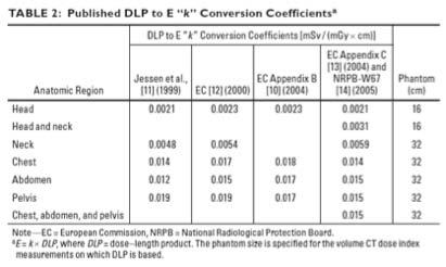
10 Weighting Factors, W R Equivalent Dose The radiation weighting factors, W R, are conversion factors between absorbed dose, D (Gy) and equivalent dose, H (Sv) H = W R x D For diagnostic x-ray energies W R is 1. H = D Effective Dose Effective dose takes into account where the radiation dose is being absorbed The effective dose attempts to reflect the equivalent whole-body dose that would result in an equivalent stochastic risk (e.g. cancer or genetic risk) from the actual absorbed dose to those tissues irradiated in a non-uniform, partial body irradiation The unit of effective dose SI Unit is the seivert: 1 Sv = 1 J/kg The non-si unit is the rem (radiation equivalent man): 1 rem = 10 msv or 1 Sv = 100 rem Effective Dose Effective dose (E) is given by the following E = Σ T (w T x H T,R ) where, w T is the tissue weighting factor for tissue T and H T is the equivalent dose of tissue T Tissue Weighting Factors Effective Doses from Radiation Protection 118: Referral Guidelines for Imaging, Published by the European Commission, 2001 Ref: Estimating Effective Dose in CT, AJR 2010; 194:
 or rad (non-si). Deterministic effects!")
 or rem (non-si). Stochastic effects! http://scicurious.wordpress.")
11 Review Exposure ability of radiation to ionize air. Units are the roentgen (non-si). Absorbed Dose energy imparted to the irradiated tissue per unit mass. Units are the gray (SI) or rad (non-si). Deterministic effects! Equivalent Dose takes into account the radiation type Effective Dose measure of the population risk posed by radiation. Units are the sievert (SI) or rem (non-si). Stochastic effects! Tissue Weighting Factors Ref: Estimating Effective Dose in CT, AJR 2010; 194: DNAstrand11.gif 1371
12 1372


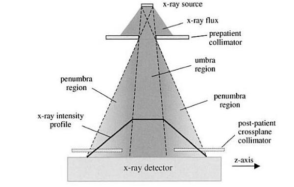
13 6 weeks 20 weeks 18 months MedicalImaging/MedicalX-Rays/ucm pdf Radiation-induced Skin Injuries from FluoroscopyThomas B. Shope, Ph.D. 1373
14
15 1375
16 1376


17 Film Dosimetry Thermoluminescent Dosimetry Bushberg, The Essential Physics of Medical Imaging /05/Thermoluminescence-Dosimetry.gif Optically Stimulated Luminescence Dosimetry Ionization Chambers Measure the charge liberated when photons interact with the gas in the chamber. That is, they measure exposure directly. Very accurate. They can be used to measure the output of x-ray tubes, as phototimers in automatic exposure control units and as dose calibrators in nuclear medicine. Geiger counters are ionization chambers operating in avalanche mode. Ionization Chamber Pocket Ionization Chambers X-ray Gas Molecules Gas Filled Chamber ma


18 Geiger-Mueller Counter Patient X-ray Tube II or Flat panel detector Scatter Table Patient X-ray Tube Scatter II or Flat panel detector Patient Scatter X-ray Tube Table Table II or Flat panel detector 1378
19 1379
20 1380
21 1381
22 1382


23 1383


24 1384


25 1385


26 1386


27 1387


28 1388


29 1389


30 $* 1390 %# (* %$ )) %% &' $ %& Table of Contents


31 1391
32 1392
33 1393
34 1394
35 1395
36 1396
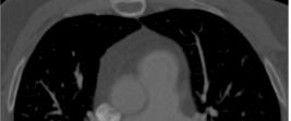
37 Routine Chest 1397


38 "Wilhelm Conrad Röntgen - Photo Gallery". Nobelprize.org. 9 Jan /rontgen-photo.html "Wilhelm Conrad Röntgen - Photo Gallery". Nobelprize.org. 9 Jan /rontgen-photo.html "Wilhelm Conrad Röntgen - Photo Gallery". Nobelprize.org. 9 Jan


39 1399


40 1400


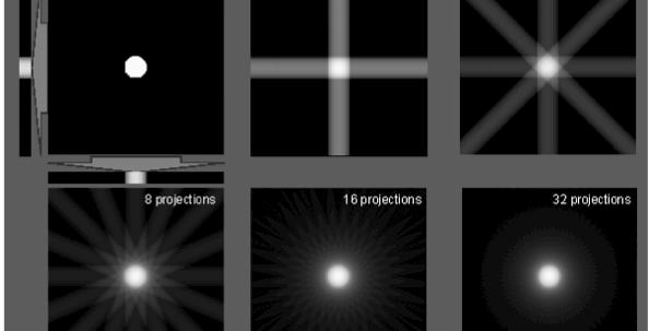


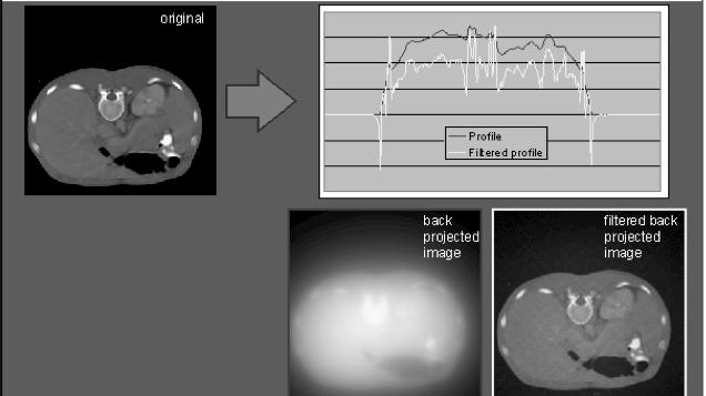
41 from RadonTransformOfSheppLoganWeb.mov Nobelprize.org 1401


42 Nobelprize.org from from from
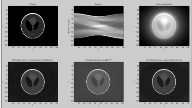


43 from I 0 μ 1,1 μ 1,2 I D,1 I 0 μ 1,1 μ 1,2 I D,1 I 0 I D,2 I 0 I D,2 μ 2,1 μ 2,2 μ 2,1 μ 2,2 d = pixel width I D,1 = I 0 e-d(μ1,1+μ1,2) 1403
44 I 0 μ 1,1 μ 1,2 I D,1 I 0 μ 1,1 μ 1,2 I D,1 I 0 I D,2 I 0 I D,2 μ 2,1 μ 2,2 μ 2,1 μ 2,2 ln(i D,1 / I 0 ) = -dμ 1,1 -dμ 1,2 (-1/d)ln(I D,1 / I 0 ) = μ 1,1 + μ 1,2 I 0 μ 1,1 μ 1,2 I D,1 I 0 μ 1,1 μ 1,2 I D,1 I 0 I D,2 I 0 I D,2 μ 2,1 μ 2,2 μ 2,1 μ 2,2 Let C 1,1 = (-1/d)ln(I D,1 / I 0 ) then C 1,1 = μ 1,1 + μ 1,2 C 1,1 = μ 1,1 + μ 1,2 C 1,2 = μ 2,1 + μ 2,2 μ 1404


45 New Image = Old Image x Correction Factor 1405


46 Scanner Computer


47

 Bow-Tie Filter http://1.bp.blogspot.")
48 Filtration Filtration Tungsten Anode Tube voltage 90 kvp Relative Output Unfiltered Filtered Energy (kev) Bow-Tie Filter collimator+ct+scan.jpg 1408


49 X-ray Xenon Gas ma CT Xenon Detector CT Scintillator Detector Gas Filled Chamber + - X-ray photon First Generation CT 1409

50 Second Generation CT

51 Third Generation CT Fourth Generation CT Fifth Generation CT 1411


52 Sixth Generation CT Slip rings used to bring power to x-ray tube on rotating gantry of a helical CT machine and, for some designs, to acquire information from the detector array. Wolbarst A B, and Hendee W R Radiology 2006;238: by Radiological Society of North America Slip rings used to bring power to x-ray tube on rotating gantry of a helical CT machine and, for some designs, to acquire information from the detector array. Wolbarst A B, and Hendee W R Radiology 2006;238: by Radiological Society of North America 1412


53 Seventh Generation Siemens Volume Zoom (4 rows) Cone-Beam CT 1413


54 Dual Source CT Dual Source CT A B Body A = Slice Thickness B= Reconstruction Intervals B-A= Gap Images 1414


 10 20")

55 Tungsten anode with tube voltage of at least kvp Tube voltage 90 kvp Relative Output Unfiltered Bremsstrahlung Relative Output L-shell K-shell Filtered Bremsstrahlung Energy (kev) Energy (kev) 1415


56 Relative Output Tube current doubled Energy (kev) X-ray Production Processes Tube voltage 90 kvp X-ray Production Processes X-ray Production Processes Relative Output Tube voltage increased from 75 to 90 kvp with tube current unchanged Energy (kev) 1416

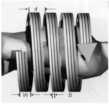
57 S d d S S S d S 1417


58 μ μ 1418


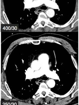
59 μ μ μ 400/ / /30 120/


60 1 mm width 6 lp/mm 8 lp/mm 12 lp/mm 15 lp/mm 22 lp/mm 34 lp/mm Ideal Resolution Good vs Poor Resolution Image of line-pair phantom Optical Density Position along line A-A Optical Density Position along line A-A 1420


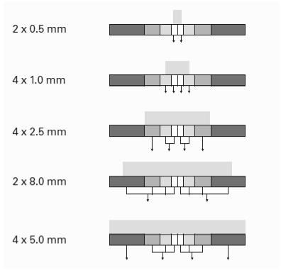
61 Matrix size 512x512 Pixel size = DFOV/Matrix size DFOV = 50cm DFOV = 36cm DFOV = 25cm 800 mas 400 mas 5 mm 2.5 mm 200 mas 100 mas 1.25 mm mm 1421


62 Matrix size = 512x512 DFOV = 50cm DFOV = 25cm B20 Smooth B40 B60 Sharp FBP 30% ASIR 50% ASIR 70% ASIR 100% ASIR ASIR = Adaptive Statistical Iterative Reconstruction 1422


63 1423


64


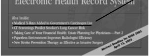

65 From the Department of Health and Human Service s 11 th Report on Carcinogens released in January, From the Department of Health and Human Service s 11 th Report on Carcinogens released in January, Quote from, Medical X-rays Added to Government Carcinogen List. RSNA News, March 2005, Vol. 15, No. 3, pp Quote from, Medical X-rays Added to Government Carcinogen List. RSNA News, March 2005, Vol. 15, No. 3, pp


66 Ref: Diagnostic CT scans: assessment of patient, physician, and radiologist awareness of radiation dose and possible risks. Radiology 2004; 231: Ref: Diagnostic CT scans: assessment of patient, physician, and radiologist awareness of radiation dose and possible risks. Radiology 2004; 231: Understand the biases of the sources of information you use to make these decisions. Why Perform CT When MRI Is Safer -- and Perhaps Better MRI is an imaging modality that is considerably safer than CT on the basis of a number of factors, of which radiation exposure is perhaps the most serious. In addition, MRI may actually be much more accurate in describing disease. Richard C. Semelka, MD, Director of MR Services; Professor of Radiology; Vice Chairman of Clinical Research, Department of Radiology, The University of North Carolina at Chapel Hill excerpt from Radiation Risk from CT Scans: A Call for Patient-Focused Imaging, Medscape Radiology 6(1),


67 from Radiation Protection 118: Referral Guidelines for Imaging, Published by the European Commission, 2001 Ref: Estimated risk of radiation induced fatal cancer from pediatric CT, AJR, Vol. 176, Feb, 2001 Ref: Estimated risk of radiation induced fatal cancer from pediatric CT, AJR, Vol. 176, Feb, 2001 Image of article obtained from


68 Ref: Risk of Cancer from diagnostic X-rays: estimates for the UK and 14 other countries, The Lancet, Vol 363, Jan 31, 2004 Image of article obtained from Ref: Effect of low doses of ionizing radiation in infancy on cognitive function in adulthood: Swedish population based cohort study, BMJ, Vol 328, Jan 3,
69 X-ray source X-ray source + - e - e - e - e - Air e - Ionization Chamber Ionization Chamber Patient Film/Screen Cassette X-ray source X-ray source X-ray source Scatter Patient s right hand Scatter Patient s left hand Scatter e - Patient e - e - Ionization Chamber Film/Screen Cassette 1429


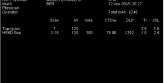
70 Σ Ref: Estimating Effective Dose in CT, AJR 2010; 194: from Radiation Protection 118: Referral Guidelines for Imaging, Published by the European Commission, 2001 Routine Head Ref: AAPM/RSNA Physics Tutorial for Residents: Topics in CT, Radiographics 2002; 22:


71 X-ray Tube Main X-ray beam Scatter Detector Ref: AAPM/RSNA Physics Tutorial for Residents: Topics in CT, Radiographics 2002; 22: Ref: AAPM/RSNA Physics Tutorial for Residents: Topics in CT, Radiographics 2002; 22: Ref: AAPM/RSNA Physics Tutorial for Residents: Topics in CT, Radiographics 2002; 22: Ref: AAPM/RSNA Physics Tutorial for Residents: Topics in CT, Radiographics 2002; 22: Radiation Dose Routine Head Fig from impactscan.org 1431


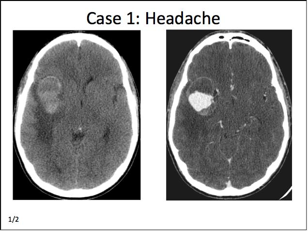
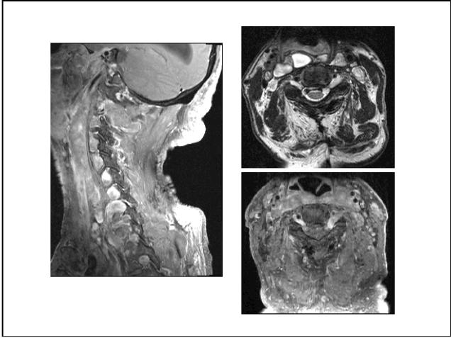
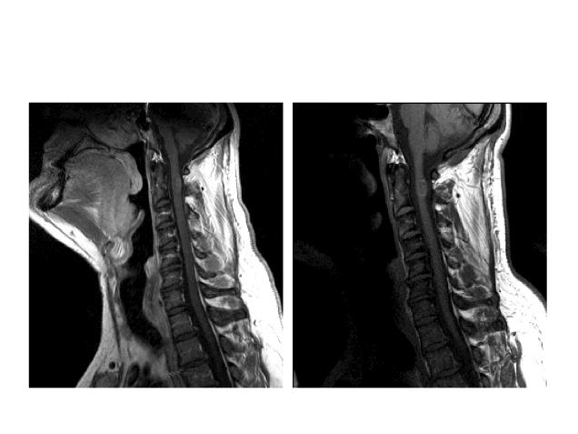
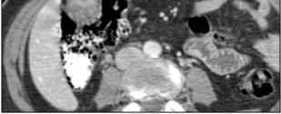
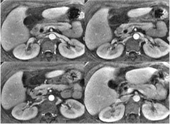
72 Radiation Dose Radiation Dose Routine Head Fig from impactscan.org Routine Chest 1432


73 Fig from impactscan.org Effective dose for various studies can be approximated via measurements with TLD anthropomorphic phantoms Effective dose for various studies can be approximated via computer simulations Ref: Estimating Effective Dose in CT, AJR 2010; 194:


74 AAPM Report no


75 1435


76 Ref: AAPM/RSNA Physics Tutorial for Residents: Topics in CT, Radiographics 2002; 22: Ref: Radiation dose and image quality in pediatric CT: effect of technical factors and phantom size and shape, Radiology 2004; 233:


77 from from Ref: CT and Computed Radiography: The Pictures are Great, but is the Radiation Dose Greater than Required?, AJR 2002; 179: Future Advances original 50% dose reduction 75% dose reduction 87.5% dose reduction 96% dose reduction 99% dose reduction Images courtesy Bruce Whiting, PhD 1437


78 McNitt-Gray 2011 AAPM Summit on CT dose 180 Ref MA 280 Ref MA 1438

79 Total Dose: 17 msv 1439
80 1440
81 1441
82 1442


83 GI Case Quiz Cynthia S Santillan How do you classify this finding? A Superficial B Intersphincteric C Transphincteric D Extrasphincteric 1443


84 Most appropriate next step? A Colonoscopy B Check AFP C Start IV antibiotics D Follow-up CT in 3 months 1444


85 What is the most likely provided history? A History of colon cancer B Elevated IgE C History of OCP D HCV, initial imaging 1445


86 1446
87 1447
88 1448
89 1449
90 1450
91 1451
92 1452
93 1453
94 1454

95 SAVE THE DATE 38 th Annual Radiology Review Course Hotel del Coronado Coronado, California April 1-7, 2018
Dosimetric Consideration in Diagnostic Radiology
 Dosimetric Consideration in Diagnostic Radiology Prof. Ng Kwan-Hoong Department of Biomedical Imaging University of Malaya ngkh@um.edu.my Radiation Dosimetry Workshop, 28-29 March 2014 2 Why do we measure
Radiologic Units: What You Need to Know
 Radiologic Units: What You Need to Know TODD VAN AUKEN M.ED. RT (R)(MR) Agenda Greys, Sieverts, Coulombs per kg, & Becquerel's Conventional Units Other Concepts (LET, Q-Factor, Effective Dose, NCRP Report
Skyscan 1076 in vivo scanning: X-ray dosimetry
 Skyscan 1076 in vivo scanning: X-ray dosimetry DOSIMETRY OF HIGH RESOLUTION IN VIVO RODENT MICRO-CT IMAGING WITH THE SKYSCAN 1076 An important distinction is drawn between local tissue absorbed dose in
Radiation physics and radiation protection. University of Szeged Department of Nuclear Medicine
 Radiation physics and radiation protection University of Szeged Department of Nuclear Medicine Radiation doses to the population 1 Radiation doses to the population 2 Sources of radiation 1 Radiation we
Why is CT Dose of Interest?
 Why is CT Dose of Interest? CT usage has increased rapidly in the past decade Compared to other medical imaging CT produces a larger radiation dose. There is direct epidemiological evidence for a an increase
Basic radiation protection & radiobiology
 Basic radiation protection & radiobiology By Dr. Mohsen Dashti Patient care & management 202 Wednesday, October 13, 2010 Ionizing radiation. Discussion issues Protecting the patient. Protecting the radiographer.
Cone Beam CT Protocol Optimisation for Prostate Imaging with the Varian Radiotherapy OBI imaging system. Dr Craig Moore & Dr Tim Wood
 Cone Beam CT Protocol Optimisation for Prostate Imaging with the Varian Radiotherapy OBI imaging system Dr Craig Moore & Dr Tim Wood Background With the increasing use of CBCT imaging alongside complex
Accounting for Imaging Dose
 Accounting for Imaging Dose High Profile Over-exposures Lead to Growing Concern FDA issues warning in October 2009-209 patients exposed to 8 times typical dose for CT brain perfusion scan (3-4 Gy) - Some
ESTABLISHING DRLs in PEDIATRIC CT. Keith Strauss, MSc, FAAPM, FACR Cincinnati Children s Hospital University of Cincinnati College of Medicine
 ESTABLISHING DRLs in PEDIATRIC CT Keith Strauss, MSc, FAAPM, FACR Cincinnati Children s Hospital University of Cincinnati College of Medicine CT Dose Indices CTDI INTRODUCTION CTDI 100, CTDI w, CTDI vol
DETERMINATION OF ENTRANCE SKIN DOSE FROM DIAGNOSTIC X-RAY OF HUMAN CHEST AT FEDERAL MEDICAL CENTRE KEFFI, NIGERIA
 DETERMINATION OF ENTRANCE SKIN DOSE FROM DIAGNOSTIC X-RAY OF HUMAN CHEST AT FEDERAL MEDICAL CENTRE KEFFI, NIGERIA Full Length Research Article 1 Ibrahim, U, 3 Daniel, I.H., 3 Ayaninola, O., 4 Ibrahim,
Patient Dose Estimates. from CT Examinations. Patient Dose Estimates
 Patient Dose Estimates from CT Examinations Patient Dose Estimates from CT Examinations John M. Boone, Ph.D. Professor and Vice Chairman of Radiology Professor of Biomedical Engineering University of California,
CT Radiation Risks and Dose Reduction
 CT Radiation Risks and Dose Reduction Walter L. Robinson, M.S. D.A.B.S.N.M., D.A.B.M.P., D.A.B.R. Consultant Certified Medical Radiation Health & Diagnostic Imaging Physicist Medical Radiation and Children
Biological Effects of Ionizing Radiation & Commonly Used Radiation Units
 INAYA MEDICAL COLLEGE (IMC) RAD 232 - LECTURE 2 & 3 Biological Effects of Ionizing Radiation & Commonly Used Radiation Units DR. MOHAMMED MOSTAFA EMAM How does radiation injure people? - High energy radiation
Patient dose assessment of CT perfusion scanning at the RSCH
 Patient dose assessment of CT perfusion scanning at the RSCH Lesley Leavesley, Emma Whitehead, Matthew Pryor, Debbie Peet Regional Radiation Protection Service Royal Surrey County Hospital, Guildford Overview
THE TUFFEST STUFF CT REGISTRY REVIEW Live Lecture Seminar SATURDAY CURRICULUM
 1. The CT Imaging Chain-10 major components & their functions a. The x-ray tube b. Generator c. Filter d. Pre-patient collimator e. Pre-detector collimator f. Detector system g. Analog to digital converter
Lecture 14 Exposure to Ionizing Radiation
 Lecture 14 Exposure to Ionizing Radiation Course Director, Conrad Daniel Volz, DrPH, MPH Assistant Professor, Environmental & Occupational Health, University of Pittsburgh, Graduate School of Public Health
3/26/2017. Personal Dosimetry Monitoring and Dose Measurements. Agenda. Dosimetric Terms and Definitions Dose Limits External Dosimetry
 Speaker David Pellicciarini, CHP, MBA Vice President, Pharmacy Safety, Practice and Technical Operations Cardinal Health Nuclear Pharmacy Services david.pellicciarini@cardinalhealth.com Personal Dosimetry
Biological Effects of Ionizing Radiation & Commonly Used Radiation Units
 INAYA MEDICAL COLLEGE (IMC) RAD 232 - LECTURE 3, 4 & 5 Biological Effects of Ionizing Radiation & Commonly Used Radiation Units DR. MOHAMMED MOSTAFA EMAM How does radiation injure people? - High energy
Managing the imaging dose during Image-guided Radiotherapy. Martin J Murphy PhD Department of Radiation Oncology Virginia Commonwealth University
 Managing the imaging dose during Image-guided Radiotherapy Martin J Murphy PhD Department of Radiation Oncology Virginia Commonwealth University Radiographic image guidance has emerged as the new paradigm
Debra Pennington, MD Director of Imaging Dell Children s Medical Center
 Debra Pennington, MD Director of Imaging Dell Children s Medical Center 1 Gray (Gy) is 1 J of radiation energy/ 1 kg matter (physical quantity absorbed dose) Diagnostic imaging doses in mgy (.001 Gy)
Radiation Safety. Bethany Gillett 14th Feb After this lecture, you should be able to:
 Radiation Safety Bethany Gillett bethany.gillett@addenbrookes.nhs.uk 14th Feb 2018 Learning Outcomes After this lecture, you should be able to: Understand different radiation protection quantities Explain
Radiation Dose in Pediatric Imaging
 Radiation Dose in Pediatric Imaging A Brief History of Radiology Dose: Why Does It Matter? Measuring Exposure and Dose Deterministic Effects Stochastic Effects Common Exams: What is the Risk? Reducing
RADIATION PROTECTION IN DIAGNOSTIC AND INTERVENTIONAL RADIOLOGY. L19: Optimization of Protection in Mammography
 IAEA Training Material on Radiation Protection in Diagnostic and Interventional Radiology RADIATION PROTECTION IN DIAGNOSTIC AND INTERVENTIONAL RADIOLOGY L19: Optimization of Protection in Mammography
Radiation Safety in the Catheterization Lab
 SCAI FALL FELLOWS COURSE - 2015 Radiation Safety in the Catheterization Lab V. Vivian Dimas, MD, FSCAI Associate Professor Pediatrics, Cardiology UT Southwestern Medical Center Dallas TX None Disclosures
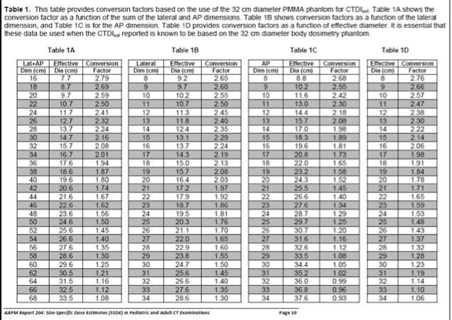
CURRENT CT DOSE METRICS: MAKING CTDI SIZE-SPECIFIC
 CURRENT CT DOSE METRICS: MAKING CTDI SIZE-SPECIFIC Keith Strauss, MSc, FAAPM, FACR Cincinnati Children s Hospital University of Cincinnati College of Medicine Acknowledgments John Boone, PhD Michael McNitt-Grey,
Measurement of organ dose in abdomen-pelvis CT exam as a function of ma, KV and scanner type by Monte Carlo method
 Iran. J. Radiat. Res., 2004; 1(4): 187-194 Measurement of organ dose in abdomen-pelvis CT exam as a function of ma, KV and scanner type by Monte Carlo method M.R. Ay 1, M. Shahriari 2, S. Sarkar 3, P.
Estimating Patient Radiation Dose from Computed Tomography
 Estimating Patient Radiation Dose from Computed Tomography C. Cagnon, J. DeMarco, E. Angel, M. McNitt-Gray UCLA David Geffen School of Medicine 1 Patient Dose from CT Advances in Technology... Helical
Upon successful completion of the course, the student should be competent in the following tasks:
 COURSE INFORMATION Course Prefix/Number: RAD 201 Course Title: Radiation Biology Lab Hours/Week: 3.0 Credit Hours/Semester: 2.0 VA Statement/Distance Learning Attendance Textbook Information Student Code
Practical Reference Dosimetry Course April 2015 PRDC Program, at a glance. Version 1.0. Day 1 Day 2 Day 3 Day 4
 Practical Reference Dosimetry Course 21-24 April 2015 PRDC 2015 Program, at a glance Version 1.0 Day 1 Day 2 Day 3 Day 4 Quantities and Units Free air chambers Uncertainties Brachytherapy traceability
Peak temperature ratio of TLD glow curves to investigate the spatial variation of LET in a clinical proton beam
 Peak temperature ratio of TLD glow curves to investigate the spatial variation of LET in a clinical proton beam University of Chicago CDH Proton Center LET study C. Reft 1, H. Ramirez 2 and M. Pankuch
Doses from pediatric CT examinations in Norway Are pediatric scan protocols developed and in daily use?
 Doses from pediatric CT examinations in Norway Are pediatric scan protocols developed and in daily use? Eva Godske Friberg * Norwegian Radiation Protection Authority, P.O. Box, Østerås, Norway Abstract.
45 Hr PET Registry Review Course
 45 HR PET/CT REGISTRY REVIEW COURSE Course Control Document Timothy K. Marshel, MBA, R.T. (R), (N)(CT)(MR)(NCT)(PET)(CNMT) The PET/CT Training Institute, Inc. SNMMI-TS 028600-028632 45hr CEH s Voice Credits
RADIATION SAFETY. Junior Radiology Course
 RADIATION SAFETY Junior Radiology Course Expectations for the Junior Radiology Course Medical School wants students to learn basic principles, factual knowledge, safety info, etc. Medical Students want
Prof. Dr. Doğan BOR Ankara University Institute of Nuclear Science
 PATIENT DOSIMETRY IN DIAGNOSTIC RADIOLOGY MODALITIES Prof. Dr. Doğan BOR Ankara University Institute of Nuclear Science Ankara University Institute of Nuclear Science USE OF RADIATION! INCREASING? Natural
8/18/2011. Acknowledgements. Managing Pediatric CT Patient Doses INTRODUCTION
 Managing Pediatric CT Patient Doses Keith J. Strauss, MSc, FAAPM, FACR President X-Ray Computations, Inc. Boston, Massachusetts Acknowledgements Marilyn Goske, MD John Boone, PhD Cynthia McCollough, PhD
Radiation Monitoring Instruments
 Radiation Monitoring Instruments This set of slides is based on Chapter 4 authored byg. Rajan, J. Izewska of the IAEA publication (ISBN 92-0-107304-6): Radiation Oncology Physics: A Handbook for Teachers
CT Dose Estimation. John M. Boone, Ph.D., FAAPM, FSBI, FACR Professor and Vice Chair of Radiology. University of California Davis Medical Center
 CT Dose Estimation John M. Boone, Ph.D., FAAPM, FSBI, FACR Professor and Vice Chair of Radiology 1 University of California Davis Medical Center CT Dose Estimation Introduction The CTDI Family of Metrics
Doses from Cervical Spine Computed Tomography (CT) examinations in the UK. John Holroyd and Sue Edyvean
 examinations in the UK. John Holroyd and Sue Edyvean") Doses from Cervical Spine Computed Tomography (CT) examinations in the UK John Holroyd and Sue Edyvean Why a new dose survey? Number of enquires received concerning the current NDRL Concern that could
Doses from Cervical Spine Computed Tomography (CT) examinations in the UK John Holroyd and Sue Edyvean Why a new dose survey? Number of enquires received concerning the current NDRL Concern that could
Introduction and Background
 CT Lung Cancer Screening and the Medical Physicist: Background, Findings and Participant Dosimetry Summary of the National Lung Screening Trial (NLST) Randell Kruger, PhD, DABR Medical Physics Section
Managing the imaging dose during image-guided radiation therapy
 Managing the imaging dose during image-guided radiation therapy Martin J Murphy PhD Department of Radiation Oncology Virginia Commonwealth University Richmond VA Imaging during radiotherapy Radiographic
Managing Patient Dose in Computed Tomography (CT) INTERNATIONAL COMMISSION ON RADIOLOGICAL PROTECTION
 INTERNATIONAL COMMISSION ON RADIOLOGICAL PROTECTION") Managing Patient Dose in Computed Tomography (CT) International Commission on Radiological Protection Information abstracted from ICRP Publication 87 Available at www.icrp.org Task Group: M.M. Rehani,
Managing Patient Dose in Computed Tomography (CT) International Commission on Radiological Protection Information abstracted from ICRP Publication 87 Available at www.icrp.org Task Group: M.M. Rehani,
Basic definitions. Dosimetry, radiation protection. Nuclear measurement techniques. Interaction of the nuclear radiation with the matter
 Dosimetry, radiation protection. Nuclear measurement techniques. properties measurement dosimetry medical applications of the nuclear radiation Basic definitions Nuclear radiation: Produced in the transition
CALCULATION OF BACKSCATTER FACTORS FOR LOW ENERGY X-RAYS USING THE TOPAS MONTE CARLO CODE
 CALCULATION OF BACKSCATTER FACTORS FOR LOW ENERGY X-RAYS USING THE TOPAS MONTE CARLO CODE Emily Hewson 1 Martin Butson 1,2 Robin Hill 1,2 1 Institute of Medical Physics, School of Physics, University of
Dianna Cody, PhD, DABR, FAAPM Professor & Clinical Operations Director Imaging Physics U.T. M.D. Anderson Cancer Center Houston, TX
 Dianna Cody, PhD, DABR, FAAPM Professor & Clinical Operations Director Imaging Physics U.T. M.D. Anderson Cancer Center Houston, TX Learning Objectives: Limitations for estimating patient dose for CT Methods
Translating Protocols Across Patient Size: Babies to Bariatric
 Translating Protocols Across Patient Size: Babies to Bariatric Cynthia H. McCollough, PhD, FACR, FAAPM Professor of Radiologic Physics Director, CT Clinical Innovation Center Department of Radiology Mayo
Reports and Activities of International Commission on Radiation units and Measurements ICRU
 Reports and Activities of International Commission on Radiation units and Measurements ICRU Hans-Georg Menzel Chairman ICRU Main Commission CERN (retired) Bethesda, 12 Oct. 2016 The definition of appropriate
PHY138Y Nuclear and Radiation
 PHY38Y Nuclear and Radiation Professor Tony Key MP40 key@physics.utoronto.ca Announcements MP problems set #4 due Sunday at midnight PS#5 WRITTEN now posted! - do in teams, no Lone Wolves!! NB correction
Staff Exposure Monitoring in Interventional Radiology
 Conference on Physics in Medicine: From Diagnosis to Treatment "Enhancing safety and quality in radiation medicine". KFMC, Riyadh 7-9 November 2017 Staff Exposure Monitoring in Interventional Radiology
Preparing for Medical Physics Components of the ABR Core Examination
 Preparing for Medical Physics Components of the ABR Core Examination The ABR core examination for radiologists contains material on medical physics. This content is based on the medical physics that is
Dosimetric characterization with 62 MeV protons of a silicon segmented detector for 2D dose verifications in radiotherapy
 Dosimetric characterization with 62 MeV protons of a silicon segmented detector for 2D dose verifications in radiotherapy C. Talamonti M. Bruzzi,M. Bucciolini, L. Marrazzo, D. Menichelli University of
A more accurate method to estimate patient dose during body CT examinations with tube current modulation
 A more accurate method to estimate patient dose during body CT examinations with tube current modulation Poster No.: C-0738 Congress: ECR 2014 Type: Scientific Exhibit Authors: A. Kawaguchi 1, Y. Matsunaga
Joint ICTP/IAEA Advanced School on Dosimetry in Diagnostic Radiology and its Clinical Implementation May 2009
 2033-4 Joint ICTP/ Advanced School on Dosimetry in Diagnostic Radiology and its Clinical Implementation 11-15 May 2009 Dosimetry for General Radiology and Clinical Uncertainty Peter Homolka EFOMP Training
Breast CT and Dosimetry
 2013 ICTP/IAEA Training Course on Radiation Protection of Patients Trieste Breast CT and Dosimetry John M. Boone, Ph.D., FAAPM, FSBI, FACR Professor and Vice Chair (Research) of Radiology Professor of
ARRT Specifications Radiation Exposure & Monitoring
 Radiation Protection Review 15% (30) 11% (22) Gina Tice, MSRS, RT(R) Gadsden State Community College ARRT Specifications Radiation Exposure & Monitoring Radiation Protection (45) Biological Aspects of
Dosimetry - Measurement of Ionising Radiation
 Dosimetry - Measurement of Ionising Radiation Assoc. Prof. Katarína Kozlíková, RN., PhD. IMPhBPhITM FM CU in Bratislava katarina.kozlikova@fmed.uniba.sk Contents Dosimetry Dose Radiation dose Absorbed
PRINCIPLES and PRACTICE of RADIATION ONCOLOGY. Matthew B. Podgorsak, PhD, FAAPM Department of Radiation Oncology
 PRINCIPLES and PRACTICE of RADIATION ONCOLOGY Matthew B. Podgorsak, PhD, FAAPM Department of Radiation Oncology OUTLINE Physical basis Biological basis History of radiation therapy Treatment planning Technology
Two-Dimensional Thermoluminescence Dosimetry System for Proton Beam Quality Assurance
 Two-Dimensional Thermoluminescence Dosimetry System for Proton Beam Quality Assurance Jan Gajewski Institute of Nuclear Physics, Kraków, Poland German Cancer Research Center, Heidelberg, Germany Existing
Managing Radiation Risk in Pediatric CT Imaging
 Managing Radiation Risk in Pediatric CT Imaging Mahadevappa Mahesh, MS, PhD, FAAPM, FACR, FACMP, FSCCT. Professor of Radiology and Cardiology Johns Hopkins University School of Medicine Chief Physicist
Medical applications round table. Radiotherapy Diagnostic radiology Nuclear medicine Radiation protection
 Medical applications round table Radiotherapy Diagnostic radiology Nuclear medicine Radiation protection Will be discussing Ion beam therapy/radiotherapy Diagnostic radiology Nuclear medicine Radiation
THE UNIVERSITY OF OKLAHOMA HEALTH SCIENCES CENTER GRADUATE COLLEGE RADIATION DOSE ESTIMATION FOR DIAGNOSTIC MODALITIES
 THE UNIVERSITY OF OKLAHOMA HEALTH SCIENCES CENTER GRADUATE COLLEGE RADIATION DOSE ESTIMATION FOR DIAGNOSTIC MODALITIES A THESIS SUBMITTED TO THE GRADUATE FACULTY in partial fulfillment of the requirements
Ionizing Radiation. Michael J. Vala, CHP. Bristol-Myers Squibb
 Ionizing Radiation Michael J. Vala, CHP Bristol-Myers Squibb michael.vala@bms.com 732-227-5096 2013 American Industrial Hygiene Association, New Jersey Section, Inc. Course Objectives At the end of this
Mammography. Background and Perspective. Mammography Evolution. Background and Perspective. T.R. Nelson, Ph.D. x41433
 - 2015 Background and Perspective 2005 (in US) Women Men Mammography Invasive Breast Cancer Diagnosed 211,240 1,690 Noninvasive Breast Cancer Diagnosed 58,940 Deaths from Breast Cancer 40,410 460 T.R.
PHYS 383: Applications of physics in medicine (offered at the University of Waterloo from Jan 2015)
") PHYS 383: Applications of physics in medicine (offered at the University of Waterloo from Jan 2015) Course Description: This course is an introduction to physics in medicine and is intended to introduce
PHYS 383: Applications of physics in medicine (offered at the University of Waterloo from Jan 2015) Course Description: This course is an introduction to physics in medicine and is intended to introduce
Radiation Units and Dosimetry 15 August Kalpana M. Kanal, Ph.D., DABR 1
 Introduction Radiation Units and Dosimetry Radiation dose quantities are used as indicators of the risk of biologic damage to patients from x-rays and thus a good knowledge of the different dose parameters
X-Ray & CT Physics / Clinical CT
 Computed Tomography-Basic Principles and Good Practice X-Ray & CT Physics / Clinical CT INSTRUCTORS: Dane Franklin, MBA, RT (R) (CT) Office hours will be Tuesdays from 5pm to 6pm CLASSROOM: TIME: REQUIRED
Quiz True/False: Large amounts of radiation to insects will cause them to mutate!
 RADS, REMS & ROENTGENS Jack L. Barr, M.S., R.T.R., F.A.S.R.T. Quiz True/False: Large amounts of radiation to insects will cause them to mutate! LARGE AMOUNTS OF RADIATION WILL CAUSE VEGETABLES TO BECOME
RADIATION MONITORING DEVICES R A D I A T I O N P R O T E C T I O N & B I O L O G Y - R H O D E S
 RADIATION MONITORING DEVICES 10-526- 1 9 7 R A D I A T I O N P R O T E C T I O N & B I O L O G Y - R H O D E S DETECTION AND MEASUREMENT OF IONIZING RADIATION Dosimeter Dose-measuring device Two classifications:
IMAGE GENTLY HOW CAN YOU HELP?
 IMAGE GENTLY HOW CAN YOU HELP? Keith J. Strauss, MSc, FAAPM, FACR Director, Radiology Physics & Engineering Children s s Hospital Boston Harvard Medical School Acknowledgment Marilyn J. Goske,, MD Robert
SCOPE OF ACCREDITATION TO ISO/IEC 17025:2005 & ANSI/NCSL Z
 SCOPE OF ACCREDITATION TO ISO/IEC 17025:2005 & ANSI/NCSL Z540-1-1994 UNIVERSITY OF WISCONSIN RADIATION CALIBRATION LABORATORY Room B1002, WIMR 1111 Highland Avenue Madison, WI 53705-2275 Larry A. DeWerd,
M. J. Maryanski, Three Dimensional BANG Polymer Gel Dosimeters AAPM'99, CE Course
 Three Dimensional BANG Polymer Gel Dosimeters Marek J. Maryanski MGS Research, Inc. Guilford, CT Educational objectives: Describe the need for high-resolution 3D dosimetry in 3D CRT. Explain the physics
QUANTIFICATION OF THE RISK-REFLECTING STOCHASTIC AND DETERMINISTIC RADIATION EFFECTS
 RAD Conference Proceedings, vol. 2, pp. 104 108, 2017 www.rad-proceedings.org QUANTIFICATION OF THE RISK-REFLECTING STOCHASTIC AND DETERMINISTIC RADIATION EFFECTS Jozef Sabol *, Bedřich Šesták Crisis Department,
IRRADIATORS. September 11, :00 am-5:00 pm Hock Plaza Auditorium Duke University Medical Center, Erwin Road
 IRRADIATORS Terry Yoshizumi,, PhD September 11, 2006 8:00 am-5:00 pm Hock Plaza Auditorium Duke University Medical Center, Erwin Road Radiation Countermeasures Center of Research Excellence 1 Acknowledgements
GATE MONTE CARLO DOSIMETRY SIMULATION OF MARS SPECTRAL CT
 GATE MONTE CARLO DOSIMETRY SIMULATION OF MARS SPECTRAL CT R Aamir, C Lowe, J Damet, P Carbonez, A P H Butler, N Schleich, N G Anderson MARFO, Emmanuel Geant4 User Workshop 2017, Wollongong Overview MARS
Measurement of Kidneys Dose in Three Hospitals during Chest and Abdomen Radiographic Examinations
 Measurement of Kidneys Dose in Three Hospitals during Chest and Abdomen Radiographic Examinations Nadia Alatta 1, Abdelrahaman Elnour 2, Ikhlas Abdelaziz 3, Tugua Tageldeen 4, Duha Abdu 5, Asma Elamin
Biological Effects of Ionizing Radiation Module 8 - AAPM/RSNA Curriculum. Basic Radiation Biology
 Biological Effects of Ionizing Radiation Module 8 - AAPM/RSNA Curriculum Basic Radiation Biology Kalpana M. Kanal, PhD, DABR Associate Professor, Radiology Director, Resident Physics Education a copy of
Neutron Interactions Part 2. Neutron shielding. Neutron shielding. George Starkschall, Ph.D. Department of Radiation Physics
 Neutron Interactions Part 2 George Starkschall, Ph.D. Department of Radiation Physics Neutron shielding Fast neutrons Slow down rapidly by scatter in hydrogenous materials, e.g., polyethylene, paraffin,
Chapter 4: Radiation Monitoring Instruments
 Chapter 4: Radiation Monitoring Instruments Set of 107 slides based on the chapter authored by G. Rajan, J. Izewska of the IAEA publication (ISBN 92-0-107304-6): Review of Radiation Oncology Physics: A
PRINCIPLES AND METHODS OF RADIATION PROTECTION
 PRINCIPLES AND METHODS OF RADIATION PROTECTION Lesson Outcomes At the end of the lesson, student should be able to: Define what is radiation protection (RP) Describe basic principles of RP Explain methods
RADIOLOGY AN DIAGNOSTIC IMAGING
 Day 2 p. 1 RADIOLOGY AN DIAGNOSTIC IMAGING Dr hab. Zbigniew Serafin, MD, PhD serafin@cm.umk.pl and Radiation Protection mainly based on: C. Scott Pease, MD, Allen R. Goode, MS, J. Kevin McGraw, MD, Don
ABSTRACT. Objectives: To investigate the radiation dose received by patients undergoing routine plain x-ray
 Radiation Dose Distribution for Patients Undergoing Routine Radiological Scans for Kidney Stone Diagnosis at the University Hospital of the West Indies DC Walker 1, WD Aiken 2, S Shah 3, MK Voutchkov 1,
ADVANCES IN RADIATION TECHNOLOGIES IN THE TREATMENT OF CANCER
 ADVANCES IN RADIATION TECHNOLOGIES IN THE TREATMENT OF CANCER Bro. Dr. Collie Miller IARC/WHO Based on trends in the incidence of cancer, the International Agency for Research on Cancer (IARC) and WHO
SPECIFIC PRINCIPLES FOR DOSE REDUCTION IN HEAD CT IMAGING. Rajiv Gupta, MD, PhD Neuroradiology, Massachusetts General Hospital Harvard Medical School
 SPECIFIC PRINCIPLES FOR DOSE REDUCTION IN HEAD CT IMAGING Rajiv Gupta, MD, PhD Neuroradiology, Massachusetts General Hospital Harvard Medical School OUTLINE 1 st Presentation: Dose optimization strategies
Bureau of Laboratory Quality Standards
 1. Condom -Lubricant ISO 4074:2002/Cor.1:2003 Annex C -Thickness ISO 4074:2002/Cor.1:2003 Annex F - Length ISO 4074:2002 / Cor1 : 2003 - Width ISO 4074 : 2002/Cor 1 : 2003 - Bursting pressure and Volume
Computed tomography Acceptance testing and dose measurements
 Computed tomography Acceptance testing and dose measurements Jonas Andersson Medical Physicist, Ph.D. Department of Radiation Sciences University Hospital of Norrland, Umeå Sweden Contents The Computed
Ultralow Dose Chest CT with MBIR
 Ultralow Dose Chest CT with MBIR Ella A. Kazerooni, M.D. Professor & Director Cardiothoracic Radiology Associate Chair for Clinical Affairs University of Michigan Disclosures Consultant: GE Healthcare
Lecture 13 Radiation Onclolgy
 Lecture 13 Radiation Onclolgy Radiation Oncology: Tumors attacked with ionizing radiation Photons (gamma rays) High Energy Electrons Protons Other particles Brachytherapy: implants of beta emitters Ionizing
Calculation of Effective Doses for Radiotherapy Cone-Beam CT and Nuclear Medicine Hawkeye CT Laura Sawyer
 Calculation of Effective Doses for Radiotherapy Cone-Beam CT and Nuclear Medicine Hawkeye CT Laura Sawyer Department of Medical Physics and Bioengineering, Royal United Hospital, Bath Overview Varian Acuity
Radiation exposure of the Yazd population from medical conventional X-ray examinations
 Iran. J. Radiat. Res., 2007; 4 (4): 195-200 Radiation exposure of the Yazd population from medical conventional X-ray examinations F. Bouzarjomehri 1*, M.H. Dashti 2, M.H. Zare 1 1 Department of Medical
Neutron dose evaluation in radiotherapy
 Neutron dose evaluation in radiotherapy Francesco d Errico University of Pisa, Italy Yale University, USA Radiation therapy with a linear accelerator (LINAC) Photoneutron production in accelerator head
Medical Physics and Informatics Original Research
 Medical Physics and Informatics Original Research Christner et al. Estimating Effective Dose for CT Medical Physics and Informatics Original Research FOCUS ON: Jodie A. Christner 1 James M. Kofler Cynthia
Automatic Patient Centering for MDCT: Effect on Radiation Dose
 Patient Centering for MDCT CT Imaging Original Research Jianhai Li 1 Unni K. Udayasankar 1 Thomas L. Toth 2 John Seamans 2 William C. Small 1 Mannudeep K. Kalra 1,3 Li J, Udayasankar UK, Toth TL, Seamans
Radiation Dose Reduction: Should You Use a Bismuth Breast Shield?
 Radiation Dose Reduction: Should You Use a Bismuth Breast Shield? Lincoln L. Berland, M.D., F.A.C.R. Michael V. Yester, Ph.D. University of Alabama at Birmingham Breast Radiation on CT Use of chest CT
PGY-1. Resident Review Session Schedule
 1. August Simulation & Treatment 1.1. Sim Setup 1.2. Sim Techniques 1.3. 4DCT 1.4. Breath Hold / Gating 1.5. Treatment Setup 1.6. Treatment Delivery 1.7. Filming 1.7.1. Port film 1.7.2. kv 1.7.3. CBCT
Radiation Quantities and Units
 Radiation Quantities and Units כולנו מתעסקים יום יום עם אמצעי דימות שונים החושפים את הנבדק לקרינה מייננת. בשנים האחרונות השימוש ב- CT עולה באופן חד ביותר בבתי החולים ובקהילה. מה אנחנו באמת יודעים לגבי
Implementation of the 2012 ACR CT QC Manual in a Community Hospital Setting BRUCE E. HASSELQUIST, PH.D., DABR, DABSNM ASPIRUS WAUSAU HOSPITAL
 Implementation of the 2012 ACR CT QC Manual in a Community Hospital Setting BRUCE E. HASSELQUIST, PH.D., DABR, DABSNM ASPIRUS WAUSAU HOSPITAL Conflict of Interest Disclaimer Employee of Aspirus Wausau
Radiation Dose Reduction Strategies in Coronary CT Angiography
 Radiation Dose Reduction Strategies in Coronary CT Angiography Noor Diyana Osman, PhD noordiyana@usm.my Contents: Introduction Radiation dosimetry in CT Radiation risk associated with coronary CT angiography
Managing Patient Dose in Computed Tomography (CT)
") Managing Patient Dose in Computed Tomography (CT) International Commission on Radiological Protection Information abstracted from ICRP Publication 87 Available at www.icrp.org Task Group: M.M. Rehani,
Managing Patient Dose in Computed Tomography (CT) International Commission on Radiological Protection Information abstracted from ICRP Publication 87 Available at www.icrp.org Task Group: M.M. Rehani,
COMPUTED TOMOGRAPHY COURSE
 CT Radiography RAD 421 4 th year semester 2 Course Lecture Tutorial Practical Credit hours CT Radiography 2-1 2 Course Description The course explores the basic physical and technical principles of CT
Radiation Exposure and Image Quality in X-Ray Diagnostic Radiology
 Horst Aichinger Joachim Dierker Sigrid Joite-Barfuß Manfred Säbel Radiation Exposure and Image Quality in X-Ray Diagnostic Radiology Physical Principles and Clinical Applications Second Edition Springer
Using Monte Carlo Method for Evaluation of kvp & mas variation effect on Absorbed Dose in Mammography
 Using Monte Carlo Method for Evaluation of kvp & mas variation effect on Absorbed Dose in Mammography Poster No.: C-2078 Congress: ECR 2011 Type: Authors: Keywords: DOI: Scientific Exhibit F. Salmani Rezaei,