組織學 Historlogy 台北醫學大學 / 解剖學科教授 : 邱瑞珍分機號碼 :3261. 電子郵件信箱
|
|
|
- Eugene Jackson
- 5 years ago
- Views:
Transcription
1 組織學 Historlogy 台北醫學大學 / 解剖學科教授 : 邱瑞珍分機號碼 :3261 電子郵件信箱 :rueijen@tmu.edu.tw 1
2 The Circulatory System 台北醫學大學 / 解剖學科教授 : 邱瑞珍分機號碼 :3261 電子郵件信箱 :rueijen@tmu.edu.tw 2
3 學習目的 The structure of the arteries The structure of the veins The structure of the lymphatic vessels 3
4 參考資料 Junqueira's Basic Histology, twelfth edition, text and atlas, Anthony L. Mescher, McGraw-Hill Companies 4
5 Summary HEART STRUCTURAL PALN OF BLOOD VESSELS LYMPHATIC VASCULAR SYSTEM 5

6 Circulatory system: blood & lymphatic vascular systems Blood vascular system: composed heart : pump blood arteries: efferent vessels, carry blood with nutrients & oxygen tissue capillaries: smallest blood vessels, interchange veins: convergence of capillaries, convey blood heart 6
7 Lymphatic vascular system: begin with lymphatic capillaries closed-ended tubules larger vessels blood vascular system return fluid of tissue space blood 7

8 Internal surface: endothelium Circulatory system: macrovasculature: vessels more than 0.1mm in diameter large arterioles, muscular & elastic arteries, muscular veins microvasculature: arterioles, capillaries, post-capillary venules functional important: site of interchange 8
9 HEART A muscular organ contract rhythmically, pumping blood R & L ventricles: blood lung & rest of body R& L atria: receive blood from body & lung Walls of heart: internal endocardium middle mycardium external epicardium 9

10 Endocardium A single layer of squamous endothelial cells Thin layer of loose connective tissue: elastic & collagen fibers & smooth muscle cells Subendocardial layer: veins & nerves, branch of impulse-conducting system 10
 support by a thin layer of connective tissue Subepicardial layer: veins, nerves, adipocytes")
11 Myocardium Thickest, consist of cardiac muscle cells Arranged in layers: surround heart chambers complex spiral Ventricles thicker than atria Epicardium Simple squamous epithelium (mesothelium) support by a thin layer of connective tissue Subepicardial layer: veins, nerves, adipocytes 11

12 Heart valves: central core: dense fibrous connective tissue (containing elastic & collagen fibers) both sides: endothelial layers bases: attach to strong fibrous rings part of fibrous skeleton site of origin & insertion of cardiac muscle fibers 12
13 Heart: specialized system generate a rhythmic stimulus for contraction Consist: 2 nodes located in right atrium: sinoatrial (SA) node: pacemaker atrioventricular (AV) node Atrioventricular bundle (of His) 13
14 SA node: a small mass of modified cardiac muscle cells fusiform, smaller & fewer myofibrils AV node: similar to SA node cytoplasmic projections branch in various directions AV bundle: originate from node pass along interventricular septum split into L & R bundles branches to both ventricles 14

15 Distal fibers of AV bundles: larger than ordinary cardiac muscle fibers conducting myofibers or Prukinje fibers 1 or 2 nuclei cytoplasm: rich in mitochondria, glycogen myofibrils: sparse, restricted to periphery Both parasympathetic & sympathetic neural components heart Ganglionic nerve cells & nerve fibers: present close to SA & AV nodes affect heart rate & rhythm 15
16 Parasympathetic: slow heartbeat Sympathetic: accelerate rhythm Between muscle fibers: afferent free nerve endings sensibility & pain Partial obstruction of coronary arteries: reduce supply of oxygen cause pain (angina pectoris) 16

 allow")
17 STRUCTURAL PALN OF BLOOD VESSELS 3 layers of vascular wall: Tunica intima One layer of squamous epithelial cells Subendothelial layer: loose connective tissue smooth muscle found in In arteries: internal elastic lamina most external component of intima composed of elastin holes (fenestrated) allow diffusion 17
18 Tunica media Middle layer Concentric layers of helically arranged smooth muscle cells Among smooth muscle cells: elastic fibers & lamellae, reticular fibers, proteoglycans, glycoprotein In arteries: external elastic lamina separate from adventitia 18
19 Tunica adventitia (tunica externa) Consist of type I collagen & elastic fibers Continuous with stromal connective tissue of organs Large vessels: vasa vasorum consist of arterioles, capillaries, venules in tunica adventitia & outer part of media provide metabolites to cells of those layers large vessels: walls too thick to be nourished by diffusion from lumen 19
20 Larger vessels: supplied with sympathetic nerve fibers norepinephrine vasoconstriction nerve not enter media of arteries: neurotransmitter diffusion smooth muscle : gap junctions Thinner walled veins: nerve ending: in adventitia & media overall density : less than encountered arteries 20
21 In skeletal muscle: arteries: also receive cholinergic vasodilator nerve supply Acetylcholine: act on endothelium produce nitric oxide diffuse to smooth muscle cells muscle relax lumen dilated 21
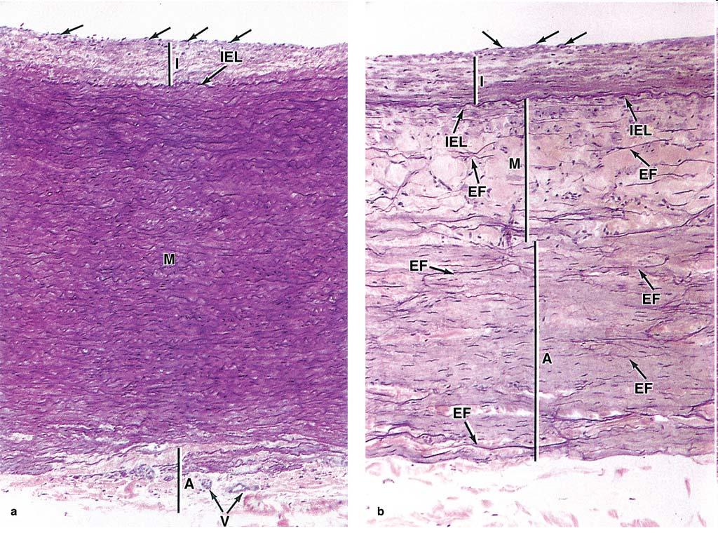
22 22

23 Large Elastic Arteries Help to stabilize blood flow Include: aorta & its large branches Fleshly: yellowish color from elastin in media Intima: thicker than muscular arteries internal elastic lamina: present may not easily discerned Media: elastic fibers & concentrically arranged elastic laminae smooth muscle cells, reticular fibers, proteoglycans, glycoproteins Adventitia: relatively underdeveloped 23

24 Muscular Arteries Control blood flow organs Intima: very thin subendothelial layer prominent internal elastic lamina Media: up to 40 layers of more prominent smooth muscle cells intermingled with elastic lamellae, reticular fibers, proteoglycans external elastic lamina: only in larger muscular arteries Adventitia: connective tissue contain lymphatic capillaries, vasa vasorum, nerves 24

25 Arterioles Muscular arteries branch repeatedly smaller and smaller 2 or 3 medial layers of muscle Arterioles: smallest arteries 1 or 2 layers of smooth muscle layers generally less than 0.5 mm in diameter lumen as wide as wall is thick subendothelial layer: very thin elastic laminae: absent media: circularly arranged smooth muscle cells adventitia: very thin & inconspicuous 25
26 Capillaries Permit different levels of metabolic exchange between blood & tissues Composed of : a single layer of endothelial cells Endothelial cells: form a tube Capillaries: 5-10 μm average diameter length: 50 μm comprise over 90% of all blood vessels total length: 96000km total diameter: 800 times larger than aorta 26

27 Velocity of blood: in aorta: 320 mm/s in capillaries: 0.3 mm/s because their thin wall & slow flow a favorable place for exchange water, solutes, macromolecules In general: endothelial cells: polygonal & elongated in the direction of blood flow nucleus: bulge into lumen junctions of tight zonula occludentes type present 27

28 Pericytes: enclosed by a basal lamina may fuse with that of endothelial cells well-developed networks of myosin, actin, tropomyosin : contractile function After tissue injuries: pericytes proliferate & differentiate form tunica media & cells with various other functions in re-establishing microvasculature & its ECM 28
 capillaries allow regular exchange of material")

29 Capillary: structural variations 3 types 1. continuous (tight) capillaries allow regular exchange of material most common type found in muscles, connective tissue, exocrine glands & nervous tissue pinocytotic vesicles : luminal & basal surface transport of material 29


30 2.Fenestrated capillaries: allow more extensive molecular exchange presence of small circular fenestrae covered by a very thin diaphragm containing heparan proteoglycans no lipid bilayer basal lamina: continuous voering fenestrae found in kidney, intestine, choroid plexus, endocrine glands 30


31 3. Sinusoid (Discontinuous) capillaries : permit maximal exchange endothelial cells: large fenestrae without diaphragm form a discontinuous layer separate from one another:wide space basal lamina: also discontinuous irregularly shaped diameter: 30-40μm found in liver, spleen, some endocrine organs, bone marrow 31

32 Venules Transition from capillaries to venules: gradually Postcapillary venules: similar structurally to capillaries with pricytes but diameter: from 15-20μm participate in exchange converge larger collecting venules 2 or 3 smooth muscle layers muscular venules 32

33 Veins Blood entering vein: very low pressure move forward heart: contraction of tunica media external compression from surrounding muscles or organs Valve: projection from tunica intima to prevent back-flow of blood Most veins: small or medium veins diameter less than 1 cm located in parallel with muscular arteries 33
34 Intima thin subendothelial layer Media small bundles of smooth muscle cells intermixed with reticular fibers & a delicate network of elastic fibers Adventitia collagenous well-developed 34

35 Big venous trunks, paired with elastic arteries: large veins Well developed intima Media: relative thin few layers of smooth muscle cells abundant connective tissue Adventitia: thick in large veins frequently contain longitudinal bundles of smooth muscle Both media & adventitia: contain elastic fibers not like those of elastic arteries 35

36 LYMPHATIC VASCULAR SYSTEM Collect excess interstitial fluid from tissue space return to blood Fluid: lymph Flow in only one direction: toward the heart Lymphatic capillaries originate in various tissue thin, closed-ended vessels a single layer of endothelium + an incomplete basal lamina held open by bundles of anchoring filaments of elastic fiber system also bind vessels to connective tissue 36

37 Lymphatic capillaries larger lymphatic vessels Interposed in path : lymph nodes Lymphatic: found in almost all organs except CNS & bone marrow Larger lymphatics: similar to veins thinner walls lack a clear-cut separation between tunics more numerous internal valves 37
38 Lymphatic vessels: dilated assumed a nodular or beaded, between valves Lymphatic circulation: aided by external forces contraction of skeletal muscles unidirectional flow: many valves contraction of smooth muscles of larger lymphatic vessels: also help to propel End up as 2 large trunk: thoracic duct & right lymphatic trunk empty lymph veins 38
39 Structure of lymphatic ducts: similar to that of large veins reinforced smooth muscle in middle layer muscle bundles: longitudinally & circularly arranged former predominating adventitia: relatively undeveloped contain vasa vasorum & neural network Lymphatic vessels: also a major distributor of lymphocytes, antibodies, immune components 39
The Circulatory System
 The Circulatory System Dr. Sami Zaqout The circulatory system Circulatory system Blood vascular systems Lymphatic vascular systems Blood vascular systems Blood vascular systems The circulatory system Circulatory
The cardiovascular system
 The cardiovascular system Components of the Cardiovascular system Heart Vessels: Arteries Capillaries Veins Functions of CVS: Transportation system where blood is the transporting vehicle Carries oxygen,
Cardiovascular (Circulatory) System
 System") Cardiovascular (Circulatory) System Piryaei May 2011 Circulatory System Heart Blood Vessels Macrovasculature (More than 0.1mm) Elastic Artery Muscular (Distributing) Artery Large Arteriol Small Vein Muscular
Cardiovascular (Circulatory) System Piryaei May 2011 Circulatory System Heart Blood Vessels Macrovasculature (More than 0.1mm) Elastic Artery Muscular (Distributing) Artery Large Arteriol Small Vein Muscular
Histology of the Cardiac System. Dr. Nabil Khoury Anatomy Department
 Histology of the Cardiac System Dr. Nabil Khoury Anatomy Department Objectives 1. Identify the 3 layers of the heart endocardium, myocardium, epicardium 2. Differentiate cardiacmuscle 3. Define intercalated
The Circulatory System
 The Circulatory System This system comprises both the blood and lymphatic vascular system. Blood vascular system is composed from; The heart: an organ whose function is to pump the blood. The arteries:
Practical Histology. Cardiovascular System. Dr Narmeen S. Ahmad
 Practical Histology Cardiovascular System Dr Narmeen S. Ahmad The Cardiovascular System A closed system of the heart and blood vessels Functions of cardiovascular system: Transport nutrients, hormones
CVS HISTOLOGY. Dr. Nabil Khouri.
 CVS HISTOLOGY Dr. Nabil Khouri http://anatomy.kmu.edu.tw/blockhis/block3/slides/block4_24.html The Heart Wall Contract as a single unit Cardiac Muscle Simultaneous contraction due to depolarizing at the
SCPA602 Cardiovascular System
 SCPA602 Cardiovascular System Associate Professor Dr. Wannee Jiraungkoorskul Department of Pathobiology, Faculty of Science, Mahidol University Tel: 02-201-5563, E-mail: wannee.jir@mahidol.ac.th 1 Objectives
Histology of the myocardium and blood vessels. Prof. Abdulameer Al-Nuaimi
 Histology of the myocardium and blood vessels Prof. Abdulameer Al-Nuaimi E-mail: a.al-nuaimi@sheffield.ac.uk E-mail: abdulameerh@yahoo.com Histology of blood vessels The walls of arteries and veins are
Copyright 2010 Pearson Education, Inc. Blood Vessel Structure
 Blood Vessel Structure Structure of Blood Vessel Walls Arteries and veins Tunica intima, tunica media, and tunica externa Lumen Central blood-containing space Capillaries Endothelium with sparse basal
Lecture name: blood 2 & The Circulatory System Edited by: Buthainah Al masaeed & Yousef Qandeel
 Lecture name: blood 2 & The Circulatory System Edited by: Buthainah Al masaeed & Yousef Qandeel Now we will take about A granulocytes : Lymphocyte Monocytes 1- Lymphocyte - The second major type of presence
Remember: the CVS system is deriving from the mesenchyma and is lined by simple squamous epithelium called the endothelium
 Lecture 10 General med_2nd semester Microscopic anatomy and embryology of cardiovascular system Microscopic structure of the heart, excitomotoric system - its structural peculiarities Blood vessels - arteries
Microscopic Anatomy CARDIOVASCULAR SYSTEM
 Microscopic Anatomy CARDIOVASCULAR SYSTEM I. Introduction The cardiovascular system is a closed system consisting of a pump, the heart, and a series of tubular blood vessels that interconnect all body
UNIT 4: BLOOD VESSELS
 UNIT 4: BLOOD VESSELS Dr. Moattar Raza Rizvi NRS237, Physiology Generalized Structure of Blood Vessels 1 Tunica interna (tunica intima) Endothelial layer that lines the lumen of all vessels In vessels
Cardiovascular System Blood Vessels
 Cardiovascular System Blood Vessels Structure of Blood Vessels The three layers (tunics) Tunica intima composed of simple squamous epithelium Tunica media sheets of smooth muscle Contraction vasoconstriction
Extra notes for lab- 1 histology. Slide 1 : cross section in the elastic artery ( aortic arch, ascending aorta, descending aorta )
") Extra notes for lab- 1 histology Slide 1 : cross section in the elastic artery ( aortic arch, ascending aorta, descending aorta ) - twin of ascending aorta is the pulmonary trunk. Ascending aorta represents
Extra notes for lab- 1 histology Slide 1 : cross section in the elastic artery ( aortic arch, ascending aorta, descending aorta ) - twin of ascending aorta is the pulmonary trunk. Ascending aorta represents
Blood Vessels. Types of Blood Vessels Arteries carry blood away from the heart Capillaries smallest blood vessels. Veins carry blood toward the heart
 C H A P T E R Blood Vessels 20 Types of Blood Vessels Arteries carry blood away from the heart Capillaries smallest blood vessels The site of exchange of molecules between blood and tissue fluid Veins
2. capillaries - allow exchange of materials between blood and tissue fluid
 Chapter 19 - Vascular System A. categories and general functions: 1. arteries - carry blood away from heart 2. capillaries - allow exchange of materials between blood and tissue fluid 3. veins - return
Cardiac Conduction System
 Cardiac Conduction System What causes the Heart to Beat? Heart contracts by electrical signals! Cardiac muscle tissue contracts on its own an electrical signal is sent out by the heart so that all cells
a) Endocardium The endocardium is the innermost layer of the heart wall. It forms the internal lining of the atria and ventricles.
 Endocardium The endocardium is the innermost layer of the heart wall. It forms the internal lining of the atria and ventricles.") Chapter 11 Circulatory System The circulatory system consists of a muscular, pulsing heart and a system of blood vessels.the blood vessels include: Arteries which will carry the blood and it's dissolved
Chapter 11 Circulatory System The circulatory system consists of a muscular, pulsing heart and a system of blood vessels.the blood vessels include: Arteries which will carry the blood and it's dissolved
Major Function of the Cardiovascular System. Transportation. Structures of the Cardiovascular System. Heart - muscular pump
 Structures of the Cardiovascular System Heart - muscular pump Blood vessels - network of tubes Blood - liquid transport vehicle brachiocephalic trunk superior vena cava right pulmonary arteries right pulmonary
Cardiovascular System
 Cardiovascular System Purpose Transport oxygen and nutrients Take waste products away from tissues & organs Things we learned Blood pressure: the force of blood pushing against the walls of blood vessels
Anatomy Review: The Heart Graphics are used with permission of A.D.A.M. Software, Inc. and Benjamin/Cummings Publishing Co.
 Anatomy Review: The Heart Graphics are used with permission of A.D.A.M. Software, Inc. and Benjamin/Cummings Publishing Co. Anatomy Views Label the diagrams of the heart below: Interactive Physiology Study
The Cardiovascular and Lymphatic Systems Cardiovascular System Blood Vessels Blood Vessels Arteries Arteries Arteries
 CH 12 The Cardiovascular and s The Cardiovascular and s OUTLINE: Cardiovascular System Blood Vessels Blood Pressure Cardiovascular System The cardiovascular system is composed of Blood vessels This system
Chapter 14. The Cardiovascular System
 Chapter 14 The Cardiovascular System Introduction Cardiovascular system - heart, blood and blood vessels Cardiac muscle makes up bulk of heart provides force to pump blood Function - transports blood 2
Ch. 12 The Circulatory System. The heart. The heart is a double pump. A quick note on arteries vs. veins. = the muscular pump of the CV system
 Ch. 12 The Circulatory System The heart A.k.a. the cardiovascular system Blood was discussed in Ch. 11 Focus of Ch. 12: heart and blood vessels = the muscular pump of the CV system ~ 100,000 heartbeats/day!
10. Thick deposits of lipids on the walls of blood vessels, called, can lead to serious circulatory issues. A. aneurysm B. atherosclerosis C.
 Heart Student: 1. carry blood away from the heart. A. Arteries B. Veins C. Capillaries 2. What is the leading cause of heart attack and stroke in North America? A. alcohol B. smoking C. arteriosclerosis
Heart. Heart 2-Tunica media: middle layer (media ='middle') muscle fibers (smooth or cardiac).
 muscle fibers (smooth or cardiac).") t. innermost lumenal General Circulatory system heart and blood vessels walls have 3 layers (inside to outside) 1-Tunica interna: aka tunica intima layer--lumenal layer epithelium--endothelium simple squamous
t. innermost lumenal General Circulatory system heart and blood vessels walls have 3 layers (inside to outside) 1-Tunica interna: aka tunica intima layer--lumenal layer epithelium--endothelium simple squamous
2. right heart = pulmonary pump takes blood to lungs to pick up oxygen and get rid of carbon dioxide
 A. location in thorax, in inferior mediastinum posterior to sternum medial to lungs superior to diaphragm anterior to vertebrae orientation - oblique apex points down and to the left 2/3 of mass on left
Cardiovascular Anatomy Dr. Gary Mumaugh
 Cardiovascular Anatomy Dr. Gary Mumaugh Location of Heart Approximately the size of your fist Location o Superior surface of diaphragm o Left of the midline in mediastinum o Anterior to the vertebral column,
組織學 Historlogy 台北醫學大學 / 解剖學科教授 : 邱瑞珍分機號碼 :3261. 電子郵件信箱
 組織學 Historlogy 台北醫學大學 / 解剖學科教授 : 邱瑞珍分機號碼 :3261 電子郵件信箱 :rueijen@tmu.edu.tw 1 Connective Tissue(II) 台北醫學大學 / 解剖學科教授 : 邱瑞珍分機號碼 :3261 電子郵件信箱 :rueijen@tmu.edu.tw 2 學習目的 Cell types Types of the fibers Types
Lab Activity 25. Blood Vessels & Circulation. Portland Community College BI 232
 Lab Activity 25 Blood Vessels & Circulation Portland Community College BI 232 Artery and Vein Histology Walls have 3 layers: Tunica intima Tunica media Tunica externa 2 Tunica Intima Is the innermost layer
The Cardiovascular and Lymphatic Systems
 BIOLOGY OF HUMANS Concepts, Applications, and Issues Fifth Edition Judith Goodenough Betty McGuire 12 The Cardiovascular and Lymphatic Systems Lecture Presentation Anne Gasc Hawaii Pacific University and
STRUCTURES OF THE CARDIOVASCULAR SYSTEM
 STRUCTURES OF THE CARDIOVASCULAR SYSTEM CARDIOVASCULAR SYSTEM Also called the circulatory system Consists of the heart, arteries, veins, and capillaries Main function is to pump/circulate oxygenated blood
The Cardiovascular System
 The Cardiovascular System The Cardiovascular System A closed system of the heart and blood vessels The heart pumps blood Blood vessels allow blood to circulate to all parts of the body The function of
The Circulatory System. Lesson Overview. Lesson Overview The Circulatory System
 33.1 THINK ABOUT IT More than one-third of the 1.2 million Americans who suffer a heart attack each year die. This grim evidence shows that the heart and the circulatory system it powers are vital to life.
Approximately the size of your fist Location Superior surface of diaphragm Left of the midline in mediastinum Anterior to the vertebral column,
 Dr. Gary Mumaugh Approximately the size of your fist Location Superior surface of diaphragm Left of the midline in mediastinum Anterior to the vertebral column, posterior to the sternum Posteriorly the
Circulation. Sinoatrial (SA) Node. Atrioventricular (AV) Node. Cardiac Conduction System. Cardiac Conduction System. Linked to the nervous system
 Node. Atrioventricular (AV) Node. Cardiac Conduction System. Cardiac Conduction System. Linked to the nervous system") Circulation Cardiac Conduction System AHS A H S Your body resembles a large roadmap. There are routes or arteries that take you downtown to the heart of the city and veins that take you to the outskirts
Circulation Cardiac Conduction System AHS A H S Your body resembles a large roadmap. There are routes or arteries that take you downtown to the heart of the city and veins that take you to the outskirts
1. What kind of blood is found in the rt. atrium? (oxygenated or deoxygenated)
") Carl Christennsen, PhD Chap. 19, 20, & 21 - Circulatory System Bio. 2304 Human Anatomy HEART 1. What kind of blood is found in the rt. atrium? (oxygenated or deoxygenated) Where does this blood come from?
Carl Christennsen, PhD Chap. 19, 20, & 21 - Circulatory System Bio. 2304 Human Anatomy HEART 1. What kind of blood is found in the rt. atrium? (oxygenated or deoxygenated) Where does this blood come from?
Health Science 20 Circulatory System Notes
 Health Science 20 Circulatory System Notes Functions of the Circulatory System The circulatory system functions mainly as the body s transport system. It transports: o Oxygen o Nutrients o Cell waste o
Cardivascular System Module 5: Structure and Function of Blood Vessels *
 OpenStax-CNX module: m49689 1 Cardivascular System Module 5: Structure and Function of Blood Vessels * Donna Browne Based on Structure and Function of Blood Vessels by OpenStax This work is produced by
Human Anatomy, First Edition
 Human Anatomy, First Edition McKinley & O'Loughlin Chapter 22 : Heart 1 Functions of the Heart Center of the cardiovascular system, the heart. Connects to blood vessels that transport blood between the
Cardiovascular System. I. Structures of the heart A. : Pericardium sack that surrounds the heart
 Cardiovascular System I. Structures of the heart A. : Pericardium sack that surrounds the heart 1. : Pericardial Cavity serous fluid filled space between the heart and the pericardium B. Heart Wall 1.
Structure and organization of blood vessels
 The cardiovascular system Structure of the heart The cardiac cycle Structure and organization of blood vessels What is the cardiovascular system? The heart is a double pump heart arteries arterioles veins
Sinusoids and venous sinuses
 LYMPHOID SYSTEM General aspects Consists of organs that are made of lymphoid tissue; Immune defense Breakdown of red blood cells. 1 Sinusoids In place of capillaries Endothelium; often fenestrated More
Human Anatomy, First Edition
 Human Anatomy, First Edition McKinley & O'Loughlin Chapter 23 : Vessels and Circulation 23-1 Blood Vessels An efficient style of transport for oxygen, nutrients, and waste products to and from body tissues.
Circulatory System: Introduction. Dr. Carmen E. Rexach Anatomy 35 Mt. San Antonio College
 Circulatory System: Introduction Dr. Carmen E. Rexach Anatomy 35 Mt. San Antonio College Components Cardiovascular system Lymphatic system Cardiovascular system Heart, blood vessels, blood Functions: transport
Blood flows away from the heart in arteries, to the capillaries and back to the heart in the veins
 Cardiovascular System Summary Notes The cardiovascular system includes: The heart, a muscular pump The blood, a fluid connective tissue The blood vessels, arteries, veins and capillaries Blood flows away
Cardiovascular System. Blood Vessel anatomy Physiology & regulation
 Cardiovascular System Blood Vessel anatomy Physiology & regulation Path of blood flow Aorta Arteries Arterioles Capillaries Venules Veins Vena cava Vessel anatomy: 3 layers Tunica externa (adventitia):
d) Cardiovascular System Higher Human Biology
 Cardiovascular System Higher Human Biology") d) Cardiovascular System Higher Human Biology What can your remember about the heart and blood vessels? What is the Cardiovascular System? The cardiovascular system, also known as the circulatory system,
d) Cardiovascular System Higher Human Biology What can your remember about the heart and blood vessels? What is the Cardiovascular System? The cardiovascular system, also known as the circulatory system,
Warm Up- Monday -AND- Setup Cornell Notes.
 Warm Up- Monday Brainstorm in your notebook: If the heart sends blood to all organs, how and where does the heart get blood to provide oxygen for its muscles? -AND- Setup Cornell Notes. Announcements Unit
Cardiovascular Physiology
 Cardiovascular Physiology Lecture 1 objectives Explain the basic anatomy of the heart and its arrangement into 4 chambers. Appreciate that blood flows in series through the systemic and pulmonary circulations.
Anatomy & Physiology of Cardiovascular System. Chapter 18 & 19
 Anatomy & Physiology of Cardiovascular System Chapter 18 & 19 Objectives..cont 1. Discuss the physiological stages of cardiac muscle contraction. 2. Trace a typical ECG and label each wave or complex 3.
Figure ) The specific chamber of the heart that is indicated by letter A is called the. Diff: 1 Page Ref: 364
 The specific chamber of the heart that is indicated by letter A is called the. Diff: 1 Page Ref: 364") Essentials of Anatomy and Physiology, 9e (Marieb) Chapter 11 The Cardiovascular System Short Answer Figure 11.1 Using Figure 11.1, identify the following: 1) The Purkinje fibers are indicated by label.
Essentials of Anatomy and Physiology, 9e (Marieb) Chapter 11 The Cardiovascular System Short Answer Figure 11.1 Using Figure 11.1, identify the following: 1) The Purkinje fibers are indicated by label.
The Circulatory System. The Heart, Blood Vessels, Blood Types
 The Circulatory System The Heart, Blood Vessels, Blood Types The Closed Circulatory System Humans have a closed circulatory system, typical of all vertebrates, in which blood is confined to vessels and
Cardiovascular System
 Cardiovascular System I. Structure of the Heart A. Average adult heart is 14 cm long and 9 cm wide. B. Lies in the mediastinum. C. Enclosed in the pericardium. 1. Fibrous pericardium- Outer, tough connective
Chapter 21 Peripheral circulation and Regulation
 Chapter 21 Peripheral circulation and Regulation I. Blood vessel structure A. Blood flows from large arteries to small capillaries 1. Large arteries contain large amounts of elastic tissue and little smooth
Topic 6: Human Physiology
 Topic 6: Human Physiology 6.2 The Blood System D.4 The Heart Essential Questions: 6.2 The blood system continuously transports substances to cells and simultaneously collects waste products. D.3 The chemical
Cardiovascular system
 BIO 301 Human Physiology Cardiovascular system The Cardiovascular System: consists of the heart plus all the blood vessels transports blood to all parts of the body in two 'circulations': pulmonary (lungs)
The Heart. The Heart A muscular double pump. The Pulmonary and Systemic Circuits
 C H A P T E R 19 The Heart The Heart A muscular double pump circuit takes blood to and from the lungs Systemic circuit vessels transport blood to and from body tissues Atria receive blood from the pulmonary
The Cardiovascular System: Vessels and Routes. Pulmonary Circulation H E A R T. Systemic Circulation
 The Cardiovascular System: Vessels and Routes 1. Overview of Blood Circulation A. Pulmonary Circulation Lung Arterioles Pulmonary Artery Capillaries Pulmonary Circulation Venules Pulmonary Veins H E A
Cardiovascular System. Biology 105 Lecture 15 Chapter 12
 Cardiovascular System Biology 105 Lecture 15 Chapter 12 Outline I. Functions of cardiovascular system II. Components of the cardiovascular system: I. Blood vessels II. Heart III. Regulation of the heartbeat
Cardiovascular System Notes: Physiology of the Heart
 Cardiovascular System Notes: Physiology of the Heart Interesting Heart Fact Capillaries are so small it takes ten of them to equal the thickness of a human hair. Review What are the 3 parts of the cardiovascular
Cardiovascular. Function of the cardiovascular system is to transport blood containing: Nutrients Waste Hormones Immune cells Oxygen
 Cardiovascular The Cardiovascular System - Arteries Arteries Cardiovascular System Function of the cardiovascular system is to transport blood containing: Carry blood away from heart Carotid arteries Deliver
Heart. Large lymphatic vessels Lymph node. Lymphatic. system Arteriovenous anastomosis. (exchange vessels)
") Venous system Large veins (capacitance vessels) Small veins (capacitance vessels) Postcapillary venule Thoroughfare channel Heart Large lymphatic vessels Lymph node Lymphatic system Arteriovenous anastomosis
Venous system Large veins (capacitance vessels) Small veins (capacitance vessels) Postcapillary venule Thoroughfare channel Heart Large lymphatic vessels Lymph node Lymphatic system Arteriovenous anastomosis
Circulatory System 10.1
 1 Circulatory System 10.1 2 ARTERIES Arteries-blood vessels that carry blood away from the heart Thick walls Inner & Outer layers: connective tissue Middle layers are muscle and elastic connective tissue
Any of these questions could be asked as open question or lab question, thus study them well
 Any of these questions could be asked as open question or lab question, thus study them well describe the factors which regulate cardiac output describe the sympathetic and parasympathetic control of heart
Anatomy and Physiology, Spring 2015 Exam II: Form A April 9, Name Student Number
 Anatomy and Physiology, Spring 2015 Exam II: Form A April 9, 2015 Name Student Number For Questions 1 2 refer to the following table. 1 Ventricular pressure is greater than aortic 6 AV valve is open 2
Microcirculation. Lecture Block 11 (contributions from Brett Burton)
") Lecture Block 11 (contributions from Brett Burton) Elements of Arterioles, capillaries, venules Structure and function: transport Fluid balance Lymph system Vessels of the Circulatory System Diameter Aorta
Lecture Block 11 (contributions from Brett Burton) Elements of Arterioles, capillaries, venules Structure and function: transport Fluid balance Lymph system Vessels of the Circulatory System Diameter Aorta
Chapter 21 (1) An Introduction to Blood Vessels and Circulation
 An Introduction to Blood Vessels and Circulation") Chapter 21 (1) An Introduction to Blood Vessels and Circulation Lecture Objectives Compare and contrast the structure of an artery, arteriole, vein, venule, and capillary Discuss the structure and function
Chapter 21 (1) An Introduction to Blood Vessels and Circulation Lecture Objectives Compare and contrast the structure of an artery, arteriole, vein, venule, and capillary Discuss the structure and function
CIRCULATORY SYSTEM BLOOD VESSELS
 Name: Block: CIRCULATORY SYSTEM Multicellular organisms (above the level of roundworms) rely on a circulatory system to bring nutrients to, and take wastes away from, cells. In higher organisms such as
Function: Transportation of. Oxygen Nutrients Waste Hormones gases
 Function: Transportation of Oxygen Nutrients Waste Hormones gases Pericardium: double sac of serous membrane filled with fluid (pericardial fluid to be exact) that surrounds the heart. Parietal pericardium:
The heart & Cardiovascular system
 The heart & Cardiovascular system The heart s continuous pulse create a base for our understanding of rhythms in everyday life. Bonnie Bainbridge Cohen The heart constantly beats throughout our lives never
KEY - Sample Exam Biology 2050 Circulatory and Lymphatic Systems - KEY
 KEY - Sample Exam Biology 2050 Circulatory and Lymphatic Systems - KEY Note: Not all of the lymphatic system will be on the actual exam next Monday so disregard any questions that deal with something that
Chapter 20 (1) The Heart
 The Heart") Chapter 20 (1) The Heart Learning Objectives Describe the location and structure of the heart Describe the path of a drop of blood from the superior vena cava or inferior vena cava through the heart out
Chapter 20 (1) The Heart Learning Objectives Describe the location and structure of the heart Describe the path of a drop of blood from the superior vena cava or inferior vena cava through the heart out
Sample Exam Biology 2050 Circulatory and Lymphatic Systems
 Sample Exam Biology 2050 Circulatory and Lymphatic Systems Note: Not all of the lymphatic system will be on the actual exam next Monday so disregard any questions that deal with something that wasn t covered
Blood Vessels. Over view. We have about 60,000 miles of blood vessels!
 Blood Vessels Over view 3 types of blood vessels arteries - carry blood away from heart "branch", "diverge", and "fork" veins - carry blood toward heart "join", "merge", and "converge" capillaries - site
Chapter 20: Cardiovascular System: The Heart
 Chapter 20: Cardiovascular System: The Heart I. Functions of the Heart A. List and describe the four functions of the heart: 1. 2. 3. 4. II. Size, Shape, and Location of the Heart A. Size and Shape 1.
Chapter 13 The Cardiovascular System: Cardiac Function
 Chapter 13 The Cardiovascular System: Cardiac Function Overview of the Cardiovascular System The Path of Blood Flow through the Heart and Vasculature Anatomy of the Heart Electrical Activity of the Heart
Cardiovascular System
 Cardiovascular System The Heart Cardiovascular System The Heart Overview What does the heart do? By timed muscular contractions creates pressure gradients blood moves then from high pressure to low pressure
The Heart. Happy Friday! #takeoutyournotes #testnotgradedyet
 The Heart Happy Friday! #takeoutyournotes #testnotgradedyet Introduction Cardiovascular system distributes blood Pump (heart) Distribution areas (capillaries) Heart has 4 compartments 2 receive blood (atria)
Cardiovascular System, Blood, and Blood Cell Formation
 Cardiovascular System, Blood, and Blood Cell Formation 7 CONTENTS OVERVIEW OF THE CARDIOVASCULAR SYSTEM GENERAL STRUCTURE OF THE HEART Heart Valves HEART AS A PUMP Contractile Properties of Cardiac Myocytes
The Circulatory System (p )
") The Circulatory System (p. 268-281) How Does Gravity Affect Blood Circulation? As with all land animals, the giraffe and the corn snake are constantly subject to the force of gravity The circulatory system
The Circulatory System (p. 268-281) How Does Gravity Affect Blood Circulation? As with all land animals, the giraffe and the corn snake are constantly subject to the force of gravity The circulatory system
Levels of Organization. Chapter 19 6/11/2012. Homeostasis & Organization of the animal body. 4 Primary Tissues
 Levels of Organization Chapter 19 Homeostasis & Organization of the animal body Chemical Cellular Tissue Organs System Level Organismic 1-2 4 Primary Tissues 1. Epithelial Tissue: covers surfaces lines
Blood Vessels. Chapter 20
 Blood Vessels Chapter 20 Summary of the Characteristics of Arteries and Veins Characteristic Artery Vein Wall thickness thick thin Shape in cross section round flattened Thickest tunic media externa Collagen
11/10/2014. Muscular pump Two atria Two ventricles. In mediastinum of thoracic cavity 2/3 of heart's mass lies left of midline of sternum
 It beats over 100,000 times a day to pump over 1,800 gallons of blood per day through over 60,000 miles of blood vessels. During the average lifetime, the heart pumps nearly 3 billion times, delivering
Collin County Community College. ! BIOL Anatomy & Physiology! WEEK 5. The Heart
 Collin County Community College! BIOL. 2402 Anatomy & Physiology! WEEK 5 The Heart 1 (1578-1657) A groundbreaking work in the history of medicine, English physician William Harvey s Anatomical Essay on
CIRCULATION. Cardiovascular & lymphatic systems Functions. Transport Defense / immunity Homeostasis
 CIRCULATION CIRCULATION Cardiovascular & lymphatic systems Functions Transport Defense / immunity Homeostasis 2 Types of Circulatory Systems Open circulatory system Contains vascular elements Mixing of
The Cardiovascular System
 Essentials of Human Anatomy & Physiology Elaine N. Marieb Slides 11.1 11.19 Seventh Edition Chapter 11 The Cardiovascular System Functions of the Cardiovascular system Function of the heart: to pump blood
Muscle Tissue. General concepts. Classification of muscle. I. Functional classification is based on the type of neural control.
 Muscle Tissue LEARNING OBJECTIVES 1. Identify the three types of muscle tissue at the light microscopic level. 2. List and compare the structural and functional features of each of the three muscle fiber
1. Which of the following blood vessels has a thin elastic layer? A. Aorta. B. Pulmonary artery. C. Posterior vena cava. D. Mesenteric capillary.
 CIRCULATORY SYSTEM 1. Which of the following blood vessels has a thin elastic layer? A. Aorta. B. Pulmonary artery. C. Posterior vena cava. D. Mesenteric capillary. 2. Capillary beds are equipped with
The Heart and Cardiovascular System
 The Heart and Cardiovascular System What you will learn The location of the heart 3 layers and covering of the heart Explain the function of the heart as 2 separate pumps Identify the 4 chambers of the
Anatomy of the Heart. Figure 20 2c
 Anatomy of the Heart Figure 20 2c Pericardium & Myocardium Remember, the heart sits in it s own cavity, known as the mediastinum. The heart is surrounded by the Pericardium, a double lining of the pericardial
Chp. 5 The cardiovascular system. What are the function of the cardiovascular system? Arteries and arterioles:
 5.1 Overview of the cardiovascular system Chp. 5 The cardiovascular system Includes the heart and blood vessels Brings nutrients to cells and helps get rid of wastes Blood is refreshed in the lung, kidneys,
Lec #2 histology. Bronchioles:
 Lec #2 histology. Last lecture we talked about the upper respiratory tract histology, this one is about the lower part histology. We will discuss the histology of: -bronchioles -respiratory bronchioles
Ch 19: Cardiovascular System - The Heart -
 Ch 19: Cardiovascular System - The Heart - Give a detailed description of the superficial and internal anatomy of the heart, including the pericardium, the myocardium, and the cardiac muscle. Trace the
Muscle tissues. Dr. Hersh Abdul Ham-Karim BVM&S, PG Dip, MSc and PhD
 Muscle tissues Dr. Hersh Abdul Ham-Karim BVM&S, PG Dip, MSc and PhD Muscle tissue is a soft tissue that composes muscles in animal bodies, and gives rise to muscles' ability to contract. Muscle tissue
Tissues 10/21/2016. Epithelial Tissue
 Tissues This is a generalized cell diagram. It shows the anatomy of a cell, but most cells do not actually look like this. Cells can have a wide variety of shapes and sizes, depending on their function.
Skeletal muscle. General features :
 Muscular tissues In the first embryonic life the muscular tissues arise from mesoderm, The function of movement in multicellular organisms is usually assumed by specialized cells called muscle fibers which
: thick middle layer; cardiac muscles : thin inner layer; endothelial lining
 1 2 3 4 Bio 1102 Lecture 5 (guided) Chapter 8: Heart & Blood Vessels Functions of Circulatory System: To carry from lungs to all cells, tissues, and organs of body To carry from digestive system to all