Neuroanatomy. Dr. Maha ELBeltagy. Assistant Professor of Anatomy Faculty of Medicine The University of Jordan
|
|
|
- Sheila Lewis
- 5 years ago
- Views:
Transcription
1 Neuroanatomy Dr. Maha ELBeltagy Assistant Professor of Anatomy Faculty of Medicine The University of Jordan 2018


2 THE BRAIN STEM It includes: Midbrain Pons Medulla oblongata Anterior view Anterior view 1. Optic chiasma 2. Optic nerve 3. Optic tract 4. Medial sulcus of the crus cerebri 5. Oculomotor nerve 6. Pons 7. Pyramidal eminence of the pons 8. Retroolivary fossa 9. Olive 10. Posterolateral sulcus 11. Decusssation of the pyramids 12. Anterolateral sulcus 13. Lateral funiculus 14. Pyramid 15. Foramen caecum 16. Middle cerebellar pedunculus 17. Trigeminal nerve 18. Crus cerebri 19. Posterior perforated substance 20. Mammillary body 21. Tuber cinereum 22. Infundibulum
3 The midbrain The cerebral aqueduct divides the midbrain into : Tectum (behind the duct). 2 cerebral peduncles: each one consist itof Crus cerebri (2), Substantia nigra & Tegmentum. Anterior surface of midbrain: Tegmentum Tectum Substantia nigra Is formed of the 2 crura enclosing interpeduncular fossa in between them. The oculomotor nerve (1) emerges from the medial aspect of the crus cerebri. Posterior surface: Is formed of : 2 superior colliculi (3): connected to the lateral 1 2 geniculate body (6) by superior brachium (5). 5 2 inferior colliculi (4) : connected to the medial 3 geniculate body (8) by inferior brachium (7). 6 The trochlear nerve emerges from the back of midbrain
4 The interpeduncular Fossa Boundaries: Anterior: Optic chiasma (1). Posterior: Pons Anterolateral: Optic tract t& crura cerebri bi Crus cerebri Contents: Tuber cinereum (2). Mammillary bodies (3) Posterior perforated substance (4) Oculomotor nerve (5) emerges from the medial aspect of crus cerebri bi. 5 Pons 3 4
5 The Pons Anterior surface: 15 Has a median basilar groove for basilar artery (15). 16 Pyramidal eminence (16) on either side of basilar groove with transverse pontine fibers. MCP Middle cerebellar peduncle (MCP). Trigeminal nerve (17) between the pons & MCP. Posterior surface: Forms the upper part of the floor of the 4 th ventricle. It is separated from the medulla by medullarystria (15). It has a posterior median sulcus (8) with medial eminence (9) on either side. Facial colliculus (16) is a small swelling in the lower part of the medial eminence Superior fovea (17) is a depression lateral to facial colliculus Anterior surface 9 9 Posterior surface MCP
6 The Medulla Oblongata Its upper ½ has no central canal & is known as open medulla. Its lower ½ has central canal & is known as open medulla. Anterior surface: Has: Anterior median fissure which is obliterated below by pyramidal decussation (11). Pyramid (14) on either side of the median fissure Lateral surface: Has Olive (9) separated from the pyramid by anterolateral sulcus (12) to which rootlets of hypoglossal nerve are attached. Inferior cerebellar peduncle (ICP) separated from the olive by posterolateral lsulcus (10) to which h rootlets of glossopharyngeal, vagus & accessory nerves are attached. Pyramid 12 Olive 10 Lateral surface
7 Posterior surface: Upper part: formslowerpartof the floor of the 4 th ventricle & has the following features: Posterior median sulcus. Inferior fovea ((19) which is an inverted V shaped sulcus dividing the back of medulla into; Hypoglossal triangle (14), vagal triangle (13) & vestibular area (18). Lower part: back of closed medulla which has the following features: Posterior mediansulcus. On either side of the sulcus Gracile & Cuneate tracts ascend & expand above to form Gracile (10) & Cuneate tubercle (9)
8 The fourth ventricle It is a diamond shaped cavity of the hindbrain. It lies behind the pons & open medulla & in front of the cerebellum. Its superior angle is continuous with the cerebral aqueduct of midbrain & its inferior angle is continuous with the central canal of closed medulla (at the obex). It has 2 lateral recesses which curve around the inferior cerebellar peduncle & open by lateral apertures in the subarachnoid space at the flocculus. The roof: Is tent shaped & is formed of The superior cerebellar peduncles (SCPs). the superior medullary velum (SMV) stretching between the 2 SCPs. The inferior medullary velum (IMV) which has a median aperture (of Magendie) connecting the 4 th ventricle to the subarachnoid space. SMV IMV Cerebellum Median aperture
9 The floor: Is formed of : The posterior surface of the pons: with posterior median sulcus, medial eminence (1), facial MCP colliculus (2) & superior fovea (3). Vestibular Medullary stria (4) The posterior surface of the open medulla: with inferior fovea, hypoglossal triangle (5), vagal triangle (6) & vestibular area, SCP area
10 Superficial Attachment of cranial nerves 1) Olfactory (I) Nerve is formed of about 20 rootlets which pass through the cribriform plate of ethmoid to end in the olfactory bulb. 2) Optic (II) Nerve is attached to the optic chiasma. 3) Oculomotor (III) Nerve is attached to the medial side of crus cerebri of the midbrain. 4) Trochlear (IV) Nerve emerges from the back of midbrain below the inferior colliculus then it winds around the midbrain to appear on its anterior aspect. 5) Trigeminal (V) Nerve is attached to the pons at its junction with the middle cerebellar peduncle. 6) Abducent (VI) Nerve arises from the pons at the pontomedullary junction between pons & the pyramid of the medulla. 7) Facial (VII) Nerve arises from the pons at the ponto cerebellar angle. 8) Auditory or Stato Acoust or Vestibulocochlear (VIII) Nerve arises from the pons at the pontocerebellar angle above the olive ofthe medulla. 9) Glossopharyngeal (IX), Vagus (X) & Accessory (XI) Nerves are attached to the posterolateral sulcus of the medulla. 10) Hypoglossal (XII) Nerve is attached to the anterolateral sulcus of the medulla between the pyramid & olive.


11 The Cerebellum It is formed of median vermis & 2 cerebellar hemispheres. h It has 2 surfaces: Superior surface: facing the midbrain & tentorium cerebelli. Inferior surface: divided into anterior & posterior parts. The surfaces have many parallel folds called folia It has 2 notches: th Anterior notch occupied by the brain stem. Posterior notch (vellecula) occupied by falx cerebelli. The main fissures of the cerebellum: Primary fissure: separates the anterior & posterior lobes. Horizontal lfissure: Et Extends bt between the middle cerebellar peduncles. Posterolateral fissure: Separates the flocculus & nodule from the rest of the cerebellum. Cerebellar tonsil : on either sides of uvula of inferior vermis Retrotonsillar fissure : separates tonsil from the rest of cerebellum. Anterior notch Cerebellar Hemisphere Posterior notch Tonsil 1ry fissure V E R M Cerebellar I Hemisphere S Superior surface Nodule Inferior surface Flocculusl


12 Lobes of the cerebellum: (Horizontal division) Anterior lobe: in front of the primary fissure. Posterior lobe: behind the primary fissure. Flocculo nodular lobe: Consists of the flocculus & nodule. 1ry Fissure Anterior Lobe Posterior Lobe Paravermis hemispheres
13 Cerebellar nuclei: Dentate nucleus Interposed nuclei: Emboliform Globose Fastigial "Don't Eat Greasy Food"

 represents head, neck,")
 represents muscles of upper and lower")
14 Arbor vitae In latin tree of life it is the white matter of the white matter of cerebellum. It is so called because of the tree like appearance. It brings sensory and motor sensation to and from cerebellum Vertical subdivisions of the cerebellum 1- vermis (central part on superior and inferior surfaces) represents head, neck, trunk, shoulders and hips). Projects to Fastigeal N 2- Paravemis (lateral to vermis) represents muscles of upper and lower limbs Projects to Globose and Emboliform N 3- Rest of cerebellar hemispheres Project to Dentate N Fourth ventricle Arbor vitae
15 Cerebellar peduncles: Superior cerebellar peduncle (SCP): Connects the cerebellum with the midbrain. Middle cerebellar peduncle (MCP): Connects the cerebellum with the pons. Inferior cerebellar peduncle (ICP): Connects the cerebellum with the medulla oblongata. MCP ICP
16
17
18 Cerebellar lobules Superior view
19 Cerebellar lobules Superior view
20 Cerebellar Lobules (inferior view)
21
22 Functional Subdivisions of The Human Cerebellum Archicerebellum (Vestibulocerebellum): Flocculo Nodular Lobe, Lingual Lobule+ fastigeal N It receives afferent Fibres. From vestibular apparatus of internal ear Via vestibulocerebellar tracts.. Function: Equilibrium, Vestibulo Ocular Reflex (VOR). It has connections with vestibular & reticular nuclei of brain stem through the inferior cerebellar peduncle. Efferents: to the L.M.system bilaterally ll via descending di vestibulo spinal & reticulospinal tracts.
23 Paleocerebellum (Spino cerebelllum): it is formed of Anterior lobe+ midline vermis + surrounding paravermis + globose & emboliform nuclei. It receives afferent proprio ceptive impulses from Ms.& tendons Via spinocerebellar tracts (dorsal & ventral) mainly. it sends efferents to : VL nuclei of thalamus which project to precenteral gyrus (trunk area) from which anterior corticospinal fibers arise (control axial and proximal limb muscles) red nucleus of midbrain and lateral corticospinal tracts (controlling distal limb muscles) Globose Emboliform Rubral pathway Function: it is concerned with muscle tone and postural Control.

.")
 2- Regulation of force and timing Of Movement.")
24 Neocerebellum (cerebro cerebellum) It is the remaining largest part of cerebellum. It includes the most 2-cerebellar hemispheres h + dendate d nuclei. It receives afferent impulses from the cerebral cortex+pons Via cerebro-ponto- cerebellar pathway. it sends efferents to red nucleus and Ventro lateral nucleus of thalamus (VL). Function: 1- controls voluntary movements, planning of sequence of intended movements (even before execution of motor activity) 2- Regulation of force and timing Of Movement. The whole pathway Cortico-ponto-cerebellar- cerebellar Dentato-rubro-thalamo-cortical
25

 Inner Granular Layer")
 3- Deep")
26 Structure of the cerebellum Cerebellar Cortex Outer Molecular Layer (stellate and basket cells) Middle Purkinje Cell Layer (inhibitory to all other cells) Inner Granular Layer (excitatory) Include 2G cells (granule and golgi) 2- Corpus Medullare (Medullary Center) 3- Deep Cerebellar Nuclei
27


. C.")
28 White matter of the cerebellum Consists of three types of nerve fibres in the white matter A. Axons of purkinje cells (efferent The only axons to leave cerebellar cortex to end in deep cerebellar nuclei specially dendate nucleus. B. Mossy fibres (afferent) They end in the granular layer first then purkinje layer (indirect activation of purkinje). C. Climbing fibres They end directly in purkinje (direct activation) or molecular layer (olivocerebellar tracts mainly)
 Fibres entering the cerebellum")

 Cerebello-reticular fibres (Fastigeal")
29 Fibres entering and leaving through cerebellar peduncles Superior cerebellar peduncle (major efferent) Fibres entering the cerebellum Ventral spino-cerebellar tractt Tecto-cerebellar fibres Fibres leaving the cerebellum Cerebello-rubral rubral fibres (Globose-Emboliform-rubro-spinal) Cerebello-thalamic fibres (Dentato-thalamo-cortical) Cerebello-reticular fibres (Fastigeal nucleus) Cerebello-olivary fibres (Dentate nucleus)
30 Middle cerebellar peduncle Pontocerebellar fibres (cortico-ponto-cerebellum) to dentate nucleus) Inferior cerebellar peduncle (afferent) Fibres entering cerebellum Posterior spino cerebellar tract Cuneo-cerebellar tract Olivo-cerebellar fibres Reticulo-cerebellar Vestibulo-cerebellar fibres Trigemino-cerebellar fibres Anterior external arcuate fibers Fibres Leaving the cerebellum Cerebello-olivary fibres Cerebello-vestibular fibres cerebello reticular fibres
31 Blood Supply of the Cerebellum It is supplied by 3 cerebellar arteries Superior cerebellar artery: from the basilar artery Anterior inferior cerebellar artery: from the basilar artery Posterior inferior cerebellar artery: from the vertebral artery


32 Basal ganglia and cerebellum Cerebellum and basal nuclei are the 2 major subcortical centers which affect cortical activity Both receive input from cortex Both projet output to the cortex via the thalamus Cerebellm Receive cortico-ponto-cerebellar from contralateral cerebral cortex Projects to VL nucleus of the contralateral thalamus projects directly to areas 4,6 Controls movement of the ipsilateral half of the body Basal ganglia g Receives cortico-striate fibres from ipsilateral cerebral cortex Projects to VA nucleus of the ipsilateral thalamus which projects first to SMA then to areas 4,6 Controls movement of the contralateral half of the body


")

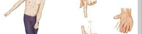
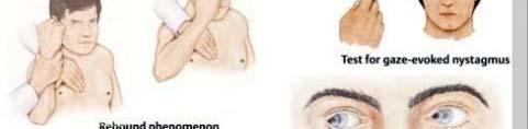
33 Syndromes Ataxia: incoordination of movement decomposition of movement dysmetria, past-pointing dysartheria dysdiadochokinesia rebound phenomenon of Holmes gait ataxia, truncal ataxia Intention Tremor Hypotonia, Nystagmus Archicerebellar Lesion: Medulloblastoma (see later) Paleocerebellar Lesion: gait disturbance dstuba Neocerebellar Lesion: hypotonia, ataxia, tremor


34

35 Cerebellar Medulloblastoma Cerebellar tumors on vermis Truncal Ataxia Frequent Falling The child in this picture: - would not try to stand unsupported would not let go of the bed rail if she was stood on the floor.
36 THANK YOU
Neuroanatomy Dr. Maha ELBeltagy Assistant Professor of Anatomy Faculty of Medicine The University of Jordan 2018
 Neuroanatomy Dr. Maha ELBeltagy Assistant Professor of Anatomy Faculty of Medicine The University of Jordan 2018 Dr Maha ELbeltagy THE BRAIN STEM Dr Maha ELbeltagy It includes: Midbrain - Pons - Medulla
Abdullah AlZibdeh. Dr. Maha ElBeltagy. Maha ElBeltagy
 19 Abdullah AlZibdeh Dr. Maha ElBeltagy Maha ElBeltagy Introduction In this sheet, we discuss the cerebellum; its lobes, fissures and deep nuclei. We also go into the tracts and connections in which the
Located below tentorium cerebelli within posterior cranial fossa. Formed of 2 hemispheres connected by the vermis in midline.
 The Cerebellum Cerebellum Located below tentorium cerebelli within posterior cranial fossa. Formed of 2 hemispheres connected by the vermis in midline. Gray matter is external. White matter is internal,
DEVELOPMENT OF BRAIN
 Ahmed Fathalla OBJECTIVES At the end of the lecture, students should: List the components of brain stem. Describe the site of brain stem. Describe the relations between components of brain stem & their
Brainstem. Amadi O. Ihunwo, PhD School of Anatomical Sciences
 Brainstem Amadi O. Ihunwo, PhD School of Anatomical Sciences Lecture Outline Constituents Basic general internal features of brainstem External and Internal features of Midbrain Pons Medulla Constituents
Cerebellum John T. Povlishock, Ph.D.
 Cerebellum John T. Povlishock, Ph.D. OBJECTIVES 1. To identify the major sources of afferent inputs to the cerebellum 2. To define the pre-cerebellar nuclei from which the mossy and climbing fiber systems
Lecture 4 The BRAINSTEM Medulla Oblongata
 Lecture 4 The BRAINSTEM Medulla Oblongata Introduction to brainstem 1- Medulla oblongata 2- Pons 3- Midbrain - - - occupies the posterior cranial fossa of the skull. connects the narrow spinal cord
By Dr. Saeed Vohra & Dr. Sanaa Alshaarawy
 By Dr. Saeed Vohra & Dr. Sanaa Alshaarawy 1 By the end of the lecture, students will be able to : Distinguish the internal structure of the components of the brain stem in different levels and the specific
Developmental sequence of brain
 Cerebellum Developmental sequence of brain Fourth week Fifth week Location of cerebellum Lies above and behind the medullar and pons and occupies posterior cranial fossa Location of cerebellum External
Connection of the cerebellum
 CEREBELLUM Connection of the cerebellum The cerebellum has external layer of gray matter (cerebellar cortex ), & inner white matter In the white matter, there are 3 deep nuclei : (a) dentate nucleus laterally
Unit VIII Problem 5 Physiology: Cerebellum
 Unit VIII Problem 5 Physiology: Cerebellum - The word cerebellum means: the small brain. Note that the cerebellum is not completely separated into 2 hemispheres (they are not clearly demarcated) the vermis
Done by : Areej Al-Hadidi
 Brainstem &diencephalon Done by : Areej Al-Hadidi Brainstem Functions Ascending and descending tracts Reflex centers Cardiovascular and respiratory centers Coughing, sneezing, swallowing Nuclei of the
Brainstem. By Dr. Bhushan R. Kavimandan
 Brainstem By Dr. Bhushan R. Kavimandan Development Ventricles in brainstem Mesencephalon cerebral aqueduct Metencephalon 4 th ventricle Mylencephalon 4 th ventricle Corpus callosum Posterior commissure
Fig. 1.4 (A B). The brain stem. Anterior view. Bar: 5 mm.
. The brain stem. Anterior view. Bar: 5 mm.") Section I A Fig. 1.4 (A B). The brain stem. Anterior view. Bar: 5 mm. Medulla The boundaries between the spinal cord and medulla are noted on Fig. 1.30. The pontomedullary sulcus (1) separates the medulla
Section I A Fig. 1.4 (A B). The brain stem. Anterior view. Bar: 5 mm. Medulla The boundaries between the spinal cord and medulla are noted on Fig. 1.30. The pontomedullary sulcus (1) separates the medulla
PHYSIOLOHY OF BRAIN STEM
 PHYSIOLOHY OF BRAIN STEM Learning Objectives The brain stem is the lower part of the brain. It is adjoining and structurally continuous with the spinal cord. 1 Mid Brain 2 Pons 3 Medulla Oblongata The
Brainstem. Telencephalon Diencephalon Cerebellum Brain stem
 Brainstem Brainstem 脑 脊髓 Brainstem Telencephalon Diencephalon Cerebellum Brain stem Ventral view Lateral view 10 pairs of the cranial nerves are attached to the brain stem The brainstem Midbrain Pons Medulla
b. The groove between the two crests is called 2. The neural folds move toward each other & the fuse to create a
 Chapter 13: Brain and Cranial Nerves I. Development of the CNS A. The CNS begins as a flat plate called the B. The process proceeds as: 1. The lateral sides of the become elevated as waves called a. The
Unit VIII Problem 3 Neuroanatomy: Brain Stem, Cranial Nerves and Scalp
 Unit VIII Problem 3 Neuroanatomy: Brain Stem, Cranial Nerves and Scalp - Brain stem: It is connected to the cerebellum and cerebral hemispheres. Rostral end of brain stem: diencephalon is the area which
Copy Right- Hongqi ZHANG-Department of Anatomy-Fudan University. Systematic Anatomy. Nervous system Cerebellum. Dr.Hongqi Zhang ( 张红旗 )
") Systematic Anatomy Nervous system Cerebellum Dr.Hongqi Zhang ( 张红旗 ) Email: zhanghq58@126.com 1 The Cerebellum Cerebellum evolved and developed with the complication of animal movement. Key points about
Systematic Anatomy Nervous system Cerebellum Dr.Hongqi Zhang ( 张红旗 ) Email: zhanghq58@126.com 1 The Cerebellum Cerebellum evolved and developed with the complication of animal movement. Key points about
The Cerebellum. The Little Brain. Neuroscience Lecture. PhD Candidate Dr. Laura Georgescu
 The Cerebellum The Little Brain Neuroscience Lecture PhD Candidate Dr. Laura Georgescu Learning Objectives 1. Describe functional anatomy of the cerebellum - its lobes, their input and output connections
M555 Medical Neuroscience Lab 1: Gross Anatomy of Brain, Crainal Nerves and Cerebral Blood Vessels
 M555 Medical Neuroscience Lab 1: Gross Anatomy of Brain, Crainal Nerves and Cerebral Blood Vessels Anatomical Directions Terms like dorsal, ventral, and posterior provide a means of locating structures
The Cerebellum. Little Brain. Neuroscience Lecture. Dr. Laura Georgescu
 The Cerebellum Little Brain Neuroscience Lecture Dr. Laura Georgescu Learning Objectives 1. Describe functional anatomy of the cerebellum- its lobes, their input and output connections and their functions.
Doctor Osama Asa ad Khader. Mohammad Alsalem
 6 Doctor 2015 Osama Asa ad Khader Mohammad Alsalem A quick revision for the spinal cord blood supply: Arterial Blood supply of spinal cord The spinal cord got its arterial supply by two ways: Longitudinal
Omar Sami. Aseel Abdeen. Muhammad Al-Salem. 1 P a g e
 Omar Sami Aseel Abdeen Muhammad Al-Salem 1 P a g e Using only section 2 record, I wrote this sheet; as the video is not ready yet. Despite pointing the structures, I ve tried to include all the scientific
Medial View of Cerebellum
 Meds 5371 System Neuroscience D. L. Oliver CEREBELLUM Anterior lobe (spinal) Posterior lobe (cerebral) Flocculonodular lobe (vestibular) Medial View of Cerebellum 1 Ventral View of Cerebellum Flocculus
Brainstem: Medulla oblongata and pons
 Brainstem: Medulla oblongata and pons 1. Overview of the brainstem subdivisions 2. Embryonic development of the brainstem 3. Medulla oblongata external features 4. Internal structure of the medulla oblongata
Cranial Nerve VIII (The Vestibulo-Cochlear Nerve)
") Cranial Nerve VIII (The Vestibulo-Cochlear Nerve) Please view our Editing File before studying this lecture to check for any changes. Color Code Important Doctors Notes Notes/Extra explanation Objectives
Cranial Nerve VIII (The Vestibulo-Cochlear Nerve) Please view our Editing File before studying this lecture to check for any changes. Color Code Important Doctors Notes Notes/Extra explanation Objectives
{Important Notes} *visual association Cortex lesion Visual agnosia: able to see but unable to understand.
 {Important Notes} *visual association Cortex lesion Visual agnosia: able to see but unable to understand. *Lateral to medial: Posterior ramus of the lateral fissure Insula lentiform nucleus Thalamus 3
Cerebellum. Steven McLoon Department of Neuroscience University of Minnesota
 Cerebellum Steven McLoon Department of Neuroscience University of Minnesota 1 Anatomy of the Cerebellum The cerebellum has approximately half of all the neurons in the central nervous system. The cerebellum
Non-cranial nerve nuclei
 Brainstem Non-cranial nerve nuclei Nucleus Gracile nucleus Cuneate nucleus Infeiro olivary nucleus Pontine nucleus inferior colliculus superior colliculus Red nucleus Substantia nigra Pretectal area Site
Motor System Hierarchy
 Motor Pathways Lectures Objectives Define the terms upper and lower motor neurons with examples. Describe the corticospinal (pyramidal) tract and the direct motor pathways from the cortex to the trunk
Brain and Cranial Nerves (Ch. 15) Human Anatomy lecture. caudal = toward the spinal cord)
 Human Anatomy lecture. caudal = toward the spinal cord)") Insight: Some cranial nerve disorders Brain and Cranial Nerves (Ch. 15) Human Anatomy lecture I. Overview (Directional terms: rostral = toward the forehead caudal = toward the spinal cord) A. 3 Major parts
Insight: Some cranial nerve disorders Brain and Cranial Nerves (Ch. 15) Human Anatomy lecture I. Overview (Directional terms: rostral = toward the forehead caudal = toward the spinal cord) A. 3 Major parts
FUNCTION: It COORDINATES movement HOW IT WORKS
 CEREBELLUM Chris Cohan, Ph.D. Dept. of Pathology/Anat Sci University at Buffalo Objectives: Describe the anatomy of the cerebellum, its 3 functions and associated regions Describe how the cerebellum influences
Ch 13: Central Nervous System Part 1: The Brain p 374
 Ch 13: Central Nervous System Part 1: The Brain p 374 Discuss the organization of the brain, including the major structures and how they relate to one another! Review the meninges of the spinal cord and
Faculty of Dental Medicine and Surgery. Sem 4 Cerebellum Dr. Abbas
 Faculty of Dental Medicine and Surgery Sem 4 Cerebellum Dr. Abbas Anatomy of the cerebellum Cerebellum Configurations External - located in posterior cranial fossa - communicate with other structure via
 Spinal Cord Tracts DESCENDING SPINAL TRACTS: Are concerned with somatic motor function, modification of ms. tone, visceral innervation, segmental reflexes. Main tracts arise form cerebral cortex and others
Internal Organisation of the Brainstem
 Internal Organisation of the Brainstem Major tracts and nuclei of the brainstem (Notes) The brainstem is the major pathway for tracts and houses major nuclei, that contain sensory, motor and autonomics
The Cerebellum. Outline. Lu Chen, Ph.D. MCB, UC Berkeley. Overview Structure Micro-circuitry of the cerebellum The cerebellum and motor learning
 The Cerebellum Lu Chen, Ph.D. MCB, UC Berkeley 1 Outline Overview Structure Micro-circuitry of the cerebellum The cerebellum and motor learning 2 Overview Little brain 10% of the total volume of the brain,
THE CEREBELLUM SUDIVISIONS, STRUCTURE AND CONNECTIONS
 THE CEREBELLUM Damage to the cerebellum produces characteristic symptoms primarily with respect to the coordination of voluntary movements. The cerebellum receives information from the skin, joints, muscles,
Blood supply to the brain Blood brain barrier isolates neural tissue from general circulation
 The Brain and Cranial Nerves Objectives Name the major regions of the brain and describe their functions. Discuss the formation, circulation, and functions of the CSF. List the main components of the medulla
Chapter 14: The Brain and Cranial Nerves. Copyright 2009, John Wiley & Sons, Inc.
 Chapter 14: The Brain and Cranial Nerves Development of the Brain Three to four-week embryo: prosencephalon, mesencephalon and rhombencephalon. Five-week embryo: telencephalon (cerebrum), diencephalon
The Cerebellum. Outline. Overview Structure (external & internal) Micro-circuitry of the cerebellum Cerebellum and motor learning
 Micro-circuitry of the cerebellum Cerebellum and motor learning") The Cerebellum P.T Ji Jun Cheol Rehabilitation Center 1 HansarangAsan Hospital. Outline Overview Structure (external & internal) Micro-circuitry of the cerebellum Cerebellum and motor learning 2 1 Cerebellum
The Cerebellum P.T Ji Jun Cheol Rehabilitation Center 1 HansarangAsan Hospital. Outline Overview Structure (external & internal) Micro-circuitry of the cerebellum Cerebellum and motor learning 2 1 Cerebellum
I: To describe the pyramidal and extrapyramidal tracts. II: To discuss the functions of the descending tracts.
 Descending Tracts I: To describe the pyramidal and extrapyramidal tracts. II: To discuss the functions of the descending tracts. III: To define the upper and the lower motor neurons. 1. The corticonuclear
Basal Nuclei (Ganglia)
") Doctor said he will not go deep within these slides because we will take them in physiology, so he will explain the anatomical structures, and he will go faster in the functions sheet in yellow Basal Nuclei
Doctor said he will not go deep within these slides because we will take them in physiology, so he will explain the anatomical structures, and he will go faster in the functions sheet in yellow Basal Nuclei
For more information about how to cite these materials visit
 Author(s): Peter Hitchcock, PH.D., 2009 License: Unless otherwise noted, this material is made available under the terms of the Creative Commons Attribution Non-commercial Share Alike 3.0 License: http://creativecommons.org/licenses/by-nc-sa/3.0/
Motor tracts Both pyramidal tracts and extrapyramidal both starts from cortex: Area 4 Area 6 Area 312 Pyramidal: mainly from area 4 Extrapyramidal:
 Motor tracts Both pyramidal tracts and extrapyramidal both starts from cortex: Area 4 Area 6 Area 312 Pyramidal: mainly from area 4 Extrapyramidal: mainly from area 6 area 6 Premotorarea: uses external
Auditory and Vestibular Systems
 Auditory and Vestibular Systems Objective To learn the functional organization of the auditory and vestibular systems To understand how one can use changes in auditory function following injury to localize
PETER PAZMANY CATHOLIC UNIVERSITY Consortium members SEMMELWEIS UNIVERSITY, DIALOG CAMPUS PUBLISHER
 PETER PAZMANY CATHOLIC UNIVERSITY SEMMELWEIS UNIVERSITY Development of Complex Curricula for Molecular Bionics and Infobionics Programs within a consortial* framework** Consortium leader PETER PAZMANY
Strick Lecture 3 March 22, 2017 Page 1
 Strick Lecture 3 March 22, 2017 Page 1 Cerebellum OUTLINE I. External structure- Inputs and Outputs Cerebellum - (summary diagram) 2 components (cortex and deep nuclei)- (diagram) 3 Sagittal zones (vermal,
Brainstem. Steven McLoon Department of Neuroscience University of Minnesota
 Brainstem Steven McLoon Department of Neuroscience University of Minnesota 1 Course News Change in Lab Sequence Week of Oct 2 Lab 5 Week of Oct 9 Lab 4 2 Goal Today Know the regions of the brainstem. Know
I. Anatomy of the Brain A. Cranial Meninges and Ventricles of the Brain 1. Meninges a. Dura mater 1) Endosteal/Periosteal Layer - Outer 2) Meningeal
 Endosteal/Periosteal Layer - Outer 2) Meningeal") I. Anatomy of the Brain A. Cranial Meninges and Ventricles of the Brain 1. Meninges a. Dura mater 1) Endosteal/Periosteal Layer - Outer 2) Meningeal Layer - Inner 3) Falx cerebri a) Superior sagittal sinus
I. Anatomy of the Brain A. Cranial Meninges and Ventricles of the Brain 1. Meninges a. Dura mater 1) Endosteal/Periosteal Layer - Outer 2) Meningeal Layer - Inner 3) Falx cerebri a) Superior sagittal sinus
Anatomy and Physiology (Bio 220) The Brain Chapter 14 and select portions of Chapter 16
 The Brain Chapter 14 and select portions of Chapter 16") Anatomy and Physiology (Bio 220) The Brain Chapter 14 and select portions of Chapter 16 I. Introduction A. Appearance 1. physical 2. weight 3. relative weight B. Major parts of the brain 1. cerebrum 2.
Anatomy and Physiology (Bio 220) The Brain Chapter 14 and select portions of Chapter 16 I. Introduction A. Appearance 1. physical 2. weight 3. relative weight B. Major parts of the brain 1. cerebrum 2.
CN V! touch! pain! Touch! P/T!
 CN V! touch! pain! Touch! P/T! Visual Pathways! L! R! B! A! C! D! LT! E! F! RT! G! hypothalamospinal! and! ALS! Vestibular Pathways! 1. Posture/Balance!!falling! 2. Head Position! 3. Eye-Head Movements
SENSORY (ASCENDING) SPINAL TRACTS
 SPINAL TRACTS") SENSORY (ASCENDING) SPINAL TRACTS Dr. Jamila El-Medany Dr. Essam Eldin Salama OBJECTIVES By the end of the lecture, the student will be able to: Define the meaning of a tract. Distinguish between the different
SENSORY (ASCENDING) SPINAL TRACTS Dr. Jamila El-Medany Dr. Essam Eldin Salama OBJECTIVES By the end of the lecture, the student will be able to: Define the meaning of a tract. Distinguish between the different
The Nervous System: Sensory and Motor Tracts of the Spinal Cord
 15 The Nervous System: Sensory and Motor Tracts of the Spinal Cord PowerPoint Lecture Presentations prepared by Steven Bassett Southeast Community College Lincoln, Nebraska Introduction Millions of sensory
ACTIVITY 7: NERVOUS SYSTEM HISTOLOGY, BRAIN, CRANIAL NERVES
 ACTIVITY 7: NERVOUS SYSTEM HISTOLOGY, BRAIN, CRANIAL NERVES LABORATORY OBJECTIVES: 1. Histology: Identify structures indicated on three different slides or images of nervous system tissue. These images
Embryonic Brain Development
 Chapter 14 The Brain and Cranial Nerves Largest organ in the body? Brain functions in sensations, memory, emotions, decision making, behavior 19-1 19-2 Embryonic Brain Development Principal Parts of the
Brainstem: Midbrain. 1. Midbrain gross external anatomy 2. Internal structure of the midbrain:
 Brainstem: Midbrain 1. Midbrain gross external anatomy 2. Internal structure of the midbrain: cerebral peduncles tegmentum tectum (guadrigeminal plate) Midbrain Midbrain general features location between
Cerebellum: little brain. Cerebellum. gross divisions
 Cerebellum The anatomy of the cerebellum and its gross divisions Its principal input and output pathways The organization of the cerebellar cortex Role of climbing vs. mossy fibre input The parallel-fibre/
Functional Distinctions
 Functional Distinctions FUNCTION COMPONENT DEFICITS Start Basal Ganglia Spontaneous Movements Move UMN/LMN Cerebral Cortex Brainstem, Spinal cord Roots/peripheral nerves Plan Cerebellum Ataxia Adjust Cerebellum
Lecturer. Prof. Dr. Ali K. Al-Shalchy MBChB/ FIBMS/ MRCS/ FRCS 2014
 Lecturer Prof. Dr. Ali K. Al-Shalchy MBChB/ FIBMS/ MRCS/ FRCS 2014 Dorsal root: The dorsal root carries both myelinated and unmyelinated afferent fibers to the spinal cord. Posterior gray column: Long
THE CENTRAL NERVOUS SYSTE M
 THE CENTRAL NERVOUS SYSTE M Structure and Functio n THIRD EDITIO N PER BRODAL A Brief Survey, x i Studying the Structures and Function of the Nervous System, xii i Animal Experiments Crucial for Progress,
CASE 48. What part of the cerebellum is responsible for planning and initiation of movement?
 CASE 48 A 34-year-old woman with a long-standing history of seizure disorder presents to her neurologist with difficulty walking and coordination. She has been on phenytoin for several days after having
Cerebellum: little brain. Cerebellum. gross divisions
 Cerebellum The anatomy of the cerebellum and its gross divisions Its principal input and output pathways The organization of the cerebellar cortex Role of climbing vs. mossy fibre input The parallel-fibre/
Introduction to the Central Nervous System: Internal Structure
 Introduction to the Central Nervous System: Internal Structure Objective To understand, in general terms, the internal organization of the brain and spinal cord. To understand the 3-dimensional organization
The Brain and Cranial Nerves Pg. 129
 The Brain and Cranial Nerves Pg. 129 Three Main Regions of the Brain Forebrain Cerbral hemispheres Diencephalon Midbrain Brain stem Hindbrain Pons Cerebellum Medulla oblongata Forebrain Interprets sensory
Principles of Anatomy and Physiology
 Principles of Anatomy and Physiology 14 th Edition CHAPTER 14 The Brain and Cranial Nerves Introduction The purpose of the chapter is to: 1. Understand how the brain is organized, protected, and supplied
Neuroanatomy. Assistant Professor of Anatomy Faculty of Medicine The University of Jordan Dr Maha ELBeltagy
 Neuroanatomy Dr. Maha ELBeltagy Assistant Professor of Anatomy Faculty of Medicine The University of Jordan 2018 Development of the Central Nervous System Development of the nervous system Development
Brain ميهاربا لض اف دمح ا د The Meninges 1- Dura Mater of the Brain endosteal layer does not extend meningeal layer falx cerebri tentorium cerebelli
 .احمد د فاضل ابراهيم Lecture 15 Brain The Meninges Three protective membranes or meninges surround the brain in the skull: the dura mater, the arachnoid mater, and the pia mater 1- Dura Mater of the Brain
The Brain and Cranial Nerves Pg Three Main Regions of the Brain. Forebrain
 The Brain and Cranial Nerves Pg. 129 Three Main Regions of the Brain Forebrain Cerbral hemispheres Diencephalon Midbrain Brain stem Hindbrain Pons Cerebellum Medulla oblongata Interprets sensory inputs
Human Brain and Senses October 13, 2008 Page 1. Examination of the Human Brain
 Human Brain and Senses October 13, 2008 Page 1 Examination of the Human Brain With only a few hours today we can only begin to scratch the surface of a complex subject like neuroanatomy. The purpose of
Laboratory Manual for Comparative Anatomy and Physiology Figure 15.1 Transparency Master 114
 Neuron Capillary Astrocyte Microglial cell Neuron Fluid-filled cavity Process of oligodendrocyte Ependymal cells Brain or spinal cord tissue Myelin sheath Nerve fibers Figure 15.1 Transparency Master 114
Unit 18: Cranial Cavity and Contents
 Unit 18: Cranial Cavity and Contents Dissection Instructions: The calvaria is to be removed without damage to the dura mater which is attached to the inner surface of the calvaria. Cut through the outer
Sheet lab 3. Page 8B Section1 of medulla at pyramidal {motor} decussation:
 Sheet lab 3 Page 8B Section1 of medulla at pyramidal {motor} decussation: This section is at lower third of medulla and is the most close part to spinal cord and it has some characteristic of spinal cord
Lab 2. we will look into several angled horizontal sections ( orbitomeatal plane ) i.e passing from the orbit into the ear
 i.e passing from the orbit into the ear") we will look into several angled horizontal sections ( orbitomeatal plane ) i.e passing from the orbit into the ear Figure I page 76 : looking at the key on the left side this section passed through the
we will look into several angled horizontal sections ( orbitomeatal plane ) i.e passing from the orbit into the ear Figure I page 76 : looking at the key on the left side this section passed through the
-Zeina Assaf. -Omar Odeh. - Maha Beltagy
 -3 -Zeina Assaf -Omar Odeh - Maha Beltagy 1 P a g e The Inferior Surface Of The Brain The inferior surface of the brain is divide by the stem of the lateral fissure into 2 parts : The orbital surface and
PHYSIOLOGY OF THE BRAIN STEM
 PHYSIOLOGY OF THE BRAIN STEM Dr Syed Shahid Habib Professor & Consultant Clinical Neurophysiology Dept. of Physiology College of Medicine & KKUH King Saud University OBJECTIVES At the end of this lecture
A&P 1 Brain & Cranial Nerves Guide #1 - Pre-Lab Exercises
 A&P 1 Brain & Cranial Nerves Guide #1 - Pre-Lab Exercises In this "Pre-lab Guide", we will be looking at the brain & cranial nerves. This should be done before lab, so we don't waste time in lab! This
Chapter 13 Brain and Cranial Nerves
 Chapter 13 Brain and Cranial Nerves 13-1 Brain and Cranial Nerves Brain Part of CNS contained in cranial cavity Control center for many of body s functions Much like a complex computer but more Parts of
Motor Functions of Cerebral Cortex
 Motor Functions of Cerebral Cortex I: To list the functions of different cortical laminae II: To describe the four motor areas of the cerebral cortex. III: To discuss the functions and dysfunctions of
Unit 12a: The Nervous System The Brain. MDL231 Principle of Anatomy
 Unit 12a: The Nervous System The Brain MDL231 Principle of Anatomy The Brain - Overview Cerebrum T PP H midbrain Cerebellum pons m.o. Brain stem medulla oblongata (M.O.) pons midbrain (mesencephalon) Diencephalon
CNS MCQ 2 nd term. Select the best answer:
 Select the best answer: CNS MCQ 2 nd term 1) Vestibular apparatus: a) Represent the auditory part of the labyrinth. b) May help in initiating the voluntary movements. c) Contains receptors concerned with
Brain, Cranial Nerves, and Spinal Cord
 Bio101 Laboratory 13 Neuron/Spinal Cord Histology Brain Anatomy Ear & Eye Anatomy 1 Brain, Cranial Nerves, and Spinal Cord Objectives for today s lab Become familiar with the gross anatomy of the brain
Chapter 13 Lecture Outline *
 Anatomy and Physiology, Seventh Edition Rod R. Seeley Idaho State University Trent D. Stephens Idaho State University Philip Tate Phoenix College Chapter 13 Lecture Outline * *See PowerPoint Image Slides
Cranial Nerves. Steven McLoon Department of Neuroscience University of Minnesota
 Cranial Nerves Steven McLoon Department of Neuroscience University of Minnesota 1 Course News Change in Lab Sequence Week of Oct 2 Lab 5 Week of Oct 9 Lab 4 2 Sensory and Motor Systems Sensory Systems:
Brain anatomy and artificial intelligence. L. Andrew Coward Australian National University, Canberra, ACT 0200, Australia
 Brain anatomy and artificial intelligence L. Andrew Coward Australian National University, Canberra, ACT 0200, Australia The Fourth Conference on Artificial General Intelligence August 2011 Architectures
Basal nuclei, cerebellum and movement
 Basal nuclei, cerebellum and movement MSTN121 - Neurophysiology Session 9 Department of Myotherapy Basal Nuclei (Ganglia) Basal Nuclei (Ganglia) Role: Predict the effects of various actions, then make
Gross Morphology of the Brain
 Gross Morphology of the Brain Done by : Marah Marahleh & Razan Krishan *slides in bold Principal Parts of the Brain Cerebrum : largest part of the brain Diencephalon Thalamus & hypothalamus Cerebellum
The neurvous system senses, interprets, and responds to changes in the environment. Two types of cells makes this possible:
 NERVOUS SYSTEM The neurvous system senses, interprets, and responds to changes in the environment. Two types of cells makes this possible: the neuron and the supporting cells ("glial cells"). Neuron Neurons
Chapter 3. Structure and Function of the Nervous System. Copyright (c) Allyn and Bacon 2004
 Allyn and Bacon 2004") Chapter 3 Structure and Function of the Nervous System 1 Basic Features of the Nervous System Neuraxis: An imaginary line drawn through the center of the length of the central nervous system, from the
Chapter 3 Structure and Function of the Nervous System 1 Basic Features of the Nervous System Neuraxis: An imaginary line drawn through the center of the length of the central nervous system, from the
Pathways of proprioception
 The Autonomic Nervous Assess Prof. Fawzia Al-Rouq Department of Physiology College of Medicine King Saud University Pathways of proprioception System posterior column& Spinocerebellar Pathways https://www.youtube.com/watch?v=pmeropok6v8
Biological Bases of Behavior. 3: Structure of the Nervous System
 Biological Bases of Behavior 3: Structure of the Nervous System Neuroanatomy Terms The neuraxis is an imaginary line drawn through the spinal cord up to the front of the brain Anatomical directions are
BRAIN STEM AND CEREBELLUM..
 Lecture Title: BRAIN STEM AND CEREBELLUM.. (CNS Block, Radiology) Dr. Hamdy Hassan Ass.Prof. Consultant Radiology Department KKHU King Saud University Lecture Objectives.. Students at the end of the lecture
Subcortical Motor Systems: cerebellum
 Outline Subcortical Motor Systems: cerebellum 陽明大學醫學院腦科所陳昌明副教授 Anatomy Cerebellar cortex Neuronal circuitry Cerebellar connections Vestibulocerebellum Spinocerebellum Neocerebellum Other cerebellar functions
Model 3-50B or 3-88 III VIII. Olfactory Nerve. Optic Nerve. Oculomotor Nerve. Trochlear Nerve. Trigeminal Nerve. Abducens Nerve.
 Model 3-50B or 3-88 I Olfactory Nerve II Optic Nerve Oculomotor Nerve III IV Trochlear Nerve Trigeminal Nerve V VI Abducens Nerve Glossopharyngeal Nerve IX VII Facial Nerve VIII Vestibocochlear Nerve or
Voluntary Movement. Ch. 14: Supplemental Images
 Voluntary Movement Ch. 14: Supplemental Images Skeletal Motor Unit: The basics Upper motor neuron: Neurons that supply input to lower motor neurons. Lower motor neuron: neuron that innervates muscles,
General Sensory Pathways of the Trunk and Limbs
 General Sensory Pathways of the Trunk and Limbs Lecture Objectives Describe gracile and cuneate tracts and pathways for conscious proprioception, touch, pressure and vibration from the limbs and trunk.
1. The cerebellum coordinates fine movement through interactions with the following motor-associated areas:
 DENT/OBHS 131 2009 Take-home test 4 Week 6: Take-home test (2/11/09 close 2/18/09) 1. The cerebellum coordinates fine movement through interactions with the following motor-associated areas: Hypothalamus
Biology 218 Human Anatomy
 Chapter 21 Adapted form Tortora 10 th ed. LECTURE OUTLINE A. Overview of Sensations (p. 652) 1. Sensation is the conscious or subconscious awareness of external or internal stimuli. 2. For a sensation