Human Pontine Glioma Cells Can Induce Murine Tumors
|
|
|
- Sarah Hopkins
- 5 years ago
- Views:
Transcription
1 Chapter 2 Human Pontine Glioma Cells Can Induce Murine Tumors Viola Caretti, A. Charlotte P. Sewing, Tonny Lagerweij, Pepijn Schellen Marianna Bugiani, Marc H. A. Jansen, Dannis G. van Vuurden, Anna C. Navis, Ilona Horsman, W. Peter Vandertop, David P. Noske, Pieter Wesseling, Gertjan J.L. Kaspers, Javad Nazarian, Hannes Vogel, Esther Hulleman #, Michelle Monje #, Thomas Wurdinger # # These authors contributed equally Acta Neuropathologica, 2014 (6) 127:
2 30 Chapter 2 Abstract Diffuse intrinsic pontine glioma (DIPG), with a median survival of only nine months, is the leading cause of pediatric brain cancer mortality. Dearth of tumor tissue for research has limited progress in this disease until recently. New experimental models for DIPG research are now emerging. To develop preclinical models of DIPG, two different methods were adopted: cells obtained at autopsy 1) were directly xenografted orthotopically into the pons of immunodeficient mice without an intervening cell culture step or 2) were first cultured in vitro and, upon successful expansion, injected in vivo. Both strategies resulted in pontine tumors histopathologically similar to the original human DIPG tumors. However, following the direct transplantation method all tumors proved to be composed of murine and not of human cells. This is in contrast to the indirect method that included initial in vitro culture and resulted in xenografts comprised of human cells. Of note, direct injection of cells obtained post mortem from the pons and frontal lobe of human brains not affected by cancer did not give rise to neoplasms. The murine pontine tumors exhibited an immunophenotype similar to human DIPG, but were also positive for microglia/ macrophage markers, such as CD45, CD68 and CD11b. Serial orthotopic injection of these murine cells results in lethal tumors in recipient mice. Direct injection of human DIPG cells in vivo can give rise to malignant murine tumors. This represents an important caveat for xenotransplantation models of DIPG. In contrast, an initial in vitro culture step can allow establishment of human orthotopic xenografts. The mechanism underlying this phenomenon observed with direct xenotransplantation remains an open question.
3 Pontine glioma cells can induce human tumors 31 Introduction Diffuse intrinsic pontine glioma (DIPG) is a devastating brain cancer affecting mainly children 88,89. Compared to other brain malignancies, DIPG carries the worst prognosis; the majority of affected children die within one year from diagnosis 90,91. DIPG is also one of the pediatric tumors that has been the least investigated in laboratories, due to a lack of primary tissue available for research and the paucity of robust preclinical models. We have established autopsy protocols to develop experimental models of DIPG and to investigate its biology 25,31. Recent studies have shown that DIPG is molecularly distinct from adult highgrade gliomas 92,93. Although DIPG shares recurrent aberrations in histone 3 genes with supratentorial pediatric gliomas, they are molecularly distinct tumors DIPG is characterized by unique hallmarks including a remarkable spatiotemporal specificity (occurring in the ventral pons during mid-childhood) and a diffuse growth pattern. As the disease progresses, DIPG often invades the cerebellum and even supratentorial brain regions, such as the thalamus, lateral ventricles and cerebral cortex 96. DIPG infiltrative growth and the delicate anatomical location where it arises preclude surgical resection. The pontine microenvironment at mid-childhood may be of paramount importance in the biology, pathogenesis and infiltrative nature of DIPG 31. To find a cure for this disease, it is critical to identify the microenvironmental and genetic factors that allow the tumor to spread throughout the brainstem and the entire brain. Here, we present data showing that direct injection of human DIPG cells obtained from post mortem tissue produced tumors composed exclusively of murine cells, while injection of human DIPG cells, obtained at autopsy but first cultured in vitro, gave rise to human xenografts. In comparison, direct injection of cells obtained post mortem from the pons and frontal lobe of human brains not affected by cancer did not give rise to murine neoplasms. The cells of these induced murine neoplasms exhibited immunocytochemical markers consistent with DIPG and also with the microglia/ macrophage phenotype. 02
4 32 Chapter 2 Materials and Methods Tissue Donors All tumor samples were post-treatment autopsy specimens from VU University Medical Center Amsterdam (VUMC) (The Netherlands), Stanford University (SU) and Children s National Medical Center (CNMC) in Washington DC (United States). Patients were included if they had classic DIPG MRI findings and clinical presentation. All parents signed informed consent forms for the use of biological material after autopsy for research purposes; and the appropriate Institutional Review Board (IRB) approved all procedures. The autopsy protocols have been previously reported 25,31. Patients clinical characteristics are summarized in Table 1 and in Supplementary Table 1. Table 1 Patients clinical characteristics Direct Method Indirect Method Patient h-vu-dipg-3 h-vu-dipg-4 h-vu-dipg-5 h-cnmc-d1 h-su-dipg-vi Age (years) Sex F M M M F Survival time (months) Time to Progression (months) Death-Autopsy Interval (hours) H3F3A K27M status Histological diagnosis at autopsy GBM GBM GBM GBM Astrocytoma (WHO grade III) This table summarizes the clinical characteristics of patients whose DIPG post mortem tissue was employed to develop DIPG models via the direct and indirect methods. WHO refers to World Health Organization and GBM refers to glioblastoma multiforme (WHO grade IV). For clarity, the suffix h for human and m for murine will be used in front all patients and cell lines and tumor sample names. For clarity, the suffix h for human and m for murine will be used in front all patients and cell lines and tumor sample names. All human patients affected by DIPG were treated with radiotherapy and/or chemotherapy, with the exception of patient h-su-dipg-i, who received only minimal treatment for the tumor 31. The human donor brains used for control tissue were obtained from the National Development and Research Institute (NDRI) ( The donors were
5 Pontine glioma cells can induce human tumors 33 males not affected by brain cancer; they were age 53 (cause of death: cardiac arrest) and age 59 (cause of death: respiratory disease); the samples collected were named h-su- NDRI-2 and h-su-ndri-3, respectively. The death-autopsy interval was 12.5 hours for h-su-ndri-2 and 5.56 hours for h-su-ndri Direct and Indirect Xenotransplantation Methods For the direct transplantation method, immediately after rapid brain autopsy of four DIPG patients (h-vu-dipg-3, -4 and -5 and h-cnmc-d1; mean post mortem delay was 3.4 hours) 25, a single cell suspension was prepared from pontine tissue macroscopically infiltrated by tumor. Tumor material was cut in 0.5 cm 3 pieces with a sterile scalpel and mechanically dissociated using a 0μm strainer and resuspended in Optimem medium or, for injection subcutaneously, in Matrigel (BD biosciences) diluted 1:3 in phosphate buffered saline (PBS). The indirect method, previously described 31, also followed rapid brain autopsy of DIPG patients (h-su-dipg-vi; post mortem delay was 2 hours). Briefly, for chemical dissociation, minced tissue was placed in Hepes-HBSS with DNaseI (250 U/mL) and collagenase type IV (1 mg/ml) at 37 C on a Nutator (Fisher Scientific). Cells were then further mechanically dissociated using 0, 70 and 40 μm strainers in series. Next, a 30% sucrose gradient and the ACK lysis buffer (Invitrogen) were employed to deplete myelin and red blood cells, respectively. Finally, the single cells suspension was cultured in serum-free Tumor Stem Media (TSM) consisting of Neurobasal media (-A) (Invitrogen), B27 (-A) (Invitrogen), human-basic FGF (20 ng/ml; Shenandoah Biotech), human EGF (20 ng/ml; Shenandoah Biotech), human PDGF-AA and -BB (20 ng/ml; Shenandoah Biotech), and heparin ( ng/ml) 31. Primary Tumor Cell Culture Human SU-DIPG-VI cells were cultured in TSM, as described above. After extraction from subcutaneous tumors ( and -5) and from pons xenograft (CNMC-D1 cells), murine cells were cultured in NeuroCult Basal Medium (StemCell Technologies) supplemented with murine EGF (20 ng/ml; Shenandoah Biotech), murine FGF (20 ng/ ml; Shenandoah Biotech) and heparin ( ng/ml). Stereotactic DIPG Cell Injection. All animal experiments were approved and performed according to the guidelines of the VU University Ethical and Scientific Committee on Animal Experiments, of Stanford
6 34 Chapter 2 University Institutional Care and Use Committee and of CNMC Institutional Animal Use and Care Protocols (#01429, #01335). At the VU non-obese diabetic (NOD) severe combined immunodeficiency (SCID) mice and female athymic nude (age six twelve weeks; Harlan), at Stanford NOD-SCID- IL2 gamma chain deficient (NSG) mice (age P1-5 and six twelve weeks; The Jackson Laboratory) and at the CNMC nude mice J:NU (age P-5, The Jackson Laboratory) were kept under specific pathogen-free conditions in air-filtered cages and received food and water ad libitum. We have previously reported the procedures used for stereotactic tumor cell transplantation both in adult 50 and in pups 31, mice and for tumor cell injection subcutaneously 50. For surgeries in adult mice, 1x 6 human DIPG cells/5μl or 1x 5 murine VU-DIPG-3 and -5 cells/5μl were injected in the pons or striatum of immunodeficient mice. For adult mice, the stereotactic coordinates used to target the pons were: 0.8 mm posterior to lambda, 1 mm lateral to the sagittal suture and 5 mm deep; and to target the striatum were: 2 mm anterior to bregma, 0.5 mm lateral to the sagittal suture and 2 mm deep. For pup surgeries, 4x 4 cells/5μl were injected in the IV th ventricle, using the following coordinates: 3 mm posterior to lambda, 0 mm lateral to the sagittal suture and 3mm deep. Immunohistochemical Staining Immunohistochemistry (IHC), immunofluorescence (IF), and fluorescence in situ hybridization (FISH), were performed on 5 μm tissue sections of paraffin-embedded (IHC and FISH) or fresh frozen (IF) human and murine DIPG tumor samples. IHC was performed as previously described 25. The following antibodies were used: NeuN (mouse monoclonal, 1:1,600, Millipore), GFAP (rabbit polyclonal, 1:300, Dako), Synaptophysin (mouse monoclonal, 1:150, Dako), CD45 (mouse monoclonal, 1:00, Dako), CD3 (rabbit polyclonal, 1:0, Cell Marque), CD68 (monoclonal mouse, 1:1600, Dako) and CD163 (mouse monoclonal, 1:50, Novocastra) all against human and mouse antigen; Vimentin (mouse monoclonal, 1:4000, Dako) and Ki67 (mouse monoclonal, 1:160, Dako) specifically reacting with the human antigen; Ki67 (rabbit polyclonal, 1:2,000, Dianova) specifically reacting with the mouse antigen. Antigen retrieval was performed by microwave heating, in citrate buffer (ph 6) for Glut-1, Vimentin and GFAP antibodies and in TRIS- EDTA (ph 9) for NeuN and Ki67 antibodies, while Synaptophysin-staining required no antigen retrieval. For CD45, CD3 and CD163, Ventana s proprietary antigen retrieval solution (ph 8.5), and, for CD68, Leica s proprietary antigen retrieval solution (ph 6.0) were used. All antibodies were incubated for one hour at room temperature Powervision
7 Pontine glioma cells can induce human tumors 35 (Glut-1, Ki67, NeuN and Synapthophysin) and Envision (Vimentin, GFAP) were used for visualization (both from Dako). Visualization for CD3, CD68, CD45, and CD163 was performed by the Ventana iview DAB Detection Kit. IF was carried out as described in a previously published protocol 97 with the exception that antibody incubation time was 24 hours. Additional antibodies used not reported in 97 were: SOX2 antibody (rabbit polyclonal, 1:200, Millipore) and Human Nuclei (mouse monoclonal, 1:200, Millipore). FISH was performed as previously reported 98. The chromosome enumeration probe 1 (CEP1 Spectrum Orange Probe, diluted 1: in uni-44 buffer, Abbott Molecular, Abbott Park, Illinois, USA) was used. Hybridization was performed overnight at 37 C. 02 Karyotyping Metaphase preparations of and -5, m-cnmc-d1 and h-su-dipg-vi cells were obtained by standard cytogenetic procedures of mitotic arrest, hypotonic shock and cell fixation. Metaphase cells were stained by standard G-banding methodology and imaged using a Zeiss Axioskop 20 microscope with 0x plan apochromatic objective and a CytoVision imaging system (Leica Microsystems, USA). Array-Comparative Genomic Hybridization and Sanger Sequencing of Exons Human VU-DIPG-3 and -5 somatic tumor DNA was isolated from the original pontine tumor. Non-somatic DNA was isolated from areas far away from the original pontine tumor (temporal lobe) with no macroscopic signs of intraparenchymal tumor growth. Cases were independently reviewed by a senior neuropathologist (PW) according to the World Health Organization (WHO) guidelines. Histologically, both tumors were labeled as glioblastoma (GBM) (Table 1). Control murine DNA was isolated from corresponding nude or SCID mice. DNA was isolated from fresh-frozen tissue using an all prep DNA/ RNA isolation kit (Qiagen). Samples were labeled and hybridized on human and murine Array-Comparative Genomic Hybridization (acgh) oligo Microarray 4x180k (Agilent Technologies, Palo Alto, CA), as previously described 99. Image acquisition of the Agilent arrays was performed using the Agilent DNA Microarray scanner G2505C, and image analysis was performed using Feature Extraction software version (Agilent Technologies). Amplifications of EGFR, PTEN and PDGFR-α were analyzed by verifying intensity of hybridized probes in the genomic regions of these genes.

 was obtained at autopsy.")

 Low H&E magnification image of xenograft at transplant generation 1 in vivo.")
 H&E of small-cell DIPG phenotype in h-vu-dipg-3 and -5 tissues and correspondent (transplant generation 1) and -5 (transplant generation 2) pontine tissue.")
 were sequenced using Sanger fluorescent sequencing after amplification by polymerase")
8 36 Chapter 2 Figure 1 a b * Direct method ( and -5, m-cnmc-d1) Indirect method (h-su-dipg-vi) * * VUMC-DIPG M c h-vu-dipg-3 Human DIPG H&E h-vu-dipg-5 Murine Tumor H&E m-vu-dipg-5 Figure. 1 Direct and indirect xenotransplantation of human post mortem DIPG cells gives rise to tumors resembling the original DIPG neuropathology (a) Sketch summarizing the direct and indirect method adopted to develop DIPG xenograft mouse models. DIPG tumor tissue (symbolized in red) was obtained at autopsy. The direct method consisted of rapid mechanical dissociation and xenotransplantation, while the indirect method comprised an intermediate in vitro culturing step followed by in vivo injection. Using both methods, xenografts developed in the pons but not subcutaneously. In the direct method, xenografts were serially transplanted from the pons or striatum (only for ) of a mouse to the pons of another mouse and subcutaneously. Henceforth, the xenografts grew subcutaneously as well as in the pons. (b) Low H&E magnification image of xenograft at transplant generation 1 in vivo. Note the diffuse growth pattern in the ventral pons (arrow) and in the leptomeninges (asterisks). (c) H&E of small-cell DIPG phenotype in h-vu-dipg-3 and -5 tissues and correspondent (transplant generation 1) and -5 (transplant generation 2) pontine tissue. Scale bars = (B) 1mm and (C) 20 μm. Exons coding of interest on H3F3A, HIST1H3B and PI3KCA (H47R/L and E542K) were sequenced using Sanger fluorescent sequencing after amplification by polymerase chain reaction using standard methods. The primers were designed using primer BLAST from NCBI (Supplementary Table 2). PCR products were directly sequenced using BigDye Terminator v3.1 Cycle Sequencing Kit (Life technologies) and analyzed by a genetic analyzer (Applied Biosystems). DNA Fingerprinting Short tandem repeat (STR) analysis was performed according to the manufacturer
9 Pontine glioma cells can induce human tumors 37 protocol (PowerPLex 18D system, Promega). The STR fingerprint for h-su-dipg-vi control cortex is reported in Supplementary Table 3. Flow Cytometry After in vitro culture, h-su-dipg-vi and m-cnmc-d1 cells were enzymatically dissociated as previously described 31, subcutaneous and -5 tumors were mechanically dissociated as described above. The following antibodies were incubated for one hour: PerCP-Cy5.5 anti-mouse H-2K d, PE anti-human HLA-A,B,C, Brilliant Violet421 TM anti-mouse CD45 (all by BioLegend) and PE-Cyanine7 anti-mouse and human CD11b (ebioscience). Cells were gated on the basis of forward- and sidescatter profiles, and live/dead discrimination was obtained with 7-amino-actinomycin D (7-AAD) (DAPI) or Propidium Iodide. Flow cytometry was performed using a FACS Aria II (BD Biosciences). Data were analyzed with FlowJo software. 02 Results DIPG xenografts resemble the original human neoplasm histologically To establish xenograft DIPG models, we used early post mortem autopsy protocols at the VU 25 and at Stanford University School of Medicine 31. While at the VU a direct transplantation strategy was adopted, at Stanford human DIPG cells were first cultured in vitro and subsequently, upon successful expansion, injected in vivo (indirect method) (Fig. 1a). In the direct transplantation method, post mortem DIPG tissue (h-vu-dipg-3, -4 and -5) was immediately dissociated using rapid mechanical mincing and straining, without chemical processing of the sample or myelin depletion. The single cell suspension was directly injected subcutaneously (x 6 cells/0 μl/flank) and into the brain (pons and striatum; 1x 6 cells/5 μl) of immunodeficient mice (n=3/patient) (Fig. 1a, left panel). In the indirect method, post mortem DIPG tissue (h-su-dipg-vi) was dissociated enzymatically and the single cell suspension was cultured and expanded in vitro. Once neurospheres formed and expanded in vitro, cells were enzymatically dissociated and injected in the pons of NSG pups (Fig. 1a, right panel). Neither the direct or indirect method resulted in subcutaneous tumor growth. Mice that received direct cell injection into the pons were sacrificed due to neurological symptoms after three, six, and nine months for the h-vu-dipg-5, -3 and -4 xenografts,
10 38 Chapter 2 respectively. Necropsy analysis showed that human DIPG cells, injected directly into the pons, gave rise to tumors in 3 out of 3 autopsy cases (transplant generation 1); only for one autopsy case, h-vu-dipg-3, tumor developed in the striatum (transplant generation 1). Human SU-DIPG-VI xenografts produced neurological symptoms after approximately six months. Histologically, all tumors showed characteristics of the original human DIPG tissue including the diffuse growth pattern, hallmark of DIPG (Fig. 1b and Supplementary Fig.1a-j). Human DIPG tumors are highly heterogeneous 0,1, and -5 resembled the small-cell component observed in the corresponding h-vu-dipg-3 and -5 tumors (Fig. 1c and Supplementary Fig.1). Human VU-DIPG-3 and -5 presented perivascular growth, as did and -5 (Supplementary Fig.1 b, d, f, g and l). Leptomeningeal spread in human DIPG is often detected at recurrence 2 and it was found both in h-vumc-dipg-5 and in and -5 (Fig. 1b and Supplementary Fig.1c, f, and i). Perineuronal satellistosis 31 was also found both in the h-vumc-dipg-3 and -5 and in the m-vumc-dipg-3 and -5 (Supplementary Fig. 1h). Tumor invasion in the skull bone was found after direct xenotransplantation of h-vu-dipg-5 cells in the murine pons (Supplementary Fig.1k). Of note, after the and -5 were transplanted subcutaneously and thereafter in the murine pons, along with the diffuse tumor spread, areas of more compact growth were also detected (Supplementary Fig.1g). To establish VU-DIPG-3, -4 and -5 mouse models and given the low in vitro proliferation rate of VU-DIPG-3, -4 and -5 cells, serial tumor transplantations was performed. Once mice showed neurological symptoms, brain were harvested and affected areas were resected using sterile instruments. A single cell suspension was prepared (mechanical dissociation), and cells were re-injected into the pons of nude mice (transplant generation 2; n=3) and subcutaneously in Matrigel (n=2). This time, tumors developed both subcutaneously and in the pons for and m-vu-dipg-5 within three weeks post-injection (Fig. 1a, left panel), while no tumor growth could be detected for m-vu-dipg-4 nine months post-injection. Via serial transplantation in nude mice the and -5 models were established. For subsequent experiments, m-vu- DIPG-3 and -5 cells were first expanded subcutaneously and thereafter injected in the murine pons (transplant generations 3-).


 FISH for human")


 Human Nuclei")


 and 1 (m-cnmc-d1 cells were")
 PBS or a single cells suspension, derived from post")

11 Pontine glioma cells can induce human tumors 39 a b c d FISH centromere 1 h-e98-fm h-su-dipg-vi Human Vimentin h-e98-fm Ki67 α human antigen h-e98-fm Ki67 α mouse antigen h-e98-fm 02 e h-su-dipg-vi f h-su-dipg-vi g Ladder m-vu-dipg-5 m-cnmc-d1 m-dna CTRL h-su-dipg-vi h-dna CTRL Water h PBS injection h-su-ndri-2 Human Nuclei m-cnmc-d1 Human Vimentin m-cnmc-d Figure 2 Human DIPG cells injected directly from the human brain to the mouse brain induce lethal murine pontine tumors Murine VU-DIPG-3 and -5 (transplant generation 1 and 2, respectively) and h-e98fm pontine tumor tissues were subjected to (a) FISH for human centromere 1 (fucsia), (c) human Vimentin and (d) Ki67 immunostaining against the human and mouse antigen. (b) Metaphase spreads demonstrating the typical telocentric mouse karyotype in (transplant generations 3-) and classical X shape of human chromosome in h-su-dipg-vi. Human-SU-DIPG-VI cells were cultured in vitro for 6 passages. (e and f) Human-SU-DIPG-VI pontine xenografts (h-su-dipg-vi cells passaged in vitro 6 times before xenotransplantation) and m-cnmc-d1 cells were stained for antibodies directed against (e) Human Nuclei antigen (green) and (f) human Vimentin (green). Murine CNMC-D1 cells were obtained form pontine xenograft (transplant generation 1) and passaged in vitro 5 times. (g) DNA fingerprinting showing no overlap between h-su-dipg-vi and h-dna-ctrl and and-5, m-cnmc-d1 and m-dna-ctrl bands, indicating that no significant human STR is found in any of the murine tumor samples. DNA was extracted from cells obtained at transplant generations 3- ( and -5) and 1 (m-cnmc-d1 cells were then cultured in vitro for 6 passages). (h) PBS or a single cells suspension, derived from post mortem human pontine tissue not affected by brain cancer, was injected in the murine pons. The only cells positive for Ki67 staining (green) were detected in the proximity of the needle trajectory, indicating response to tissue damage. No tumor formation could be detected. (a, e, f, h) Blue = DAPI. (c and d) Blue= hematoxylin. Scale bars = (a) 5 μm; (c, e, f and h) 20 μm; (d) μm.
12 40 Chapter 2 Direct injection of human DIPG cells in the murine pons gives rise to murine tumors The tumors that developed after direct injection of h-vu-dipg-3, -4 and -5 cells into the murine pons were comprised of murine instead of human cells (Fig. 2a-d and g). Conversely, h-su-dipg-vi cells injected in the pons after in vitro passaging gave rise to human tumors (Fig. 2e and f). Several lines of evidence were used to verify that the and -5 tumors were entirely composed of murine cells. For the purpose of comparison, we used tissue of the h-e98-fm adult glioblastoma-derived pontine xenografts, known to be of human origin 50. The exclusive presence in the normal murine karyotype of telocentric chromosomes is a major morphologic distinction from the human karyotype, which has no true telocentric chromosomes. Additionally, mouse and human chromosomes are readily separable by their distinct G-band patterns and their normal diploid number, 40 and 46, respectively. Analysis of, -5 and m-cnmc-d1 cells demonstrated only chromosomes of obvious murine origin by telocentric morphology and banding pattern. No human chromosomes were observed. Conversely, h-su-dipg-vi demonstrated human chromosomes only, as assessed by morphology and banding pattern (Fig. 2b and Supplementary Fig. 2). The modal chromosome count for was 41 (range: 40-44). Murine VU-DIPG-5 demonstrated cells of 40 and 80 (tetraploid) chromosomes. Murine CNMC-D1 demonstrated bimodal populations of 40 chromosomes (range 39-40) and 81 (range 56-81). Human SU-DIPG-VI demonstrated an average 95 chromosomes (range: , near-tetraploid). Next, we performed IHC employing antibodies raised against specific human or murine antigens. First, and -5 tumors were negative for human Vimentin, in contrast to h-e98-fm tumor tissue (Fig. 2c). Second, Ki67 antibodies directed specifically against the human antigen did not react in the and -5 tissues, whereas Ki67 directed against the mouse antigen did react, again in contrast to h-e98-fm tumor tissues (Fig. 2d). Further, cross species hybridization of and h-vu-dipg-3 on both human and murine acgh platforms led to a clear decrease in the amount of DNA hybridization on the arrays of the opposite species (Supplementary Fig. 3) and neither platforms showed an analyzable profile, suggesting no large somatic human sequences were retained in the murine tumor. After correction for species-specific location of alleles on the genome, hybridization of DNA on the proper platform showed
13 Pontine glioma cells can induce human tumors 41 no corresponding gains and losses between mouse and human tumors (Table 2). In addition, via exon sequencing the presence of known DIPG mutations and amplifications was analyzed both in the human and murine tumors. The only aberration detected in the human VU-DIPG-3 and 5 (H3F3A K27M) was not present in the corresponding murine tumors (Table 2). Human SU-DIPG-VI xenograft tissue revealed Human Nuclei staining and human Vimentin staining (Fig. 2e and f, upper panels). When analyzed by flow cytometry, h-su- DIPG-VI cells cultured in vitro only exhibited, as expected, 99% positivity to HLA (marker for the human major histocompatibility complex) and no cell positivity for H-2K d (marker for the murine major histocompatibility complex in the immunodeficient mice strains used in this study) (Fig. 3e, right panels). These data indicate that human DIPG cells, when first cultured in vitro, engraft in the murine brain and give rise to tumors of human origin. The h-su-dipg-vi cells as well as the h-su-dipg-vi xenograft harbored the H3F3A K27M mutation (Table 1). 02 Table 2 Genetic characterization of VU-DIPG-3 and -5 human tumors and murine xenografts DNA from and -5 was isolated from subcutaneous tumor at transplant generation 1 and 2, respectively. n.a.: not analyzable. VU-DIPG-3 VU-DIPG-5 human murine human murine Specific mutation H3F3A K27M H3F3A G34R HIST1H3B K27M PI3KCA H47R PI3KCA H47L PI3KCA E542K Amplification EGFR - - n.a. n.a. PTEN - - Deletion n.a. PDGFR- - - n.a. n.a. Array CGH profile Human 180K Gain 1q, loss 17p n.a. n.a. n.a. (distal) Mouse 180K n.a. Loss 4 (C4-C5), gain 4 (C5-D2.2) n.a. n.a.


 Immunohistochemical analysis of PPC markers,")
 and GFAP (green) for and -5 pontine tumors and")

 and cultured")

 As in (a) for the original h-vu-dipg-3 and -5 tumor (b)")
 are negative for the neuronal markers NeuN and")
 and the neuropil rich in synapses (lower panel).")
 Murine-VU-DIPG-5 cells obtained from subcutaneous tumors")
 and SOX2 (red).")


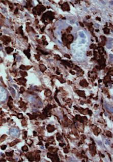

14 42 Chapter 2 Figure 3 SYNAP % 3 4 GFAP % CD11b 92% 3 4 CD % % CD11b 7% 3 HLA HLA % CD11b CD % 0 2 HLA 4 CD Human DIPG 4 12% h-su-dipg-vi h-vu-dipg f h-vu-dipg % 2 h-vu-dipg-3 m-cnmc-d1 5 h-vu-dipg-3 H-2Kd e H-2Kd h-su-dipg-vi H-2Kd m-vu-dipg-5 CD68/Human Nuclei SOX2/CD68 d Human DIPG NeuN Nestin m-cnmc-d1 m-vu-dipg-5 Olig2 m-vu-dipg-5 c Olig2 b m-cnmc-d1 Nestin/GFAP Olig2/SOX2 Murine Tumor Nestin/GFAP a % % 0 2 CD45 3 CD45 Gated agaist live cells/unstained control for each gate Figure. 3 Murine pontine tumors exhibit neural markers and microglial markers (a) Immunohistochemical analysis of PPC markers, Nestin (red and green for m-cnmc-d1), Olig2 (red), SOX2 (green) and GFAP (green) for and -5 pontine tumors and m-cnmc-d1 cells. Murine-VU-DIPG-3 and -5 images refer to transplant generation 1 and 2, respectively, while m-cnmc-d1 cells were obtained form pontine xenograft (transplant generation 1) and cultured in vitro for 5 passages. The arrow indicates reactive gliosis comprised of GFAP+ host cells surrounding the tumor. (c) As in (a) for the original h-vu-dipg-3 and -5 tumor (b) Murine VU-DIPG-3 and -5 pontine xenografts (transplant generations 3-) are negative for the neuronal markers NeuN and synaptophysin (SYNAP), in contrast to host neurons (upper panel) and the neuropil rich in synapses (lower panel). Arrows indicate the diffuse component of the tumors. (d) Murine-VU-DIPG-5 cells obtained from subcutaneous tumors (transplant generations 3-) and cultured on chamber slides resulted positive for CD68 (green) and SOX2 (red). Note the presence of multinucleated cells (arrows). Human SU-DIPG-VI xenograft (h-su-dipg-vi cells passaged in vitro 6 times before xenotransplantation) exhibit sparsely distributed cells immunopositive both for the Human Nuclei antigen (green) and for CD68 (red) (green arrow). Note the presence of a CD68 positive cell that is not positive for Human Nuclei antigen (white arrow) (e) Flow cytometry analysis for H-2Kd, HLA, CD45 and CD11b. On the y- and x-axes the logarithmic intensity of the fluorophore is depicted. Murine-VU-DIPG-3 cells were obtained from subcutaneous tumors (transplant generations 3-); m-cnmc-d1 cells were obtained form pontine xenograft (transplant generation 1) and cultured in vitro for 8 passages and h-su-dipg-vi were cultured in vitro for 12 passages. (f) Human-SUDIPG-I post mortem pontine tissue is immunopositive for CD68 and CD163 microglia/macrophage markers. (a, c, left panels and d) Blue = DAPI. (c, right panels and f) Blue= hematoxylin. Scale bars = (a, left panels) 50 μm and m-vu-dipg-5 μm; (a, central and right panels and c) μm, m-vu-dipg-5 and m-cnmc-d1 20 μm; (b) 12.5 μm and m-vudipg-5 25 μm; (d, left panel and f) μm and (d, right panel) 5 μm.
15 Pontine glioma cells can induce human tumors 43 Development of murine neoplasms using the direct transplantation method was observed independently at two institutions (at the VUMC and at the CNMC). At the CNMC, human DIPG cells gave rise to a murine tumor, named m-cnmc-d1. Murine CNMC-D1 cells were negative both for Human Nuclei and human Vimentin staining (Fig. 2e and f, lower panels). DNA fingerprinting was performed on DNA obtained from, -5, m-cnmc-d1, h-su-dipg-vi cells, and from a tail biopsy of an NSG mouse (m-dna- CTRL). On the gel, human STR are identified by bands lower than 500 base pairs (bp); while h-su-dipg-vi and h-dna-ctrl showed a clear band pattern lower than 500 bp, no bands lower than 500 bp was identified for any of the murine samples (Fig. 2g). Next,, -5 and m-cnmc-d1 cells were analyzed by flow cytometry; no HLA positive cells could be detected, while 93%, 94% and 12% of the cells resulted positive to H-2K d, respectively (Fig. 3e, left and central panels). To test if this phenomenon could have been caused by cells derived from apparently normal human brain tissue rather than from tissue affected by DIPG, we collected post mortem samples from the pons and frontal lobe of two patients not affected by brain cancer. We used the exact same extraction, tissue dissociation and direct transplantation method employed for human DIPG tissue. NSG mice were injected subcutaneously or in the pons or in the frontal lobe with human noncancerous brain cells or with PBS. After 6 to 9 months mice did not show any subcutaneous mass, weight loss or neurological symptoms, therefore mice were sacrificed and the brains analyzed. No tumor was detected in tissue slices stained with hematoxylin and eosin (H&E) (data not shown). Staining for Ki67 did not show any proliferation with the exception of cells lining the needle trajectory (Fig. 2h). Taken together, these data indicate that human DIPG cells derived from tissue collected at autopsy and injected directly into the pons of immunodeficient mice, but not after in vitro passaging, induce lethal murine brain tumors. 02 Murine tumors are positive both for immunomarkers seen in DIPG and for microglia/macrophage markers We then set out to characterize the cell identity of these pontine murine tumors. By analyzing both human and murine healthy pontine tissue at different ages, our group previously identified a specific cell population present uniquely in the ventral pons and at mid-childhood (corresponding to P21 in mice). Hence, these cells, defined as pontine precursor-like cell (PPC), could be the cells of origin of DIPG 31. PPCs express
16 44 Chapter 2 Nestin, Olig2 and SOX2 but not GFAP 31. The DIPG immunophenotype is very similar, but typically exhibits GFAP immunopositivity 31. Interestingly, the murine pontine tumors also revealed the Nestin + /Olig2 + /SOX2 + /GFAP - phenotype 31 (Fig. 3a). The and -5 tumors did not exhibit NeuN/Synaptophysin immunopositivity, arguing against a neuronal origin of these neoplasms (Fig. 3b). Consistent with previous reports 31,96 the h-vu-dipg-3 and -5 primary tumor, while exhibiting cells that are Nestin + /Olig2 +, also exhibited a high percentage of GFAP + cells (Fig. 3c). Intriguingly, almost all and -5 cells were CD68+ and CD45+, as detected by IHC (Fig. 3d) and flow cytometry analysis (Fig. 3e, left panel). In addition, approximately 7% of the and -5 cell population resulted positive for CD11b (Fig. 3e, left panel), indicating that these cells not only exhibit an immunophenotype characteristic of neural precursor cells but also characteristic of microglia/macrophages 3. Next, we tested m-cnmc-d1 cells and h-su-dipg-vi cells for the presence of microglia/macrophage markers, and, while both were negative for CD45, they did present a subpopulation of cells positive for CD11b, 16% and 19%, respectively (Fig. 3e central and right panel). In line with this evidence, h-su-dipg-vi xenografts exhibited cells positive both for Human Nuclei antibody and CD68 (Fig. 3d, right panel). Finally, we stained six human DIPG post mortem tissue samples (Supplementary Table 1) for CD45 (marker for inflammatory cells), CD68 and CD163 (marker for microglia/macrophages) and CD3 (marker for T-cells). While only few cells were positive for CD45 and CD3, a significant number of CD68 and CD163 positive cells were found within the tumor parenchyma (Fig. 3f). Discussion DIPG, with a two-year overall survival rate less than 2%, is the pediatric brain malignancy with the worst prognosis. In the last thirty years, significant improvement in survival has been achieved in other pediatric cancers, while no progress has been made for children affected by DIPG, despite numerous clinical trials 89. We have now made progress due to a focused effort to obtain tissue for research and the establishment of new experimental model systems. Recent studies have demonstrated that DIPG is molecularly distinct from not only adult but also pediatric supratentorial gliomas 29,93,95,1,4,5. Moreover, a specific, recurrent mutation in histone 3 at lysine 27 (H3 K27M) affecting the H3F3A gene (histone 3.3) or the HIST1H3B (histone 3.1) gene is found in nearly 80% of DIPG tumors 32,93,95,6. Thalamic high-grade gliomas of childhood share this specific recurrent K27M point mutation in histone 3.1 or 3.3 genes, while pediatric cortical high-grade glioma
17 Pontine glioma cells can induce human tumors 45 exhibit a similarly specific histone 3 mutation at a different site 93,95. Elegant studies have demonstrated that the recurrent histone 3 K27M mutation results in global loss of function of EZH2, a critical component of the epigenetic machinery responsible for trimethylation of lysine 27 in the N terminal tail of the histone, along with broad changes in the epigenetic landscape 34,35. Yet, further research is needed to fully understand how these epigenetic changes are linked to DIPG tumorigenesis. Strikingly, DIPG presents in a specific temporal window, peaking in incidence at 6-9 years old, and in a specific anatomical area (the ventral pons) 31. Hence, not only its genetic composition but also the pontine microenvironment at mid-childhood may account for the aggressive growth pattern and resistance to therapy this cancer exhibits. Given the urgent need for translational studies on DIPG, we set out to develop models for this disease using post mortem DIPG tissue 25,31. The direct method was attempted to avoid loss of putative important microenvironment growth factors after in vitro culture 7,8 ; the indirect method was used to allow DIPG cell expansion. Interestingly, all 02 tumors developed in the murine pons, and only in one case in the striatum, but in none subcutaneously. This observation may provide further evidences of the importance of the brain and particularly of the pontine microenvironment in facilitating DIPG tumor growth. At two independent institutions, all tumors developed after direct injection of DIPG cells (without in vitro expansion) proved to be of murine and not of human origin, signifying that human DIPG cells, via mechanisms still to be elucidated, induced transformation of healthy murine brain cells into malignant ones. Conversely, h-su- DIPG-VI cells, expanded in vitro before in vivo xenotransplantation, gave rise to tumors of human origin. To prove that the indirect method consistently allows development of human DIPG xenografts, the direct and indirect methods need to be tested in parallel on the same post mortem DIPG cells. Of note, although we performed different control experiments with post mortem tissue not affected by brain cancer, we cannot exclude that development of murine tumors could also occur after injection of other glioma or other non-brain cancer cells. While h-su-dipg-vi cells resulted negative for the H-2K d, a murine marker, not all murine neoplastic cells resulted positive; it has been reported that H-2K d is absent in murine neural stem cells 9,1. Clearly, these findings raise multiple mechanistic questions. Although not yet described for DIPG tumors, the occurrence of mouse tumors after injection of human tumor cells has been previously reported as a sporadic event , and may be attributable to viral transformation 111, human growth factor stimulation 118, cell fusion 112,119 or transference of human DNA into the mouse host cells 113. Such events may lead to
18 46 Chapter 2 single catastrophic events causing the rapid and reproducible onset of mouse DIPG tumors as observed here 120. It is unique to this study that, in all cases attempted with the direct xenotransplantation method, a murine and not a human tumor developed Different reports have demonstrated the presence of human DNA 113 and even human protein expression 112 in cancer cells with the genetic background of the host species 124. In our studies we did not find any evidence of human genes or proteins in the murine neoplasms, possibly due to limitations of the methods employed. Thus, alternative approaches, such as whole genome sequencing, and analysis of the original xenografts, that developed directly after human cell injection, are warranted. A previous study, in which human GBM cells induced transformation of host hamster cells, reported that human chromosomes were segregated within the first transplant generation 112. Finally, the murine neoplasm exhibited microglia/macrophage markers, as did approximately 20% of the h-su-dipg-vi cells. Hence, we analyzed post mortem DIPG tissue and identified microglia/macrophages in the tumor parenchyma. Although there are no reports yet evaluating these cell types in the DIPG microenvironment, studies have shown that cells expressing microglia/macrophage markers comprise 30-50% of the cells in benign and malignant gliomas 3. While the role of microglia/macrophages in glioma has been controversial 126, recent studies have demonstrated that pharmacologic and genetic inhibition of microglial/macrophage function reduces tumor growth in experimental rodent glioma models 35,127,128 and the inhibition of colony stimulating factor-1 receptor has been shown to block glioma progression in a transgenic mouse model of proneuronal GBM 129. Intriguingly, it has been hypothesized that microglia/ macrophages in the glioma microenvironment may be malignant cells themselves and, given their amoeboid properties, maybe capable of invading the surrounding parenchyma giving rise to tumor growth and even tumor metastasis 130. In conclusion, this study highlights the need for diligent precaution when employing DIPG orthotopic xenograft models, and possibly other glioma and non-brain tumor models, to exclude a murine origin of the resulting tumor. It is tempting to speculate that this phenomenon may underscore a biological process relevant to the human disease; alternatively this may be a phenomenon attributable to the altered immune function of the recipient immunodeficient mouse. The work presented here represents an important consideration for orthotopic xenograft models of DIPG, requiring careful species analysis to ensure that preclinical testing is performed in models as faithful to the human disease as possible.
19 Pontine glioma cells can induce human tumors 47 Acknowledgements We would like to thank the patients and families who so generously donated tissue for this research. We sincerely thank architect and artist Alessandra Luoni for drawing Fig. 1a. We gracefully acknowledge Alyssa Noll (Departments of Neurology and Pediatrics) for precious support with animal experiments, and Patty Lovelace (FACS Core) for her expertise with flow cytometry experiments; all from Stanford, United States. The flow cytometer used was purchased using a NIH S Shared Instrumentation Grant (1SRR ). We are thankful to Jacqueline Cloos (Department of Pediatric Oncology, VU University Medical Center, The Netherlands) and Dana Bangs (Cytogenetics Laboratory, Stanford, United States) for their mastery in analyzing metaphase spreads. We are also thankful to Sridevi Yadavilli for processing post mortem specimens and assisting in murine injections, Research Center for Genetic Medicine, Children s National Medical Center, Washington, United States. 02 Funding This work was supported by the Semmy Foundation, KiKa Children Cancer Free, Child Health Research Institute, Lucile Packard Foundation for Children s Health, as well as the Stanford CTSA -award number UL1 TR (V.C.), Stanford University School of Medicine Dean s Fellowship (V.C.), National Institutes of Neurological Disease and Stroke (NINDS grant K08NS070926), Alex s Lemonade Stand Foundation, McKenna Claire Foundation, The Cure Starts Now, Lyla Nsouli Foundation, Connor Johnson Memorial Fund, Dylan Jewett Memorial Fund, Dylan Frick Memorial Fund, Abigail Jensen Memorial Fund, Zoey Ganesh Memorial Fund, Wayland Villars Memorial Fund and Musella Foundation.


 (transplant generations 3-, except h")


 Leptomeningeal spread in h-vu-dipg-5 and in")


 Tumor cells clustered around vessels in")
 Murine intraparencymal tumors presented areas of")
20 48 Chapter 2 Supplementary figure 1 e f g h * * m-vu-dipg-5 c d i j k l h-vu-dipg-5 b h-vu-dipg-3 a Supplementary Figure 1 Histopathological characterization of human and murine VU-DIPG-3 and -5. H&E images of (a and b) h-vu-dipg-3, (c and d) h-vu-dipg-5, (e-h) (transplant generations 3-, except h = transplant generation 1) and (i-l) m-vu-dipg-5 (transplant generations 3-, except k = transplant generation 1). High magnification image of small cell phenotype in (a and d) h-vu-dipg-3 and -5 and in (e and j) the correspondent and -5. (b and f) Numerous tumor cells surrounding the tunica adventitia in h-vu-dipg-3 and. The arrow in figure b indicates a fibrin aggregate. (c, f and i) Leptomeningeal spread in h-vu-dipg-5 and in and -5. The arrow in figure C points at a leptomeningeal artery presenting thick walls. Note the tumor front invading the surrounding brain parenchyma in figure i. (d, g and l) Tumor cells clustered around vessels in h-vu-dipg-3 and and -5. Arrows indicate perivascular growth. (g) Murine intraparencymal tumors presented areas of more compact growth (asterisk) along with diffuse growth after the first 2 transplant generation. (h) Perineuronal satellistosis, the arrow points at the neuronal cell body. (k) m-vu-dipg-5 invading the skull bone. Scale bars = (a, c, d, e, g, h, I and j) μm; (b) 0 μm; (f, k and l) 20 μm.

 Murine VU-DIPG-3 abnormal metaphase with 41 chromosomes including an apparent dicentric")
 Murine VU-DIPG-5 polyploid metaphase with 80 chromosomes including two centric fusion")
 Murine CNMC-D1 diploid metaphase and (d) grossly abnormal metaphase.")


21 Pontine glioma cells can induce human tumors 49 Supplementary figure 2 a c m-cnmc-d1 e h-su-dipg-vi 02 b d m-vu-dipg-5 m-cnmc-d1 Supplementary Figure 2 Metaphase spreads in murine pontine tumors and h-su-dipg-vi. (a) Murine VU-DIPG-3 abnormal metaphase with 41 chromosomes including an apparent dicentric chromosome (arrow). (b) Murine VU-DIPG-5 polyploid metaphase with 80 chromosomes including two centric fusion chromosomes (arrows). (c) Murine CNMC-D1 diploid metaphase and (d) grossly abnormal metaphase. (e) Human SU-DIPG-VI (in vitro passage ) tetraploid metaphase spread with arrows indicating some of the abnormal chromosomes present. Murine VU-DIPG-3 and -5 cells were obtained from subcutaneous tumors at transplant generation 3-; m-cnmc-d1 cells were obtained form pontine xenograft (transplant generation 1) and cultured in vitro for 8 passages. Supplementary figure 3 a Human platform b Murine platform Supplementary Figure 3 Human and murine array-comparative Genomic Hybridization raw data Fluorescent image of hybridized with m-dna CTRL on (a) a human platform and on (b) a murine platform.
22 50 Chapter 2 Supplementary Table 1 Clinical characteristics of subjects whose DIPG tumors were evaluated for microglia/macrophage markers Patient h-su-dipg-i h-su-dipg-iii h-su-dipg-v h-su-dipg-vii h-su-dipg-viii Age (years) Sex M M F M M Histological diagnosis at autopsy Anaplatic astrocytoma (WHO grade III) GBM GBM GBM Astrocytoma (WHO grade III) WHO refers to World Health Organization and GBM refers to glioblastoma multiforme (grade IV). The clinical characteristics for SU-DIPG-VI are reported in Table 1. Supplementary Table 2 Primer sequences Gene Forward Reverse Human H3F3A GGGCAGGAGCCTCTCTTAAT ACCAAGTAGGCCTCACAAGC Hist1H3b AAGCAGAGGCTGACCAATCC TCCTTGGGCATGATGGTGAC PI3KCA H47R/L GCTCCAAACTGACCAAACTGT AATCGGTCTTTGCCTGCTGA PI3KCA E542K CATCTGTGAATCCAGAGGGGA ACATGCTGAGATCAGCCAAA Murine H3F3A GGGCAGGAGCCTCTCTTAAT ACCAAGTAGGCCTCACAAGC Hist1H3b AAGCAGAGGCTGACCAATCC TCCTTGGGCATGATGGTGAC PI3KCA H47R/L ACTAGCAGCTCACTGACCAGA GCTATCAAACCCTGCTTGCG PI3KCA E542K TCTTCCCCTTCGCCAAGTCTA ACAGGAAGAAGGTCCCTCGG Supplementary Table 3 Human SU-DIPG-VI Control Cortex DNA Short Tandem Repeat (STR) fingerprint AMEL CSF1PO1 D13S317 D16S539 D21S11 D5S818 D7S820 TH01 TPOX vwa h-su- DIPG-VI Cortex Control X/X /11 11/ 8/13 29/31 /12 8/9 7/8 8/11 17/18
23 Pontine glioma cells can induce human tumors 51 02

24
(A) PCR primers (arrows) designed to distinguish wild type (P1+P2), targeted (P1+P2) and excised (P1+P3)14-
 PCR primers (arrows) designed to distinguish wild type (P1+P2), targeted (P1+P2) and excised (P1+P3)14-") 1 Supplemental Figure Legends Figure S1. Mammary tumors of ErbB2 KI mice with 14-3-3σ ablation have elevated ErbB2 transcript levels and cell proliferation (A) PCR primers (arrows) designed to distinguish
1 Supplemental Figure Legends Figure S1. Mammary tumors of ErbB2 KI mice with 14-3-3σ ablation have elevated ErbB2 transcript levels and cell proliferation (A) PCR primers (arrows) designed to distinguish
Deceptive morphologic and epigenetic heterogeneity in diffuse intrinsic pontine glioma
 CHAPTER 5 Deceptive morphologic and epigenetic heterogeneity in diffuse intrinsic pontine glioma Veldhuijzen van Zanten SEM*, Bugiani M*, Caretti V, Schellen P, Aronica E, Noske DP, Vandertop WP, Kaspers
Enterprise Interest None
 Enterprise Interest None Heterogeneous chromosomal profiles in a unique series of DIPG in children and young adults European Congress of Pathology Amsterdam, 6 th September 2017 Charlotte Dufour, Romain
Supplemental Figure 1. Intracranial transduction of a modified ptomo lentiviral vector in the mouse
 Supplemental figure legends Supplemental Figure 1. Intracranial transduction of a modified ptomo lentiviral vector in the mouse hippocampus targets GFAP-positive but not NeuN-positive cells. (A) Stereotaxic
Anti-tumor Effects of Activated Human Natural Killer Cells in Orthotopic Human Brain Tumor Models
 Anti-tumor Effects of Activated Human Natural Killer Cells in Orthotopic Human Brain Tumor Models William Murphy, PhD Depts. of Dermatology and Internal Medicine Neal Goodwin, PhD Jackson Laboratories
Supporting Information
 Supporting Information Sasportas et al. 10.1073/pnas.0806647106 SI Methods Lentiviral Transduction. MSC and glioma cells were transduced with LVs at varying multiplicity of infection (moi) by incubating
IDH1 R132H/ATRX Immunohistochemical validation
 IDH1 R132H/ATRX Immunohistochemical validation CIQC/DSM 2016 12 June 2016 0835-0905 Stephen Yip, M.D., Ph.D., FRCPC University of British Columbia Disclosure Statement I have nothing to disclose I will
Supplementary Information
 Supplementary Information Title Degeneration and impaired regeneration of gray matter oligodendrocytes in amyotrophic lateral sclerosis Authors Shin H. Kang, Ying Li, Masahiro Fukaya, Ileana Lorenzini,
Pharmacologic inhibition of histone demethylation as a therapy for pediatric brainstem glioma
 Supplementary information for: Pharmacologic inhibition of histone demethylation as a therapy for pediatric brainstem glioma Rintaro Hashizume 1, Noemi Andor 2, Yuichiro Ihara 2, Robin Lerner 2, Haiyun
VEGFR2-Mediated Vascular Dilation as a Mechanism of VEGF-Induced Anemia and Bone Marrow Cell Mobilization
 Cell Reports, Volume 9 Supplemental Information VEGFR2-Mediated Vascular Dilation as a Mechanism of VEGF-Induced Anemia and Bone Marrow Cell Mobilization Sharon Lim, Yin Zhang, Danfang Zhang, Fang Chen,
Supplementary Figure 1: Neuregulin 1 increases the growth of mammary organoids compared to EGF. (a) Mammary epithelial cells were freshly isolated,
 Mammary epithelial cells were freshly isolated,") 1 2 3 4 5 6 7 8 9 10 Supplementary Figure 1: Neuregulin 1 increases the growth of mammary organoids compared to EGF. (a) Mammary epithelial cells were freshly isolated, embedded in matrigel and exposed
1 2 3 4 5 6 7 8 9 10 Supplementary Figure 1: Neuregulin 1 increases the growth of mammary organoids compared to EGF. (a) Mammary epithelial cells were freshly isolated, embedded in matrigel and exposed
Neuropathology Evening Session: Case 3
 Neuropathology Evening Session: Case 3 Christine E. Fuller, MD Cincinnati Children s Hospital Medical Center Disclosure of Relevant Financial Relationships USCAP requires that all faculty in a position
Supplementary Information Titles Journal: Nature Medicine
 Supplementary Information Titles Journal: Nature Medicine Article Title: Corresponding Author: Supplementary Item & Number Supplementary Fig.1 Fig.2 Fig.3 Fig.4 Fig.5 Fig.6 Fig.7 Fig.8 Fig.9 Fig. Fig.11
MOLECULAR DIAGNOSTICS OF GLIOMAS
 MOLECULAR DIAGNOSTICS OF GLIOMAS Arie Perry, M.D. Director, Neuropathology Division DIFFUSE GLIOMAS Cell types Astrocytomas (A) Oligodendrogliomas (O) Mixed oligoastrocytoma (MOA) Three WHO grades: II,
Classification of Diffuse Gliomas: Progress, Pearls and Pitfalls. Rob Macaulay Neuropathologist, MCC October 21, 2017
 Classification of Diffuse Gliomas: Progress, Pearls and Pitfalls Rob Macaulay Neuropathologist, MCC October 21, 2017 Objectives Explain why the designation high grade glioma is preferable to GBM for intraoperative
Supporting Information
 Supporting Information Chan et al. 1.173/pnas.9654916 A Patient B Xenograft C * remaining feature of normal lymph node * * * D lymphocytes Infiltrating transitional carcinoma cells E Enlarged axillary
(A) Cells grown in monolayer were fixed and stained for surfactant protein-c (SPC,
 Cells grown in monolayer were fixed and stained for surfactant protein-c (SPC,") Supplemental Figure Legends Figure S1. Cell line characterization (A) Cells grown in monolayer were fixed and stained for surfactant protein-c (SPC, green) and co-stained with DAPI to visualize the nuclei.
Supplemental Figure Legends Figure S1. Cell line characterization (A) Cells grown in monolayer were fixed and stained for surfactant protein-c (SPC, green) and co-stained with DAPI to visualize the nuclei.
Supplementary methods:
 Supplementary methods: Primers sequences used in real-time PCR analyses: β-actin F: GACCTCTATGCCAACACAGT β-actin [11] R: AGTACTTGCGCTCAGGAGGA MMP13 F: TTCTGGTCTTCTGGCACACGCTTT MMP13 R: CCAAGCTCATGGGCAGCAACAATA
Genomic analysis of childhood High grade glial (HGG) brain tumors
 brain tumors") Genomic analysis of childhood High grade glial (HGG) brain tumors Linda D Cooley Children s Mercy, Kansas City The Children s Mercy Hospital, 2017 Genomic analysis of childhood High grade glial (HGG) brain
Genomic analysis of childhood High grade glial (HGG) brain tumors Linda D Cooley Children s Mercy, Kansas City The Children s Mercy Hospital, 2017 Genomic analysis of childhood High grade glial (HGG) brain
Imaging of glycolytic metabolism in primary glioblastoma cells with
 63 Chapter 5 Imaging of glycolytic metabolism in primary glioblastoma cells with RIMChip 5.1. Introduction Glioblastoma(GBM) is one of the most common brain tumors 1. It is composed of heterogeneous subpopulations
Supplemental Figure 1. Isolation and characterization of CD133+ neurosphere-like
 SUPPLEMENTL FIGURE LEGENDS Supplemental Figure 1. Isolation and characterization of CD133+ neurosphere-like spheroids from a human brain tumor sample or glioma xenograft. () CD133+ tumor cells isolated
Five Most Common Problems in Surgical Neuropathology
 Five Most Common Problems in Surgical Neuropathology If the brain were so simple that we could understand it, we would be so simple that we couldn t Emerson Pugh What is your greatest difficulty in neuropathology?
Deceptive morphologic and epigenetic heterogeneity in diffuse intrinsic pontine glioma
 /, 2017, Vol. 8, (No. 36), pp: 60447-60452 Deceptive morphologic and epigenetic heterogeneity in diffuse intrinsic pontine glioma Marianna Bugiani 1,*, Sophie E.M. Veldhuijzen van Zanten 2,3,*, Viola Caretti
Supplementary Figure S1: Tanycytes are restricted to the central/posterior hypothalamus
 Supplementary Figure S1: Tanycytes are restricted to the central/posterior hypothalamus a: Expression of Vimentin, GFAP, Sox2 and Nestin in anterior, central and posterior hypothalamus. In the anterior
Comprehensive evaluation of human immune system reconstitution in NSG. and NSG -SGM3 mouse models toward the development of a novel ONCO-HU
 Comprehensive evaluation of human immune system reconstitution in NSG and NSG -SGM3 mouse models toward the development of a novel ONCO-HU xenograft model Aaron Middlebrook, 1 Eileen Snowden, 2 Warren
(A) RT-PCR for components of the Shh/Gli pathway in normal fetus cell (MRC-5) and a
 RT-PCR for components of the Shh/Gli pathway in normal fetus cell (MRC-5) and a") Supplementary figure legends Supplementary Figure 1. Expression of Shh signaling components in a panel of gastric cancer. (A) RT-PCR for components of the Shh/Gli pathway in normal fetus cell (MRC-5) and
Supplementary figure legends Supplementary Figure 1. Expression of Shh signaling components in a panel of gastric cancer. (A) RT-PCR for components of the Shh/Gli pathway in normal fetus cell (MRC-5) and
Gastric Carcinoma with Lymphoid Stroma: Association with Epstein Virus Genome demonstrated by PCR
 Gastric Carcinoma with Lymphoid Stroma: Association with Epstein Virus Genome demonstrated by PCR Pages with reference to book, From 305 To 307 Irshad N. Soomro,Samina Noorali,Syed Abdul Aziz,Suhail Muzaffar,Shahid
SUPPLEMENTARY FIGURES
 SUPPLEMENTARY FIGURES 1 2 3 4 SUPPLEMENTARY TABLES Supplementary Table S1. Brain Tumors used in the study Code Tumor Classification Age Gender HuTuP51 Glioblastoma 57 Male HuTuP52 Glioblastoma 53 Male
Evaluation of directed and random motility in microslides Assessment of leukocyte adhesion in flow chambers
 Evaluation of directed and random motility in microslides Motility experiments in IBIDI microslides, image acquisition and processing were performed as described. PMN, which ended up in an angle < 180
Ductal pancreatic cancer modeling and drug screening using human pluripotent stem cell and patient-derived tumor organoids
 Ductal pancreatic cancer modeling and drug screening using human pluripotent stem cell and patient-derived tumor organoids Ling Huang 1, Audrey Holtzinger 1,2,11, Ishaan Jagan 1,11, Michael BeGora 1, Ines
Supplemental figure 1. PDGFRα is expressed dominantly by stromal cells surrounding mammary ducts and alveoli. A) IHC staining of PDGFRα in
 IHC staining of PDGFRα in") Supplemental figure 1. PDGFRα is expressed dominantly by stromal cells surrounding mammary ducts and alveoli. A) IHC staining of PDGFRα in nulliparous (left panel) and InvD6 mouse mammary glands (right
Supplemental figure 1. PDGFRα is expressed dominantly by stromal cells surrounding mammary ducts and alveoli. A) IHC staining of PDGFRα in nulliparous (left panel) and InvD6 mouse mammary glands (right
Effective activity of cytokine-induced killer cells against autologous metastatic melanoma including cells with stemness features
 Effective activity of cytokine-induced killer cells against autologous metastatic melanoma including cells with stemness features Loretta Gammaitoni, Lidia Giraudo, Valeria Leuci, et al. Clin Cancer Res
The diagnostic and prognostic value of genetic aberrations in resectable distal bile duct cancer Rijken, A.M.
 UvA-DARE (Digital Academic Repository) The diagnostic and prognostic value of genetic aberrations in resectable distal bile duct cancer Rijken, A.M. Link to publication Citation for published version (APA):
Supporting Information
 Supporting Information Santagata et al. 10.1073/pnas.1404724111 Extended Description of Fig. 4A Intraoperative MS allows for significant advances in the frequency of intraoperative tissue sampling as well
Pearson r = P (one-tailed) = n = 9
 = n = 9") 8F4-Specific Lysis, % 1 UPN1 UPN3 8 UPN7 6 Pearson r =.69 UPN2 UPN5 P (one-tailed) =.192 4 UPN8 n = 9 2 UPN9 UPN4 UPN6 5 1 15 2 25 8 8F4, % Max MFI Supplementary Figure S1. AML samples UPN1-UPN9 show variable
8F4-Specific Lysis, % 1 UPN1 UPN3 8 UPN7 6 Pearson r =.69 UPN2 UPN5 P (one-tailed) =.192 4 UPN8 n = 9 2 UPN9 UPN4 UPN6 5 1 15 2 25 8 8F4, % Max MFI Supplementary Figure S1. AML samples UPN1-UPN9 show variable
Data Mining for Research and Diagnostics
 UCL DEPARTMENT OF GEOGRAPHY UCL INSTITUTE OF NEUROLOGY Data Mining for Research and Diagnostics Sebastian Brandner, UCL Institute of Neurology, London Data mining for Research and Diagnostics Our setup
Supplemental Figure 1
 Supplemental Figure 1 A S100A4: SFLGKRTDEAAFQKLMSNLDSNRDNEVDFQEYCVFLSCIAMMCNEFFEGFPDK Overlap: SF G DE KLM LD N D VDFQEY VFL I M N FF G PD S100A2: SFVGEKVDEEGLKKLMGSLDENSDQQVDFQEYAVFLALITVMCNDFFQGCPDR
CNS pathology Third year medical students. Dr Heyam Awad 2018 Lecture 12: CNS tumours 2/3
 CNS pathology Third year medical students Dr Heyam Awad 2018 Lecture 12: CNS tumours 2/3 Pilocytic astrocytoma Relatively benign ( WHO grade 1) Occurs in children and young adults Mostly: in the cerebellum
Islet viability assay and Glucose Stimulated Insulin Secretion assay RT-PCR and Western Blot
 Islet viability assay and Glucose Stimulated Insulin Secretion assay Islet cell viability was determined by colorimetric (3-(4,5-dimethylthiazol-2-yl)-2,5- diphenyltetrazolium bromide assay using CellTiter
Macrophages form functional vascular mimicry channels in vivo. SI Figures and Legend
 Macrophages form functional vascular mimicry channels in vivo Authors: *Faith H. Barnett, *Mauricio Rosenfeld, Malcolm Wood, William Kiosses, Yoshihiko Usui, Valentina Marchetti, Edith Aguilar, and Martin
Nature Genetics: doi: /ng.2995
 Supplementary Figure 1 Kaplan-Meier survival curves of patients with brainstem tumors. (a) Comparison of patients with PPM1D mutation versus wild-type PPM1D. (b) Comparison of patients with PPM1D mutation
Pediatric Brain Tumors: Updates in Treatment and Care
 Pediatric Brain Tumors: Updates in Treatment and Care Writer Classroom Rishi R. Lulla, MD MS Objectives Introduce the common pediatric brain tumors Discuss current treatment strategies for pediatric brain
Supplementary Figure 1
 Supplementary Figure 1 3 3 3 1 1 Bregma -1.6mm 3 : Bregma Ref) Http://www.mbl.org/atlas165/atlas165_start.html Bregma -.18mm Supplementary Figure 1 Schematic representation of the utilized brain slice
Understanding general brain tumor pathology, Part I: The basics. Craig Horbinski, M.D., Ph.D. Department of Pathology University of Kentucky
 Understanding general brain tumor pathology, Part I: The basics Craig Horbinski, M.D., Ph.D. Department of Pathology University of Kentucky plan of attack what IS a pathologist, anyway? what s so special
General: Brain tumors are lesions that have mass effect distorting the normal tissue and often result in increased intracranial pressure.
 1 Lecture Objectives Know the histologic features of the most common tumors of the CNS. Know the differences in behavior of the different tumor types. Be aware of the treatment modalities in the various
Supplementary Materials for
 immunology.sciencemag.org/cgi/content/full/2/16/eaan6049/dc1 Supplementary Materials for Enzymatic synthesis of core 2 O-glycans governs the tissue-trafficking potential of memory CD8 + T cells Jossef
Tumors of the Nervous System
 Tumors of the Nervous System Peter Canoll MD. PhD. What I want to cover What are the most common types of brain tumors? Who gets them? How do they present? What do they look like? How do they behave? 1
Comparison of primary tumor sections from MMTV-PyMT or MTLn3-ErbB3-
 Supplemental Data Comparison of primary tumor sections from MMTV-PyMT or MTLn3-ErbB3- GFP tumors in mice either injected with control or clodronate-containing liposomes and stained for macrophages using
In vitro bactericidal assay Fig. S8 Gentamicin protection assay Phagocytosis assay
 In vitro bactericidal assay Mouse bone marrow was isolated from the femur and the tibia. Cells were suspended in phosphate buffered saline containing.5% BSA and 2 mm EDTA and filtered through a cell strainer.
Supporting Information
 Supporting Information CD200 Expressing Human Basal Cell Carcinoma Cells Initiate Tumor Growth Chantal S Colmont 1, Antisar BenKetah 1, Simon H Reed 2, Nga Voong 3, William Telford 3, Manabu Ohyama 4,
To this end, we performed immunofluorescent staining for GSCs (Fig.3). All the spheroidforming cells showed immunoreactivity for
. All the spheroidforming cells showed immunoreactivity for") H.Yoshioka et al. lished for isolation ofneural stem cells. Within 24-48 hours of primary culture, murine brain tumors yielded a minority fraction of cells that formed neurosphere-like clusters (tumor
H.Yoshioka et al. lished for isolation ofneural stem cells. Within 24-48 hours of primary culture, murine brain tumors yielded a minority fraction of cells that formed neurosphere-like clusters (tumor
Supplementary Data 1. Alanine substitutions and position variants of APNCYGNIPL. Applied in
 Supplementary Data 1. Alanine substitutions and position variants of APNCYGNIPL. Applied in Supplementary Fig. 2 Substitution Sequence Position variant Sequence original APNCYGNIPL original APNCYGNIPL
ACMG/CAP Cytogenetics CY
 www.cap.org Cytogenetics Analytes/procedures in bold type are regulated for proficiency testing by the Centers for Medicare & Medicaid Services ACMG/CAP Cytogenetics CY Analyte CY Challenges per Shipment
Cytokine Complex Expanded Natural Killer Cells Improve Allogeneic Lung. Transplant Function via Depletion of Donor Dendritic Cells
 Cytokine Complex Expanded Natural Killer Cells Improve Allogeneic Lung Transplant Function via Depletion of Donor Dendritic Cells Wolfgang Jungraithmayr, Laura Codarri, Gregory Bouchaud,Carsten Krieg,
Author's response to reviews
 Author's response to reviews Title:The orthotopic xenotransplant of human glioblastoma successfully recapitulates glioblastoma-microenvironment interactions in a non-immunosuppressed mouse model Authors:
Nature Immunology: doi: /ni Supplementary Figure 1. Huwe1 has high expression in HSCs and is necessary for quiescence.
 Supplementary Figure 1 Huwe1 has high expression in HSCs and is necessary for quiescence. (a) Heat map visualizing expression of genes with a known function in ubiquitin-mediated proteolysis (KEGG: Ubiquitin
Ex vivo Human Antigen-specific T Cell Proliferation and Degranulation Willemijn Hobo 1, Wieger Norde 1 and Harry Dolstra 2*
 Ex vivo Human Antigen-specific T Cell Proliferation and Degranulation Willemijn Hobo 1, Wieger Norde 1 and Harry Dolstra 2* 1 Department of Laboratory Medicine - Laboratory of Hematology, Radboud University
Journal of Neurosurgery Pediatrics, 2017 May;19(5):
:") Chapter 4 Preclinical evaluation of convection-enhanced delivery with liposomal doxorubicin to treat pediatric diffuse intrinsic pontine glioma and thalamic high grade glioma. APC Sewing, MD, T Lagerweij,
Chapter 4 Preclinical evaluation of convection-enhanced delivery with liposomal doxorubicin to treat pediatric diffuse intrinsic pontine glioma and thalamic high grade glioma. APC Sewing, MD, T Lagerweij,
RNA extraction, RT-PCR and real-time PCR. Total RNA were extracted using
 Supplementary Information Materials and Methods RNA extraction, RT-PCR and real-time PCR. Total RNA were extracted using Trizol reagent (Invitrogen,Carlsbad, CA) according to the manufacturer's instructions.
Supplementary Figure 1. Nature Neuroscience: doi: /nn.4547
 Supplementary Figure 1 Characterization of the Microfetti mouse model. (a) Gating strategy for 8-color flow analysis of peripheral Ly-6C + monocytes from Microfetti mice 5-7 days after TAM treatment. Living
USCAP Neuropathology. Case No. 3 Elisabeth J. Rushing, MD Armed Forces Institute of Pathology Washington, DC
 USCAP Neuropathology Case No. 3 Elisabeth J. Rushing, MD Armed Forces Institute of Pathology Washington, DC Clinical history The patient is a 9 year-old boy who has had seizures since age 2, at which time
Genesis of cerebellar interneurons and the prevention of neural DNA damage require XRCC1.
 Genesis of cerebellar interneurons and the prevention of neural DNA damage require XRCC1. Youngsoo Lee, Sachin Katyal, Yang Li, Sherif F. El-Khamisy, Helen R. Russell, Keith W. Caldecott and Peter J. McKinnon.
Role of FISH in Hematological Cancers
 Role of FISH in Hematological Cancers Thomas S.K. Wan PhD,FRCPath,FFSc(RCPA) Honorary Professor, Department of Pathology & Clinical Biochemistry, Queen Mary Hospital, University of Hong Kong. e-mail: wantsk@hku.hk
CONTRACTING ORGANIZATION: Children s Hospital Los Angeles Los Angeles, CA 90027
 AD Award Number: TITLE: Studies of the Tumor Microenvironment in Pathogenesis of Neuroblastoma PRINCIPAL INVESTIGATOR: Shahab Asgharzadeh, M D CONTRACTING ORGANIZATION: Children s Hospital Los Angeles
Cytogenetics 101: Clinical Research and Molecular Genetic Technologies
 Cytogenetics 101: Clinical Research and Molecular Genetic Technologies Topics for Today s Presentation 1 Classical vs Molecular Cytogenetics 2 What acgh? 3 What is FISH? 4 What is NGS? 5 How can these
Superior Fluorescent Labeling Dyes Spanning the Full Visible Spectrum...1. Trademarks: HiLyte Fluor (AnaSpec, Inc.)
") Table of Contents Fluor TM Labeling Dyes Superior Fluorescent Labeling Dyes Spanning the Full Visible Spectrum....1 Fluor TM 405 Dye, an Excellent Alternative to Alexa Fluor 405 & DyLight 405....2 Fluor
Table of Contents Fluor TM Labeling Dyes Superior Fluorescent Labeling Dyes Spanning the Full Visible Spectrum....1 Fluor TM 405 Dye, an Excellent Alternative to Alexa Fluor 405 & DyLight 405....2 Fluor
Layered-IHC (L-IHC): A novel and robust approach to multiplexed immunohistochemistry So many markers and so little tissue
: A novel and robust approach to multiplexed immunohistochemistry So many markers and so little tissue") Page 1 The need for multiplex detection of tissue biomarkers. There is a constant and growing demand for increased biomarker analysis in human tissue specimens. Analysis of tissue biomarkers is key to
Page 1 The need for multiplex detection of tissue biomarkers. There is a constant and growing demand for increased biomarker analysis in human tissue specimens. Analysis of tissue biomarkers is key to
Online Appendix Material and Methods: Pancreatic RNA isolation and quantitative real-time (q)rt-pcr. Mice were fasted overnight and killed 1 hour (h)
rt-pcr. Mice were fasted overnight and killed 1 hour (h)") Online Appendix Material and Methods: Pancreatic RNA isolation and quantitative real-time (q)rt-pcr. Mice were fasted overnight and killed 1 hour (h) after feeding. A small slice (~5-1 mm 3 ) was taken
Online Appendix Material and Methods: Pancreatic RNA isolation and quantitative real-time (q)rt-pcr. Mice were fasted overnight and killed 1 hour (h) after feeding. A small slice (~5-1 mm 3 ) was taken
Nicholas Szerlip, MD Department of Neurosurgery Wayne State University Karmanos Cancer Institute
 Nicholas Szerlip, MD Department of Neurosurgery Wayne State University Karmanos Cancer Institute Many different names (Heregulin, Aria) Found when investigating ligands that activated HER2 (it actually
WHY BIOPSY? Diagnosis and Research
 WHY BIOPSY? Diagnosis and Research 9 2 4 1 3 1 1. Diagnosis only by Imaging (like no other tumor) The issue of Typical versus Atypical DIPG T1 FLAIR Gad. T2 Tractography Functional MRI Diffusion/Perfusion
Product Introduction
 Product Introduction Product Codes: HCL026, HCL027 and HCL028 Contents Introduction to HER2 2 HER2 immunohistochemistry 3 Cell lines as controls 5 HER2 Analyte Control DR IHC 7 HER2 Analyte Control DR
Spheroid-based engineering of a human vasculature in mice
 Spheroid-based engineering of a human vasculature in mice Abdullah Alajati, Anna M. Laib, Holger Weber, Anja M. Boos, Arne Bartol, Kristian Ikenberg, Thomas Korff, Hanswalter Zentgraf, Cynthia Obodozie,
Supplemental Table 1. Primer sequences for transcript analysis
 Supplemental Table 1. Primer sequences for transcript analysis Primer Sequence (5 3 ) Primer Sequence (5 3 ) Mmp2 Forward CCCGTGTGGCCCTC Mmp15 Forward CGGGGCTGGCT Reverse GCTCTCCCGGTTTC Reverse CCTGGTGTGCCTGCTC
Detection of Anaplastic Lymphoma Kinase (ALK) gene in Non-Small Cell lung Cancer (NSCLC) By CISH Technique
 gene in Non-Small Cell lung Cancer (NSCLC) By CISH Technique") Cancer and Clinical Oncology; Vol. 7, No. 1; 2018 ISSN 1927-4858 E-ISSN 1927-4866 Published by Canadian Center of Science and Education Detection of Anaplastic Lymphoma Kinase (ALK) gene in Non-Small Cell
Cancer and Clinical Oncology; Vol. 7, No. 1; 2018 ISSN 1927-4858 E-ISSN 1927-4866 Published by Canadian Center of Science and Education Detection of Anaplastic Lymphoma Kinase (ALK) gene in Non-Small Cell
Molecular Probes Introducing 14 new probes
 Molecular Probes Introducing 14 new probes Gene and Chromosome Probes Dual Colour ISH INFORM HER2 Dual ISH DNA Probe Cocktail Assay Product Part Number INFORM HER2 Dual ISH DNA Probe Cocktail 800-4422
Interferon γ regulates idiopathic pneumonia syndrome, a. Th17 + CD4 + T-cell-mediated GvH disease
 Interferon γ regulates idiopathic pneumonia syndrome, a Th17 + CD4 + T-cell-mediated GvH disease Nora Mauermann, Julia Burian, Christophe von Garnier, Stefan Dirnhofer, Davide Germano, Christine Schuett,
SUPPLEMENTARY FIGURES
 SUPPLEMENTARY FIGURES 1 Supplementary Figure 1, Adult hippocampal QNPs and TAPs uniformly express REST a-b) Confocal images of adult hippocampal mouse sections showing GFAP (green), Sox2 (red), and REST
SUPPLEMENTARY INFORMATION. Involvement of IL-21 in the epidermal hyperplasia of psoriasis
 SUPPLEMENTARY INFORMATION Involvement of IL-21 in the epidermal hyperplasia of psoriasis Roberta Caruso 1, Elisabetta Botti 2, Massimiliano Sarra 1, Maria Esposito 2, Carmine Stolfi 1, Laura Diluvio 2,
The pathogenesis of nervous distemper
 Veterinary Sciences Tomorrow - 2004 The pathogenesis of nervous distemper Marc Vandevelde Canine distemper is a highly contagious viral disease of dogs and of all animals in the Canidae, Mustellidae and
Differential diagnosis of hematolymphoid tumors composed of medium-sized cells. Brian Skinnider B.C. Cancer Agency, Vancouver General Hospital
 Differential diagnosis of hematolymphoid tumors composed of medium-sized cells Brian Skinnider B.C. Cancer Agency, Vancouver General Hospital Lymphoma classification Lymphoma diagnosis starts with morphologic
AP VP DLP H&E. p-akt DLP
 A B AP VP DLP H&E AP AP VP DLP p-akt wild-type prostate PTEN-null prostate Supplementary Fig. 1. Targeted deletion of PTEN in prostate epithelium resulted in HG-PIN in all three lobes. (A) The anatomy
PBMC from each patient were suspended in AIM V medium (Invitrogen) with 5% human
 with 5% human") Anti-CD19-CAR transduced T-cell preparation PBMC from each patient were suspended in AIM V medium (Invitrogen) with 5% human AB serum (Gemini) and 300 international units/ml IL-2 (Novartis). T cell proliferation
Anti-CD19-CAR transduced T-cell preparation PBMC from each patient were suspended in AIM V medium (Invitrogen) with 5% human AB serum (Gemini) and 300 international units/ml IL-2 (Novartis). T cell proliferation
SALSA MLPA probemix P315-B1 EGFR
 SALSA MLPA probemix P315-B1 EGFR Lot B1-0215 and B1-0112. As compared to the previous A1 version (lot 0208), two mutation-specific probes for the EGFR mutations L858R and T709M as well as one additional
Explain the laboratory diagnosis of Rabies?
 Explain the laboratory diagnosis of Rabies? The standard test for rabies testing is dfa. This test has been thoroughly evaluated for more than 40 years, and is recognized as the most rapid and reliable
mm Distance (mm)
") b a Magnet Illumination Coverslips MPs Objective 2575 µm 1875 µm 1575 µm 1075 µm 875 µm 545 µm 20µm 2 3 0.5 0.3mm 1 1000 100 10 1 0.1 1000 100 10 1 0.1 Field Induction (Gauss) 1.5 0 5 10 15 20 Distance
b a Magnet Illumination Coverslips MPs Objective 2575 µm 1875 µm 1575 µm 1075 µm 875 µm 545 µm 20µm 2 3 0.5 0.3mm 1 1000 100 10 1 0.1 1000 100 10 1 0.1 Field Induction (Gauss) 1.5 0 5 10 15 20 Distance
ALLEN Human Brain Atlas
 TECHNICAL WHITE PAPER: CASE QUALIFICATION AND DONOR PROFILES The case review process described here was employed for three components of the ALLEN Human Brain Atlas: (1) the Survey; (2) the Neurotransmitter
Supplementary Materials. for Garmy-Susini, et al, Integrin 4 1 signaling is required for lymphangiogenesis and tumor metastasis
 Supplementary Materials for Garmy-Susini, et al, Integrin 4 1 signaling is required for lymphangiogenesis and tumor metastasis 1 Supplementary Figure Legends Supplementary Figure 1: Integrin expression
Gliomas in the 2016 WHO Classification of CNS Tumors
 Gliomas in the 2016 WHO Classification of CNS Tumors Hindi N Al-Hindi, MD, FCAP Consultant Neuropathologist and Head Section of Anatomic Pathology Department of Pathology and Laboratory Medicine King Faisal
SUPPLEMENTARY FIG. S2. Representative counting fields used in quantification of the in vitro neural differentiation of pattern of dnscs.
 Supplementary Data SUPPLEMENTARY FIG. S1. Representative counting fields used in quantification of the in vitro neural differentiation of pattern of anpcs. A panel of lineage-specific markers were used
Supplementary Figure 1. Spitzoid Melanoma with PPFIBP1-MET fusion. (a) Histopathology (4x) shows a domed papule with melanocytes extending into the
 Histopathology (4x) shows a domed papule with melanocytes extending into the") Supplementary Figure 1. Spitzoid Melanoma with PPFIBP1-MET fusion. (a) Histopathology (4x) shows a domed papule with melanocytes extending into the deep dermis. (b) The melanocytes demonstrate abundant
Supplementary Figure 1. Spitzoid Melanoma with PPFIBP1-MET fusion. (a) Histopathology (4x) shows a domed papule with melanocytes extending into the deep dermis. (b) The melanocytes demonstrate abundant
Pair-fed % inkt cells 0.5. EtOH 0.0
 MATERIALS AND METHODS Histopathological analysis Liver tissue was collected 9 h post-gavage, and the tissue samples were fixed in 1% formalin and paraffin-embedded following a standard procedure. The embedded
2017 Diagnostic Slide Session Case 3
 2017 Diagnostic Slide Session Case 3 Andrew Gao, MD Lili-Naz Hazrati, MD, PhD Cynthia Hawkins, MD, PhD Hospital for Sick Children and University of Toronto, Toronto, Canada Disclosures: none Clinical History
NEW YORK STATE DEPARTMENT OF HEALTH CLINICAL LABORATORY EVALUATION PROGRAM. Crosswalk of Proposed Revisions to Cytogenetics Standards
 2014 Standard 2014 Guidance 2016 Standard 2016 Guidance Cytogenetics Standard 1 (CG S1) The laboratory shall request clinical information necessary for proper initiation of test procedures and interpretation
Personalized Medicine: Lung Biopsy and Tumor
 Personalized Medicine: Lung Biopsy and Tumor Mutation Testing Elizabeth H. Moore, MD Personalized Medicine: Lung Biopsy and Tumor Mutation Testing Genomic testing has resulted in a paradigm shift in the
SUPPLEMENTARY INFORMATION. CXCR4 inhibitors could benefit to HER2 but not to Triple-Negative. breast cancer patients
 SUPPLEMENTARY INFORMATION CXCR4 inhibitors could benefit to HER2 but not to Triple-Negative breast cancer patients Lefort S. 1,2, Thuleau A. 3, Kieffer Y. 1,2, Sirven P. 1,2, Bieche I. 4, Marangoni E.
Supplementary Figure 1
 Supplementary Figure 1 Genetic labeling of microglia Male and female 2-3 month-old CreERT2;R26-tdTomato mice or CreERT2;R26-tdTomato;Iba1-eGFP transgenic mice were treated with 1x, 2x (48 h apart), or
Hopkins University, Howard Hughes Medical Institute, USA) (27). Cells were maintained in DMEM
 (27). Cells were maintained in DMEM") Supplementary Materials and Methods Cell Culture HCT116 (TP53 +/+ and TP53 -/- ) cells were provided by Dr. Bert Vogelstein (Johns Hopkins University, Howard Hughes Medical Institute, USA) (27). Cells
Supplementary Materials and Methods Cell Culture HCT116 (TP53 +/+ and TP53 -/- ) cells were provided by Dr. Bert Vogelstein (Johns Hopkins University, Howard Hughes Medical Institute, USA) (27). Cells
Supplementary Fig. 1: ATM is phosphorylated in HER2 breast cancer cell lines. (A) ATM is phosphorylated in SKBR3 cells depending on ATM and HER2
 ATM is phosphorylated in SKBR3 cells depending on ATM and HER2") Supplementary Fig. 1: ATM is phosphorylated in HER2 breast cancer cell lines. (A) ATM is phosphorylated in SKBR3 cells depending on ATM and HER2 activity. Upper panel: Representative histograms for FACS
Supplementary Fig. 1: ATM is phosphorylated in HER2 breast cancer cell lines. (A) ATM is phosphorylated in SKBR3 cells depending on ATM and HER2 activity. Upper panel: Representative histograms for FACS
Supplementary Figure 1. Identification of tumorous sphere-forming CSCs and CAF feeder cells. The LEAP (Laser-Enabled Analysis and Processing)
") Supplementary Figure 1. Identification of tumorous sphere-forming CSCs and CAF feeder cells. The LEAP (Laser-Enabled Analysis and Processing) platform with laser manipulation to efficiently purify lung
Supplementary Figure 1. Identification of tumorous sphere-forming CSCs and CAF feeder cells. The LEAP (Laser-Enabled Analysis and Processing) platform with laser manipulation to efficiently purify lung
Supplementary Figures
 Inhibition of Pulmonary Anti Bacterial Defense by IFN γ During Recovery from Influenza Infection By Keer Sun and Dennis W. Metzger Supplementary Figures d a Ly6G Percentage survival f 1 75 5 1 25 1 5 1