Supplemental Figure 1. Isolation and characterization of CD133+ neurosphere-like
|
|
|
- Randall Morgan
- 5 years ago
- Views:
Transcription
1 SUPPLEMENTL FIGURE LEGENDS Supplemental Figure 1. Isolation and characterization of CD133+ neurosphere-like spheroids from a human brain tumor sample or glioma xenograft. () CD133+ tumor cells isolated from a human glioblastoma biopsy specimen (T3379) or a glioma xenograft (D456MG) formed neurosphere-like spheroids when cultured under stem cell conditions. () CD133+ glioma tumor spheroids isolated from a human brain tumor biopsy specimen (T3379) or a human glioma xenograft (D456MG) expressed neural stem cell markers (CD133, Nestin, Sox2 and Musashi 1). Supplemental Figure 2. CD133+ tumor cells isolated from the D456MG glioma cell line exhibit multi-lineage differentiation potential. D456MG CD133+ glioma cultures were subjected to differentiation conditions and examined for expression of differentiation markers for astrocytes (CD44H and CD81), oligodendrocytes (O4 and NG2) and neuronal progenitors (Map-2 and HNK-1) by immunofluorescent staining. Similar results were achieved with CD133+ cultures isolated from human glioblastoma specimens (data not shown). Supplemental Figure 3. Stem cell-like glioma cancer cells from human glioma xenografts express elevated levels of VEGF. (, ) The expression levels of angiogenic regulators in conditioned media from matched CD133+ and CD133- glioma cultures were compared using commercial angiogenesis antibody arrays (Panomics TranSignal Human ngiogenesis ntibody rray). were conditioned for 24 hours in short term cultures of CD133- or CD133+ tumor cells derived from () D456MG or () D54MG human glioma xenografts. Membranes were processed based on kit instructions. The intensity of VEGF expression was quantified using ImageJ
2 software. Total mean intensity was calculated based on the product of area and mean intensity (*, p <.1 CD133+ relative to CD133- samples by Student s t-test). (C) VEGF expression levels in media conditioned for 24 hours from D456MG cell populations grown in 24-well plates were measured using a VEGF Quantikine ELIS (R & D Systems) according to manufacturer s directions. Triplicate samples were collected for each sample. *, p <.1 normoxic CD133+ conditioned media relative to CD133- conditioned media by NOV; **, p =.4 hypoxic vs. normoxic conditions and p <.1 hypoxic CD133+ conditioned media relative to CD133- conditioned media by NOV. Supplemental Figure 4. Stem cell-like glioma cells induce endothelial cell migration. conditioned for 24 hours by CD133- or CD133+ tumor cells isolated from () a patient glioblastoma patient specimen or () D456MG xenografts were added to the bottom chambers of 24-well tissue culture plates. 2, human microvascular endothelial cells were added to the upper chambers of Transwell assays (D iosciences). Cells were allowed to invade through the membranes for 14 hours then fixed and stained. Representative images are displayed. *, p =.1 CD133+ conditioned media relative to CD133- conditioned media by Student s t-test; **, p =.2 CD133+ conditioned media relative to CD133- conditioned media by Student s t- test. Supplemental Figure 5. Stem cell-like glioma cells induce endothelial cell tube formation. conditioned for 24 hours by CD133- or CD133+ tumor cells isolated from () a patient glioblastoma patient specimen or () D456MG cultured cell populations sorted for CD133 expression were added human microvascular endothelial
3 cells grown at a density of 4, cells/ml with 5 μl of cells added to sextuplicate wells of Matrigel coated 96-well plates (D iosciences). Cells were incubated for 16 hours then imaged. Representative images are displayed. *, p <.1 CD133+ conditioned media relative to CD133- conditioned media by Student s t-test. Supplemental Figure 6. VEGF neutralizing antibody blocks stem endothelial cell migration and tube formation induced by SCLGC derived from glioma xenografts. () VEGF neutralizing antibody blocks SCLGC-induced endothelial cell migration. conditioned for 24 hours by CD133- or CD133+ cultures isolated from D456MG xenografts were added to the bottom chambers of 24-well tissue culture plates. evacizumab or an IgG antibody control (antibody final concentration,.5 mg/ml) was added to conditioned media 3 minutes before addition to endothelial cell cultures. 2, human microvascular endothelial cells were added to the upper chambers of Transwell assays (D iosciences). Cells were allowed to invade for 14 hours, fixed, stained, imaged, and quantified. Representative images are displayed. ***, p =.5 CD133+ conditioned media relative to CD133- media by NOV;, p =.1 bevacizumab treated CD133+ conditioned media vs. IgG control media by NOV. () VEGF neutralizing antibody inhibits SCLGC-induced endothelial cell tube formation. Cultures of CD133- or CD133+ glioma cells derived from D456MG xenografts were used to condition media for 24 hours. These media were added human microvascular endothelial cells grown at a density of 4, cells/ml with 5 ul of cells added to sextuplicate wells of Matrigel coated 96-well plates (D iosciences). evacizumab or an IgG antibody control (final concentration,.5 mg/ml) was added to conditioned media 3 minutes before addition to endothelial cell cultures. The endothelial cells were
4 incubated in the conditioned media for 16 hours then imaged. Representative images are displayed. ***, p =.3 CD133+ conditioned media relative to CD133- conditioned media by NOV;, p <.3 bevacizumab treated CD133+ conditioned media vs. IgG control media by NOV. Supplemental Figure 7. () VEGF neutralizing antibody, bevacizumab, inhibited SCLGC tumor growth and hemorrhage in mice brain. 1, CD133+ cells isolated from D456MG xenografts were implanted into the right frontal lobes of two groups of athymic nude albc mice (5 animals/group). Tumor-bearing mice were treated with either bevacizumab (5 mg/kg i.p. qd) or control IgG (5 mg/kg i.p. qd) for a total of 19 days until harvest. Mice were euthanized and brains bearing tumors were harvested, photographed and examined. Representative brains from the two groups are displayed. Mice implanted with SCLGC in the control IgG group demonstrated large hemorrhagic intracranial masses whereas the VEGF neutralizing antibody, bevacizumab, significantly suppressed SCLGC intracranial tumor growth and tumor hemorrhage. () CD133+ glioma stem cells are located in the area proximal to blood vessels. Immunohistochemistry staining of intracranial D456MG glioma tumor sections with a specific anti-cd133 rabbit antibody indicated that CD133+ tumor cells (indicated by arrows) are often located in the area proximal to blood vessels, suggesting a close relationship between CD133+ glioma stem cells and blood vessel formation in the glioma progression.

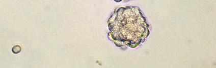
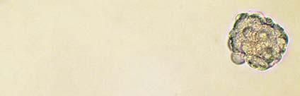

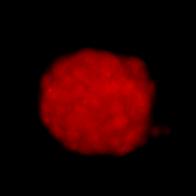
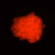
5 Supplemental Figure 1. Human Glioblastoma iopsy Specimen (T 3379) D456MG CD133 Nestin Sox2 Musashi D456MG Human Glioblastoma iopsy Specimen (T 3379)
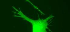
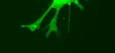


6 Supplemental Figure 2. CD44H CD81 (TP-1) strocytes Map-2 HNK-1 Oligodendrocytes Neuronal Progenitors O4 NG2 Mouse IgG Donkey anti-mouse IgG-FITC Control


![4 2 * C [VEGF] (pg/ml) 3 25 2 15 1 5](/docs-images/85/91508871/images/7-3.jpg "Normoxia Hypoxia * **")
7 Supplemental Figure 3. D456MG ngiogenin bfgf IL-6 VEGF CD133- ngiogenin bfgf IL-6 VEGF CD133+ Mean Total VEGF Intensity * D54MG ngiogenin bfgf IL-6 VEGF CD133- ngiogenin bfgf IL-6 VEGF CD133+ Mean Total VEGF Intensity * C [VEGF] (pg/ml) Normoxia Hypoxia * **
8 Supplemental Figure 4. Human Glioblastoma Specimen CD133- Conditioned Medium Human Glioblastoma Specimen CD133+ Conditioned Medium Human Microvascular Endothelial Cell Migration Number of Migrating Cells * D456MG CD133- Conditioned Medium D456MG CD133+ Conditioned Medium Human Microvascular Endothelial Cell Migration Number of Migrating Cells **
9 Supplemental Figure 5. Human Glioblastoma Specimen CD133- Conditioned Medium Human Glioblastoma Specimen CD133+ Conditioned Medium Human Microvascular Endothelial Tube Formation Tube Length (% of Control) * D456MG CD133- Conditioned Medium D456MG CD133+ Conditioned Medium Human Microvascular Endothelial Tube Formation Tube Length (% of Control) **

 Number of Migrating")


 45 4 35 3 25")

10 Supplementary Figure 6. D456MG CD133- Conditioned D456MG CD133+ Conditioned + Control IgG + nti-vegf (mb) Number of Migrating Cells Control IgG +nti-vegf *** Human Microvascular Endothelial Cell Migration D456MG CD133- Conditioned D456MG CD133+ Conditioned + nti-vegf (mb) + Control IgG Tube Length (% of Control) Control IgG +nti-vegf *** Human Microvascular Endothelial Tube Formation


11 Supplemental Figure 7. + Control IgG + nti-vegf ntibody (1, CD133+ cells / animal) nti-vegf ntibody: 5 mg/kg/d i.p. x 19 days CD133 Staining Negative Control
Reduction of metastatic and angiogenic potency of malignant cancer by Eupatorium. fortunei via suppression of MMP-9 activity and VEGF production
 Supplementary Information Reduction of metastatic and angiogenic potency of malignant cancer by Eupatorium fortunei via suppression of MMP-9 activity and VEGF production Aeyung Kim, Minju Im, Nam-Hui Yim
Supplementary Figure 1.TRIM33 binds β-catenin in the nucleus. a & b, Co-IP of endogenous TRIM33 with β-catenin in HT-29 cells (a) and HEK 293T cells
 and HEK 293T cells") Supplementary Figure 1.TRIM33 binds β-catenin in the nucleus. a & b, Co-IP of endogenous TRIM33 with β-catenin in HT-29 cells (a) and HEK 293T cells (b). TRIM33 was immunoprecipitated, and the amount of
Supplementary Figure 1.TRIM33 binds β-catenin in the nucleus. a & b, Co-IP of endogenous TRIM33 with β-catenin in HT-29 cells (a) and HEK 293T cells (b). TRIM33 was immunoprecipitated, and the amount of
SUPPLEMENTARY FIG. S2. Representative counting fields used in quantification of the in vitro neural differentiation of pattern of dnscs.
 Supplementary Data SUPPLEMENTARY FIG. S1. Representative counting fields used in quantification of the in vitro neural differentiation of pattern of anpcs. A panel of lineage-specific markers were used
Supplementary Information and Figure legends
 Supplementary Information and Figure legends Table S1. Primers for quantitative RT-PCR Target Sequence (5 -> 3 ) Target Sequence (5 -> 3 ) DAB2IP F:TGGACGATGTGCTCTATGCC R:GGATGGTGATGGTTTGGTAG Snail F:CCTCCCTGTCAGATGAGGAC
A549 and A549-fLuc cells were maintained in high glucose Dulbecco modified
 Cell culture and animal model A549 and A549-fLuc cells were maintained in high glucose Dulbecco modified Eagle medium supplemented with 10% fetal bovine serum at 37 C in humidified atmosphere containing
Supplementary Figure 1. Validation of astrocytes. Primary astrocytes were
 Supplementary Figure 1. Validation of astrocytes. Primary astrocytes were separated from the glial cultures using a mild trypsinization protocol. Anti-glial fibrillary acidic protein (GFAP) immunofluorescent
Supplemental Figure 1. Intracranial transduction of a modified ptomo lentiviral vector in the mouse
 Supplemental figure legends Supplemental Figure 1. Intracranial transduction of a modified ptomo lentiviral vector in the mouse hippocampus targets GFAP-positive but not NeuN-positive cells. (A) Stereotaxic
Supplementary Figure (OH) 22 nanoparticles did not affect cell viability and apoposis. MDA-MB-231, MCF-7, MCF-10A and BT549 cells were
 22 nanoparticles did not affect cell viability and apoposis. MDA-MB-231, MCF-7, MCF-10A and BT549 cells were") Supplementary Figure 1. Gd@C 82 (OH) 22 nanoparticles did not affect cell viability and apoposis. MDA-MB-231, MCF-7, MCF-10A and BT549 cells were treated with PBS, Gd@C 82 (OH) 22, C 60 (OH) 22 or GdCl
Supplementary Figure 1. Gd@C 82 (OH) 22 nanoparticles did not affect cell viability and apoposis. MDA-MB-231, MCF-7, MCF-10A and BT549 cells were treated with PBS, Gd@C 82 (OH) 22, C 60 (OH) 22 or GdCl
p = formed with HCI-001 p = Relative # of blood vessels that formed with HCI-002 Control Bevacizumab + 17AAG Bevacizumab 17AAG
 A.. Relative # of ECs associated with HCI-001 1.4 1.2 1.0 0.8 0.6 0.4 0.2 0.0 ol b p < 0.001 Relative # of blood vessels that formed with HCI-001 1.4 1.2 1.0 0.8 0.6 0.4 0.2 0.0 l b p = 0.002 Control IHC:
supplementary information
 DOI: 10.1038/ncb2153 Figure S1 Ectopic expression of HAUSP up-regulates REST protein. (a) Immunoblotting showed that ectopic expression of HAUSP increased REST protein levels in ENStemA NPCs. (b) Immunofluorescent
Supplemental Table 1. Primer sequences for transcript analysis
 Supplemental Table 1. Primer sequences for transcript analysis Primer Sequence (5 3 ) Primer Sequence (5 3 ) Mmp2 Forward CCCGTGTGGCCCTC Mmp15 Forward CGGGGCTGGCT Reverse GCTCTCCCGGTTTC Reverse CCTGGTGTGCCTGCTC
Supplemental Data. Integrin Alpha 6 Regulates Glioblastoma Stem Cells. Supplemental Data Inventory Supplemental Data
 Cell Stem Cell, Volume 6 Supplemental Data Integrin Alpha 6 Regulates Glioblastoma Stem Cells Justin D. Lathia, Joseph Gallagher, John M. Heddleston, Jialiang Wang, Christine E. Eyler, Jennifer MacSwords,
Reviewers' comments: Reviewer #1 (Remarks to the Author):
:") Reviewers' comments: Reviewer #1 (Remarks to the Author): This is a well written and well executed study describing a novel mechanism of pro-angiogenic signalling which may, potentially, help to explain
Reviewers' comments: Reviewer #1 (Remarks to the Author): This is a well written and well executed study describing a novel mechanism of pro-angiogenic signalling which may, potentially, help to explain
Supplementary Materials. for Garmy-Susini, et al, Integrin 4 1 signaling is required for lymphangiogenesis and tumor metastasis
 Supplementary Materials for Garmy-Susini, et al, Integrin 4 1 signaling is required for lymphangiogenesis and tumor metastasis 1 Supplementary Figure Legends Supplementary Figure 1: Integrin expression
SUPPLEMENTARY FIGURES AND TABLE
 SUPPLEMENTARY FIGURES AND TABLE Supplementary Figure S1: Characterization of IRE1α mutants. A. U87-LUC cells were transduced with the lentiviral vector containing the GFP sequence (U87-LUC Tet-ON GFP).
Macrophages form functional vascular mimicry channels in vivo. SI Figures and Legend
 Macrophages form functional vascular mimicry channels in vivo Authors: *Faith H. Barnett, *Mauricio Rosenfeld, Malcolm Wood, William Kiosses, Yoshihiko Usui, Valentina Marchetti, Edith Aguilar, and Martin
Supplemental Material
 Supplemental Material Supplementary Fig. 1. EETs stimulate primary tumor growth. a) Schematic presentation of genetic and pharmacological tools used to manipulate endogenous EET levels. b) Endothelial
Figure S1. The PDE5 inhibitor sildenafil interacts with celecoxib to kill cancer cell lines. (A) Hepatoma
 Hepatoma") Figure S1. The PDE5 inhibitor sildenafil interacts with celecoxib to kill cancer cell lines. (A) Hepatoma cells were treated with celecoxib ( 5.0 M) and/or sildenafil (, 2.0 M). ls were isolated after
Figure S1. The PDE5 inhibitor sildenafil interacts with celecoxib to kill cancer cell lines. (A) Hepatoma cells were treated with celecoxib ( 5.0 M) and/or sildenafil (, 2.0 M). ls were isolated after
Supplementary Figure 1
 Supplementary Figure 1 3 3 3 1 1 Bregma -1.6mm 3 : Bregma Ref) Http://www.mbl.org/atlas165/atlas165_start.html Bregma -.18mm Supplementary Figure 1 Schematic representation of the utilized brain slice
PREPARED FOR: U.S. Army Medical Research and Materiel Command Fort Detrick, Maryland
 AD Award Number: W81XWH-04-1-0618 TITLE: Are Breast Tumor Stem Cells Responsible for Metastasis and Angiogenesis PRINCIPAL INVESTIGATOR: Quintin Pan, Ph.D. CONTRACTING ORGANIZATION: University of Michigan
Extended Neurosphere Culture of Brain Tumor Stem Cells with the PromoCell 3D Tumorsphere Medium XF
 Extended Neurosphere Culture of Brain Tumor Stem Cells with the PromoCell 3D Tumorsphere Medium XF Application Note The PromoCell 3D Tumorsphere Medium XF While adherent cultures of brain tumor cells in
(a) Significant biological processes (upper panel) and disease biomarkers (lower panel)
 Significant biological processes (upper panel) and disease biomarkers (lower panel)") Supplementary Figure 1. Functional enrichment analyses of secretomic proteins. (a) Significant biological processes (upper panel) and disease biomarkers (lower panel) 2 involved by hrab37-mediated secretory
Supplementary Figure 1. Functional enrichment analyses of secretomic proteins. (a) Significant biological processes (upper panel) and disease biomarkers (lower panel) 2 involved by hrab37-mediated secretory
Sema3C Promotes the Survival and Tumorigenicity of Glioma Stem Cells through Rac1 Activation
 Cell Reports, Volume 9 Supplemental Information Sema3C Promotes the Survival and Tumorigenicity of Glioma Stem Cells through Rac1 Activation Jianghong Man, Jocelyn Shoemake, Wenchao Zhou, Xiaoguang Fang,
CD34 + VEGFR-3 + progenitor cells have a potential to differentiate towards lymphatic endothelial cells
 CD34 + VEGFR-3 + progenitor cells have a potential to differentiate towards lymphatic endothelial cells Tan YZ et al. J Cell Mol Med. (2014 Mar;18(3):422-33) Denise Traxler-Weidenauer April 2014 Introduction
Essential Medium, containing 10% fetal bovine serum, 100 U/ml penicillin and 100 µg/ml streptomycin. Huvec were cultured in
 Supplemental data Methods Cell culture media formulations A-431 and U-87 MG cells were maintained in Dulbecco s Modified Eagle s Medium. FaDu cells were cultured in Eagle's Minimum Essential Medium, containing
Real-time imaging reveals the single steps of brain metastasis fo mation r
 Real-time imaging reveals the single steps of brain metastasis fo mation r Yvonne Kienast, Louisa von Baumgarten, Martin Fuhrmann, Wolfgang E.F. Klinkert, Roland Goldbrunner, Jochen Herms and Frank Winkler
SUPPLEMENTARY FIGURES
 SUPPLEMENTARY FIGURES 1 2 3 4 SUPPLEMENTARY TABLES Supplementary Table S1. Brain Tumors used in the study Code Tumor Classification Age Gender HuTuP51 Glioblastoma 57 Male HuTuP52 Glioblastoma 53 Male
VEGFR2-Mediated Vascular Dilation as a Mechanism of VEGF-Induced Anemia and Bone Marrow Cell Mobilization
 Cell Reports, Volume 9 Supplemental Information VEGFR2-Mediated Vascular Dilation as a Mechanism of VEGF-Induced Anemia and Bone Marrow Cell Mobilization Sharon Lim, Yin Zhang, Danfang Zhang, Fang Chen,
Blocking antibodies and peptides. Rat anti-mouse PD-1 (29F.1A12, rat IgG2a, k), PD-
, PD-") Supplementary Methods Blocking antibodies and peptides. Rat anti-mouse PD-1 (29F.1A12, rat IgG2a, k), PD- L1 (10F.9G2, rat IgG2b, k), and PD-L2 (3.2, mouse IgG1) have been described (24). Anti-CTLA-4 (clone
Supplementary Methods Blocking antibodies and peptides. Rat anti-mouse PD-1 (29F.1A12, rat IgG2a, k), PD- L1 (10F.9G2, rat IgG2b, k), and PD-L2 (3.2, mouse IgG1) have been described (24). Anti-CTLA-4 (clone
Supplemental Table 1. Biochemical and Cellular Potency and Selectivity of PF
 Supplemental Table 1. Biochemical and Cellular Potency and Selectivity of PF- 02341066 Assay IC 50 nm Selectivity Ratio d Biochemical Activity In Vitro c-met/hgfr enzyme (Ki, nm) a 4 NA Cellular Activity
Supplementary Figure 1. EC-specific Deletion of Snail1 Does Not Affect EC Apoptosis. (a,b) Cryo-sections of WT (a) and Snail1 LOF (b) embryos at
 Cryo-sections of WT (a) and Snail1 LOF (b) embryos at") Supplementary Figure 1. EC-specific Deletion of Snail1 Does Not Affect EC Apoptosis. (a,b) Cryo-sections of WT (a) and Snail1 LOF (b) embryos at E10.5 were double-stained for TUNEL (red) and PECAM-1 (green).
Supplementary Figure 1. EC-specific Deletion of Snail1 Does Not Affect EC Apoptosis. (a,b) Cryo-sections of WT (a) and Snail1 LOF (b) embryos at E10.5 were double-stained for TUNEL (red) and PECAM-1 (green).
Plasma exposure levels from individual mice 4 hours post IP administration at the
 Supplemental Figure Legends Figure S1. Plasma exposure levels of MKC-3946 in mice. Plasma exposure levels from individual mice 4 hours post IP administration at the indicated dose mg/kg. Data represent
Simple and Easy Monitoring of Tube Formation and Migration Assays with the CytoSMART Live Cell Imaging System
 ioscience Solutions Simple and Easy Monitoring of Tube Formation and Migration ssays with the CytoSMRT Live Cell Imaging System Stefanie uesch, Sabine Schaepermeier, odo Ortmann, Claudia Schwartz, Jenny
hexahistidine tagged GRP78 devoid of the KDEL motif (GRP78-His) on SDS-PAGE. This
 on SDS-PAGE. This") SUPPLEMENTAL FIGURE LEGEND Fig. S1. Generation and characterization of. (A) Coomassie staining of soluble hexahistidine tagged GRP78 devoid of the KDEL motif (GRP78-His) on SDS-PAGE. This protein was expressed
SUPPLEMENTAL FIGURE LEGEND Fig. S1. Generation and characterization of. (A) Coomassie staining of soluble hexahistidine tagged GRP78 devoid of the KDEL motif (GRP78-His) on SDS-PAGE. This protein was expressed
Motility of glioblastoma cells is driven by netrin-1 induced gain of stemness
 Ylivinkka et al. Journal of Experimental & Clinical Cancer Research (2017) 36:9 DOI 10.1186/s13046-016-0482-0 RESEARCH Open Access Motility of glioblastoma cells is driven by netrin-1 induced gain of stemness
Human and mouse T cell regulation mediated by soluble CD52 interaction with Siglec-10. Esther Bandala-Sanchez, Yuxia Zhang, Simone Reinwald,
 Human and mouse T cell regulation mediated by soluble CD52 interaction with Siglec-1 Esther Bandala-Sanchez, Yuxia Zhang, Simone Reinwald, James A. Dromey, Bo Han Lee, Junyan Qian, Ralph M Böhmer and Leonard
EPIGENETIC RE-EXPRESSION OF HIF-2α SUPPRESSES SOFT TISSUE SARCOMA GROWTH
 EPIGENETIC RE-EXPRESSION OF HIF-2α SUPPRESSES SOFT TISSUE SARCOMA GROWTH Supplementary Figure 1. Supplementary Figure 1. Characterization of KP and KPH2 autochthonous UPS tumors. a) Genotyping of KPH2
Overview of methodology, tools and reagents for evaluating cell proliferation and invasion using multicellular tumor spheroids.
 The Next Step in the Evolution of 3D Culture: Utilizing Extracellular Matrix to Enhance Multicellular Tumor Spheroid Models for Proliferation and Invasion Overview of methodology, tools and reagents for
well for 2 h at rt. Each dot represents an individual mouse and bar is the mean ±
 Supplementary data: Control DC Blimp-1 ko DC 8 6 4 2-2 IL-1β p=.5 medium 8 6 4 2 IL-2 Medium p=.16 8 6 4 2 IL-6 medium p=.3 5 4 3 2 1-1 medium IL-1 n.s. 25 2 15 1 5 IL-12(p7) p=.15 5 IFNγ p=.65 4 3 2 1
mir-509-5p and mir-1243 increase the sensitivity to gemcitabine by inhibiting
 mir-509-5p and mir-1243 increase the sensitivity to gemcitabine by inhibiting epithelial-mesenchymal transition in pancreatic cancer Hidekazu Hiramoto, M.D. 1,3, Tomoki Muramatsu, Ph.D. 1, Daisuke Ichikawa,
Stem cells in endometriosis: pathogenetic factors and target for new medical treatments? Alberto Revelli MD PhD
 Stem cells in endometriosis: pathogenetic factors and target for new medical treatments? Alberto Revelli MD PhD Gyn/Obst 1U, Physiopathology of Reproduction and IVF Unit Dept. Surgical Sciences, S. Anna
Type of file: PDF Size of file: 0 KB Title of file for HTML: Supplementary Information Description: Supplementary Figures
 Type of file: PDF Size of file: 0 KB Title of file for HTML: Supplementary Information Description: Supplementary Figures Supplementary Figure 1 mir-128-3p is highly expressed in chemoresistant, metastatic
TITLE: Notch in Pathological Angiogenesis and Lymphangiogenesis
 Award Number: W81XWH-10-1-0304 TITLE: Notch in Pathological Angiogenesis and Lymphangiogenesis PRINCIPAL INVESTIGATOR: Minji Kim CONTRACTING ORGANIZATION: Columbia University New York, NY 10032 REPORT
SREBP-2 promotes stem cell-like properties and metastasis by transcriptional activation of c-myc in prostate cancer
 SREBP-2 promotes stem cell-like properties and metastasis by transcriptional activation of c-myc in prostate cancer Supplementary Material Supplementary Methods Supplementary References Supplementary Figure
TABLE OF CONTENTS 6.1. INTRODUCTION 6.2. MATERIALS AND METHODS
 CHAPTER 6 EFFECT OF VERNOLIDE-A ON RADIATION INDUCED HYPOXIA MEDIATED TUMOUR ANGIOGENESIS TABLE OF CONTENTS 6.1. INTRODUCTION 6.2. MATERIALS AND METHODS 6.2.1. Animals 6.2.2. Cell line 6.2.3. Chemicals
Control Paz pre-treatment withdrawal long Paz
 1. 1.. Paz pre-treatment g Paz 1 1 4 3 1 g ev mount of ascites (m) 8 6 4 g ev Suppementa Figure 1. ffect of versus g-term anti-angiogenic therapy. Mean aggregate tumor weight () and number of tumor nodues
Primary Mouse Cerebral Cortex Neurons V: 80% TE: 70%
 Primary Mouse Cerebral Cortex Neurons V: 80% TE: 70% Pictures: 9 days after electroporation Red: MAP2 Blue: GFAP Green: GFP The cells were from Embryonic Day 14 Mouse Cerebral Cortex Primary Mouse Hippocampal
injected subcutaneously into flanks of 6-8 week old athymic male nude mice (LNCaP SQ) and body
 and body") SUPPLEMENTAL FIGURE LEGENDS Figure S1: Generation of ENZR Xenografts and Cell Lines: (A) 1x10 6 LNCaP cells in matrigel were injected subcutaneously into flanks of 6-8 week old athymic male nude mice (LNCaP
SUPPLEMENTAL FIGURE LEGENDS Figure S1: Generation of ENZR Xenografts and Cell Lines: (A) 1x10 6 LNCaP cells in matrigel were injected subcutaneously into flanks of 6-8 week old athymic male nude mice (LNCaP
Supplementary Figure S1: Tanycytes are restricted to the central/posterior hypothalamus
 Supplementary Figure S1: Tanycytes are restricted to the central/posterior hypothalamus a: Expression of Vimentin, GFAP, Sox2 and Nestin in anterior, central and posterior hypothalamus. In the anterior
Figure S1. ERBB3 mrna levels are elevated in Luminal A breast cancers harboring ERBB3
 Supplemental Figure Legends. Figure S1. ERBB3 mrna levels are elevated in Luminal A breast cancers harboring ERBB3 ErbB3 gene copy number gain. Supplemental Figure S1. ERBB3 mrna levels are elevated in
Supplementary Figure 1. Deletion of Smad3 prevents B16F10 melanoma invasion and metastasis in a mouse s.c. tumor model.
 A B16F1 s.c. Lung LN Distant lymph nodes Colon B B16F1 s.c. Supplementary Figure 1. Deletion of Smad3 prevents B16F1 melanoma invasion and metastasis in a mouse s.c. tumor model. Highly invasive growth
To this end, we performed immunofluorescent staining for GSCs (Fig.3). All the spheroidforming cells showed immunoreactivity for
. All the spheroidforming cells showed immunoreactivity for") H.Yoshioka et al. lished for isolation ofneural stem cells. Within 24-48 hours of primary culture, murine brain tumors yielded a minority fraction of cells that formed neurosphere-like clusters (tumor
H.Yoshioka et al. lished for isolation ofneural stem cells. Within 24-48 hours of primary culture, murine brain tumors yielded a minority fraction of cells that formed neurosphere-like clusters (tumor
% cells forming Neurospheres 81 ± 6 % 0 % 2.6 ± 0.7 % 76 ± 8 % 0 % 3.4 ± 0.6 % 83 ± 5 % 0 % 2.4 ± 0.9 % 89 ± 5 % 3 ± 1.5 % Total 10, ± 6 % 0 %
 Bo et l., Suppl. Tle 1 Supplementl Tle 1. Neurosphere formtion nd tumorigencity is enriched within the tumour cell popultions derived from humn primry glioms nd gliom xenogrfts. GBM smples or Gliom xenogrfts
Targeted therapy of glioblastoma stem-like cells and tumor non-stem cells using cetuximab-conjugated iron-oxide nanoparticles
 Targeted therapy of glioblastoma stem-like cells and tumor non-stem cells using cetuximab-conjugated iron-oxide nanoparticles Milota Kaluzova, Emory University Alexandros Bouras, Emory University Revaz
Targeting tumour associated macrophages in anti-cancer therapies. Annamaria Gal Seminar Series on Drug Discovery Budapest 5 January 2018
 Targeting tumour associated macrophages in anti-cancer therapies Annamaria Gal Seminar Series on Drug Discovery Budapest 5 January 2018 Macrophages: Professional phagocytes of the myeloid lineage APC,
SUPPLEMENTARY INFORMATION
 DOI: 1.138/ncb3355 a S1A8 + cells/ total.1.8.6.4.2 b S1A8/?-Actin c % T-cell proliferation 3 25 2 15 1 5 T cells Supplementary Figure 1 Inter-tumoral heterogeneity of MDSC accumulation in mammary tumor
PLGA Foundation Grant Proposal March 31, Pediatric Low Grade Astrocytoma: Dedicated Tumor Banking And Establishment Of Cell Lines And Xenografts
 PLG Foundation Grant Proposal March 31, 2009 Pediatric Low Grade strocytoma: Dedicated Tumor anking nd Establishment Of Cell Lines nd Xenografts Charles G. Eberhart M.D., Ph.D., Kenneth Cohen M.D., Eli
ANGPTL2 increases bone metastasis of breast cancer cells through. Tetsuro Masuda, Motoyoshi Endo, Yutaka Yamamoto, Haruki Odagiri, Tsuyoshi
 Masuda et al. Supplementary information for ANGPTL2 increases bone metastasis of breast cancer cells through enhancing CXCR4 signaling Tetsuro Masuda, Motoyoshi Endo, Yutaka Yamamoto, Haruki Odagiri, Tsuyoshi
Low Cell Binding Property of LIPIDURE -COAT
 Technical Note_1 ver.1 Low Cell Binding Property of LIPIDURE -COAT 1. LIPIDURE -COAT MULTI DISH A-6MD (Cat. No. 51011617) 2. Cell; NIH 3T3 (Fibroblast, mouse) 1. 10 %CS-DMEM; DMEM (Dulbecco's Modified
Glioma cells enhance angiogenesis and inhibit endothelial cell apoptosis through the release of exosomes that contain long non-coding RNA CCAT2
 ONCOLOGY REPORTS 38: 785-798, 2017 Glioma cells enhance angiogenesis and inhibit endothelial cell apoptosis through the release of exosomes that contain long non-coding RNA CCAT2 Hai-li Lang 1*, Guo-wen
Monoclonal antibody targeting of N-cadherin inhibits prostate cancer growth, metastasis and castration resistance
 Monoclonal antibody targeting of N-cadherin inhibits prostate cancer growth, metastasis and castration resistance Tanaka H, Kono E, Tran CP, Miyazaki H, Yamashiro J, Shimomura T, Ladan F, Wada R, Huang
Supplementary Figure 1. IDH1 and IDH2 mutation site sequences on WHO grade III
 Supplementary Materials: Supplementary Figure 1. IDH1 and IDH2 mutation site sequences on WHO grade III patient samples. Genomic DNA samples extracted from punch biopsies from either FFPE or frozen tumor
Supplemental Information. CD4 + CD25 + Foxp3 + Regulatory T Cells Promote. Th17 Cells In Vitro and Enhance Host Resistance
 Immunity, Volume 34 Supplemental Information D4 + D25 + + Regulatory T ells Promote Th17 ells In Vitro and Enhance Host Resistance in Mouse andida albicans Th17 ell Infection Model Pushpa Pandiyan, Heather
Cancer Stem Cells & Glioblastoma
 Cancer Stem Cells & Glioblastoma JP Hugnot «Brain plasticity, Neural stem cells and Glial tumors» INSERM U1051-UM2 Institut des Neurosciences de Montpellier Montpellier 1-Stem cells and Brain Stem Cells
Cellular Physiology and Biochemistry
 Original Paper 2015 The Author(s). 2015 Published The Author(s) by S. Karger AG, Basel Published online: November 27, 2015 www.karger.com/cpb Published by S. Karger AG, Basel 2194 1421-9778/15/0376-2194$39.50/0
Cancer Tumor Therapy Drug Vicrostatin Shows. Promising Inhibition of Glioma Growth and. Angiogenesis in Vivo. Rupan Bose
 Bose p. 1 of 23. Cancer Tumor Therapy Drug Vicrostatin Shows Promising Inhibition of Glioma Growth and Angiogenesis in Vivo Rupan Bose B.A. Neuroscience 2011 Candidate Senior Thesis Dana and David Dornsife
Caffeine Modulates Hyperoxia - Induced Angiogenesis in Newborn Mice
 Caffeine Modulates Hyperoxia - Induced Angiogenesis in Newborn Mice Vikramaditya Dumpa, MD Lori C Nielsen, MS Huamei Wang, MD Vasanth HS Kumar, MD Supported by AAP Marshall Klaus Perinatal Research Grant
Hopkins University, Howard Hughes Medical Institute, USA) (27). Cells were maintained in DMEM
 (27). Cells were maintained in DMEM") Supplementary Materials and Methods Cell Culture HCT116 (TP53 +/+ and TP53 -/- ) cells were provided by Dr. Bert Vogelstein (Johns Hopkins University, Howard Hughes Medical Institute, USA) (27). Cells
Supplementary Materials and Methods Cell Culture HCT116 (TP53 +/+ and TP53 -/- ) cells were provided by Dr. Bert Vogelstein (Johns Hopkins University, Howard Hughes Medical Institute, USA) (27). Cells
Department of Orthopaedic Surgery, Tohoku University Graduate School of Medicine, Sendai, Japan, 2
 Low-energy Extracorporeal Shock Wave Therapy Promotes VEGF Expression and Angiogenesis and Improve Locomotor and Sensory Functions after spinal cord injury Kenichiro Yahata 1, Hiroshi Ozawa, M.D., Ph.D.
Imaging of glycolytic metabolism in primary glioblastoma cells with
 63 Chapter 5 Imaging of glycolytic metabolism in primary glioblastoma cells with RIMChip 5.1. Introduction Glioblastoma(GBM) is one of the most common brain tumors 1. It is composed of heterogeneous subpopulations
CRIPTO-1 A POSSIBLE NEW BIOMARKER IN GLIOBLASTOMA MULTIFORME PIA OLESEN, MD, PHD STUDENT
 CRIPTO-1 A POSSIBLE NEW BIOMARKER IN GLIOBLASTOMA MULTIFORME PIA OLESEN, MD, PHD STUDENT Glioblastoma WHO Grade IV Glioma Heterogenic Undiffenrentiated phenotype 50% of all Gliomas Around 600 patients
Supplemental Information. Differential Effects of EGFL6 on Tumor. versus Wound Angiogenesis
 Cell Reports, Volume 21 Supplemental Information Differential Effects of EGFL6 on Tumor versus Wound Angiogenesis Kyunghee Noh, Lingegowda S. Mangala, Hee-Dong Han, Ningyan Zhang, Sunila Pradeep, Sherry
Neural Stem Cell-based Cell Carriers Enhance Therapeutic Efficacy of an Oncolytic Adenovirus in an Orthotopic Mouse Model of Human Glioblastoma
 original article Neural Stem Cell-based Cell Carriers Enhance Therapeutic Efficacy of an Oncolytic Adenovirus in an Orthotopic Mouse Model of Human Glioblastoma Atique U Ahmed 1, Bart Thaci 1, Nikita G
Supplementary Information
 Supplementary Information Lymphatic endothelial cells support tumor growth in breast cancer Esak Lee a, Niranjan B. Pandey a, and Aleksander S. Popel a,b a Department of Biomedical Engineering, Johns Hopkins
Endothelial Cell Transmigration and Invasion Assay
 Endothelial Cell Transmigration and Invasion Assay Application Note Background Angiogenesis is the physiological development of new blood vessels from existing ones, a process that is essential for embryonic
Correlation between glioblastoma stem-like cells and tumor vascularization
 ONCOLOGY REPORTS 27: 45-50, 2012 Correlation between glioblastoma stem-like cells and tumor vascularization HU HE 1, CHAO SHI NIU 1,2 and MING WU LI 1 1 Department of Neurosurgery, Anhui Provincial Hospital
Effect of Propecia on the Hair Follicle in Male Androgenetic Alopecia: A Confocal Laser Scanning Microscopy and Video Imaging Study
 Effect of Propecia on the Hair Follicle in Male Androgenetic Alopecia: A Confocal Laser Scanning Microscopy and Video Imaging Study Investigators: Department of Dermatology, University of Minnesota, Minneapolis,
Supplementary Figures for
 mirns regulate s Supplementary igures for MicroRNs Reprogram Normal ibroblasts into Cancer ssociated ibroblasts in Ovarian Cancer nirban K. Mitra, Marion Zillhardt, Youjia Hua, Payal iwari, ndrea E. Murmann,
Supplementary Table 1. Characterization of HNSCC PDX models established at MSKCC
 Supplementary Table 1. Characterization of HNSCC PDX models established at MSKCC Supplementary Table 2. Drug content and loading efficiency estimated with F-NMR and UV- Vis Supplementary Table 3. Complete
Neocortex Zbtb20 / NFIA / Sox9
 Neocortex / NFIA / Sox9 Supplementary Figure 1. Expression of, NFIA, and Sox9 in the mouse neocortex at. The lower panels are higher magnification views of the oxed area. Arrowheads indicate triple-positive
Spheroid-based engineering of a human vasculature in mice
 Spheroid-based engineering of a human vasculature in mice Abdullah Alajati, Anna M. Laib, Holger Weber, Anja M. Boos, Arne Bartol, Kristian Ikenberg, Thomas Korff, Hanswalter Zentgraf, Cynthia Obodozie,
HIF-1-mediated metabolic reprogramming reduces ROS levels and facilitates
 HIF-1-mediated metabolic reprogramming reduces ROS levels and facilitates the metastatic colonization of cancers in lungs Authors: Tao ZHAO 1,2,3, Yuxi ZHU 1,2,4, Akiyo MORINIBU 1,2, Minoru KOBAYASHI 1,2,
The Contribution Of Tie2-Lineage Cells To rhbmp-2 Induced Bone Formation
 The Contribution Of Tie2-Lineage Cells To rhbmp-2 Induced Bone Formation Mille P. Kolind, Ph.D 1, Alastair Aiken 1, Kathy Mikulec 1, Lauren Peacock 1, David Little 1,2, Aaron Schindeler, PhD 1,2. 1 Orthopaedic
Hypoxia Enhances Tumor Stemness by Increasing the Invasive and Tumorigenic Side Population Fraction
 CANCER STEM CELLS Hypoxia Enhances Tumor Stemness by Increasing the Invasive and Tumorigenic Side Population Fraction BIKUL DAS, a,b,c RIKA TSUCHIDA, b DAVID MALKIN, b,c,d GIDEON KOREN, c,e SYLVAIN BARUCHEL,
For personal use only
 ASX and Media release 6 April 211 Circadian s Inhibits Tumour Growth in Models of Lung, Ovarian and Prostate Cancer Data demonstrates efficacy of with other therapeutic agents in mouse models of lung,
Glioblastoma Multiforme
 Glioblastoma Multiforme Highly malignant, invasive, difficult-to-treat primary brain tumor" " Frequency: 9,000 cases/year (peak age, 55 65 years)" " Recurrence: rapid growth; size may double every 10 days"
Electron micrograph of phosphotungstanic acid-stained exosomes derived from murine
 1 SUPPLEMENTARY INFORMATION SUPPLEMENTARY FIGURES Supplementary Figure 1. Physical properties of murine DC-derived exosomes. a, Electron micrograph of phosphotungstanic acid-stained exosomes derived from
Erzsebet Kokovay, Susan Goderie, Yue Wang, Steve Lotz, Gang Lin, Yu Sun, Badrinath Roysam, Qin Shen,
 Cell Stem Cell, Volume 7 Supplemental Information Adult SVZ Lineage Cells Home to and Leave the Vascular Niche via Differential Responses to SDF1/CXCR4 Signaling Erzsebet Kokovay, Susan Goderie, Yue Wang,
Blockade of Prolymphangiogenic VEGF-C suppresses Dry Eye Disease. Sunali Goyal MD
 Blockade of Prolymphangiogenic VEGF-C suppresses Dry Eye Disease Sunali Goyal MD Mentor: Reza Dana, MD, MPH, MSc Claes Dohlman Chair in Ophthalmology Director, Cornea & Refractive Surgery Massachusetts
Electronic Supplementary Information (ESI) for Lab on a Chip. This journal is The Royal Society of Chemistry 2012
 for Lab on a Chip. This journal is The Royal Society of Chemistry 2012") Electronic Supplementary Information (ESI) for Lab on a Chip Electronic Supplementary Information Construction of oxygen and chemical concentration gradients in a single microfluidic device for studying
Electronic Supplementary Information (ESI) for Lab on a Chip Electronic Supplementary Information Construction of oxygen and chemical concentration gradients in a single microfluidic device for studying
CytoSelect Tumor- Endothelium Adhesion Assay
 Product Manual CytoSelect Tumor- Endothelium Adhesion Assay Catalog Number CBA- 215 100 assays FOR RESEARCH USE ONLY Not for use in diagnostic procedures Introduction Cancer metastasis comprises several
Appendix Figure S1 A B C D E F G H
 ppendix Figure S1 C D E F G H ppendix Figure S1. RT and chemotherapy alter PD-L1 expression in PDC cells. Flow cytometric analysis of PD-L1 expression in () KPC and () Pan02 cells following treatment with
MicroRNA 132 mediated loss of p120rasgap activates endothelium to facilitate pathological angiogenesis
 MicroRNA 132 mediated loss of p12rasgap activates endothelium to facilitate pathological angiogenesis Sudarshan Anand, Bharat K. Majeti, Lisette M. Acevedo, Eric A. Murphy, Rajesh Mukthavaram, Lea Scheppke,
Inhibition of E-Selectin or E-selectin together with CXCR4 Re-sensitizes Multiple Myeloma to Treatment
 Inhibition of E-Selectin or E-selectin together with CXCR4 Re-sensitizes Multiple Myeloma to Treatment 1, Ph.D. Henna Bazai 1, Anita Sekula 1, William Fogler 2, Ted Smith 2, John Magnani 2 and Abdel Kareem
Nature Neuroscience: doi: /nn Supplementary Figure 1. MADM labeling of thalamic clones.
 Supplementary Figure 1 MADM labeling of thalamic clones. (a) Confocal images of an E12 Nestin-CreERT2;Ai9-tdTomato brain treated with TM at E10 and stained for BLBP (green), a radial glial progenitor-specific
Endothelial PGC 1 - α 1 mediates vascular dysfunction in diabetes
 Endothelial PGC-1α mediates vascular dysfunction in diabetes Reporter: Yaqi Zhou Date: 04/14/2014 Outline I. Introduction II. Research route & Results III. Summary Diabetes the Epidemic of the 21st Century
Integrin v 3 targeted therapy for Kaposi s sarcoma with an in vitro evolved antibody 1
 Integrin v 3 targeted therapy for Kaposi s sarcoma with an in vitro evolved antibody 1 CHRISTOPH RADER, 2 MIKHAIL POPKOV, JOHN A. NEVES, AND CARLOS F. BARBAS III 2 Department of Molecular Biology and The
SUPPLEMENTARY FIGURES
 SUPPLEMENTARY FIGURES 1 Supplementary Figure 1, Adult hippocampal QNPs and TAPs uniformly express REST a-b) Confocal images of adult hippocampal mouse sections showing GFAP (green), Sox2 (red), and REST
(A) RT-PCR for components of the Shh/Gli pathway in normal fetus cell (MRC-5) and a
 RT-PCR for components of the Shh/Gli pathway in normal fetus cell (MRC-5) and a") Supplementary figure legends Supplementary Figure 1. Expression of Shh signaling components in a panel of gastric cancer. (A) RT-PCR for components of the Shh/Gli pathway in normal fetus cell (MRC-5) and
Supplementary figure legends Supplementary Figure 1. Expression of Shh signaling components in a panel of gastric cancer. (A) RT-PCR for components of the Shh/Gli pathway in normal fetus cell (MRC-5) and
SUPPLEMENTARY INFORMATION
 DOI: 10.1038/ncb3021 Supplementary figure 1 Characterisation of TIMPless fibroblasts. a) Relative gene expression of TIMPs1-4 by real time quantitative PCR (RT-qPCR) in WT or ΔTimp fibroblasts (mean ±