Spheroid-based engineering of a human vasculature in mice
|
|
|
- Isaac Cross
- 5 years ago
- Views:
Transcription
1 Spheroid-based engineering of a human vasculature in mice Abdullah Alajati, Anna M. Laib, Holger Weber, Anja M. Boos, Arne Bartol, Kristian Ikenberg, Thomas Korff, Hanswalter Zentgraf, Cynthia Obodozie, Ralph Graeser, Sven Christian, Günter Finkenzeller, G Björn Stark, Mélanie Héroult & Hellmut G Augustin Supplementary figures and text: Supplementary Figure 1 Responsiveness of the grafted human vasculature to inflammatory stimulation. Supplementary Figure 2 Spheroids of different human EC populations give rise to a human vasculature when transplanted into SCID mice. Supplementary Figure 3 Three dimensional analysis of mural cell coverage following VEGF and FGF-2 stimulation. Supplementary Figure 4 Effect of VEGF in combination with PDGF-BB, HUASMC or NHDF on the formation of a human neovasculature originating from xenotransplanted EC spheroids. Supplementary Figure 5 Co-implantation of HUVEC spheroids with osteoblasts. Supplementary Figure 6 Schematic diagram summarizing different applications of the spheroid-based in vivo angiogenesis assay Supplementary Methods

 were mixed with Matrigel-fibrin containing VEGF and")
 into the left flank and saline as")
 were intravenously")

 Fluorescence microscopy images of sections, double stained for hicam-1 and or hvcam-1 and,")

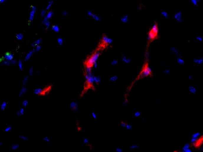
2 Suppl. Figure 1 a Control TNF-α ICAM-1 VCAM-1 b & U937 Control TNF-α Supplementary Figure 1a,b: Responsiveness of the human grafted vasculature to inflammatory stimulation. HUVEC spheroids (1000 of 100 cells each) were mixed with Matrigel-fibrin containing VEGF and FGF-2 (500ng/ml each) and injected subcutaneously into SCID mice. After 20 days, mice were injected with TNF-α (2ng/µl) into the left flank and saline as control in the right flank of the body. Dye-labelled human U937 monocytes (red fluorescence; Vybrant CM-Dil) were intravenously injected into the tail vein 3 hours after TNF-α stimulation. Mice were sacrificed 1h later and the harvested plugs were analyzed morphologically. Mice were sacrificed 4 h later and the harvested plugs were analyzed morphologically. (a) Fluorescence microscopy images of sections, double stained for hicam-1 and or hvcam-1 and, respectively. The positive human vasculature responded to TNF-α by upregulation of ICAM-1 and VCAM-1 expression. (b) Fluorescence microscopy images of a staining. Following the injection of labelled U937 cells, the recruitment and extravasation of attracted monocytic cells were directly visualized by detection of red-labelled cells in the vessels and in the matrix. All sections were stained with Hoechst for nuclear detection. Scale bar, 100 µm.


, respectively, were mixed with Matrigel-fibrin matrix")


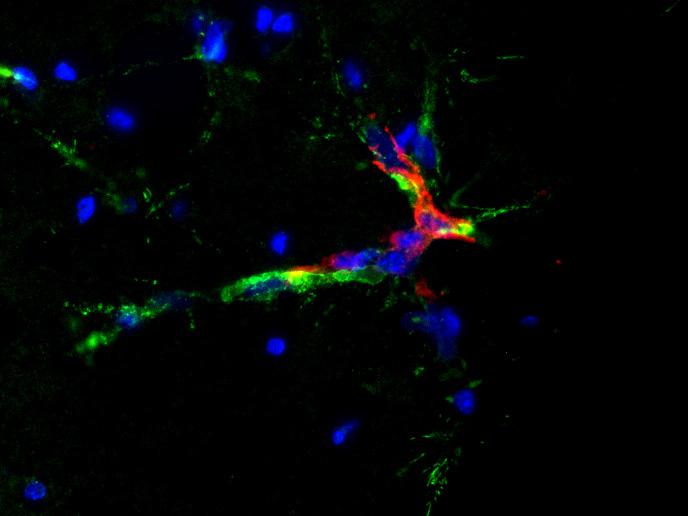
3 Suppl. Figure 2 HDMEC HSVEC HUAEC & αsma Supplementary Figure 2: Spheroids of different human EC populations give rise to a human vasculature when transplanted into SCID mice. HDMEC-, HSVEC- and HUAEC-spheroids (1000 spheroids, 100 cells each), respectively, were mixed with Matrigel-fibrin matrix containing VEGF and FGF- 2 (500 ng each) and injected subcutaneously into SCID mice. Plugs were resected 20 days after implantation. Fluorescence microscopy images of a double staining for and α-smooth muscle actin (αsma; recruited murine mural cells), counterstained with Hoechst for nuclear detection. All tested ECs developed a human capillary network that anastomosed with the mouse vascular system and was covered by mural cells 20 days after implantation. Scale bar, 100 µm. Light microscopy images of an immunohistochemical staining for and counterstaining with hemalaun revealed capillary networks carrying erythrocytes (arrows). Scale bar 50 µm.
 in a Matrigel-fibrin matrix containing VEGF or FGF-2 into SCID mice.")
 demonstrated that the newly formed human vasculature was only partly covered by mural cells upon VEGF stimulation.")
4 Suppl. Figure 3 VEGF FGF-2 hcd31 & αsma Supplementary Figure 3: Three dimensional analysis of mural cell coverage following VEGF and FGF-2 stimulation. Morphological 3D analysis of human neovessel formation after subcutaneous injection of HUVEC spheroids (1000 of 100 cells each) in a Matrigel-fibrin matrix containing VEGF or FGF-2 into SCID mice. Plugs were resected 20 days after implantation. Sections (20 µm) were immunofluorescence double stained with anti-hcd31 and α-smooth muscle actin (αsma; recruited murine mural cells). Three dimensional analysis by confocal microscopy, using Imaris 3.1 (3D-software) demonstrated that the newly formed human vasculature was only partly covered by mural cells upon VEGF stimulation. In contrast, FGF-2 stimulation resulted in a nearly complete mural cell coverage of human neovessels. Scale bar, 25 µm.


 60 40")

5 Suppl. Figure 4 a & αsma & dextran VEGF PDGF & VEGF SMC & VEGF NHDF & VEGF b MVD (vessels per mm²) PDGF SMC NHDF c percentage coverage (%) PDGF SMC NHDF d percentage perfusion (%) PDGF SMC NHDF
6 Suppl. Figure 4 Supplementary Figure 4a: Effect of VEGF in combination with PDGF-BB, HUASMC or NHDF on the formation of a human neovasculature originating from xenotransplanted EC spheroids. HUVEC spheroids (1000 of 100 cells each) were subcutaneously injected in a Matrigel-fibrin matrix containing VEGF, VEGF and PDGF-BB, VEGF and HUASMC, or VEGF and NHDF (500ng/ml for VEGF and PDGF- BB, 10 5 cells for HUASMC and NHDF) into SCID mice. Plugs were resected 20 days after implantation. (a) Representative fluorescence microscopy images of a staining revealed a human vascular network 20 days after subcutaneous injection in all groups. Fluorescence microscopy images of a double staining for (grafted HUVEC) and α-smooth muscle actin (αsma; recruited murine mural cells) revealed only little mural cell coverage in the VEGF stimulated group, whereas VEGF in combination with PDGF, SMC or NHDF resulted in a high degree of mural cell vessel coverage; right: Intravenous injection of FITC-Dextran in combination with immunofluorescence staining of sections for revealed a perfused human vasculature in the presence of PDGF, SMC and NHDF. In contrast, stimulation with VEGF alone resulted in non perfused, rarely mural cell covered human vessels. All sections were stained with Hoechst for nuclear detection. Scale bars, 100 µm. (b) Quantification of MVD. MVD was quantified by counting the number of -positive vessels per mm². (c) Mural cell coverage was assessed by counting the percentage of -positive vessels that co-localized with αsma-positive host mural cells. (c) Perfusion was analyzed by counting the percentage of hcd31-positive vessels, containing host RBC. Mural cell coverage as well as perfusion were significantly increased in the presence of PDGF, SMC or NHDF compared to the VEGF alone stimulated group. Stimulation of EC spheroids with VEGF alone was not sufficient to develop a mature human vascular network. The results are mean values ± s.d. (n = 3).


 HUVEC spheroids (500 spheroids; 400 cells per")
 spheroids (500 spheroids; 800 cells per")
 were mixed in a Matrigel-fibrin matrix")


.")


7 Suppl. Figure 5 EC hcd31 & αsma c b hob & EC MVD (vessels per mm²) a EC hob & EC EC hob EC hob hob Vimentin & osteonectin alizarin Supplementary Figure 5: Co-implantation of HUVEC spheroids with osteoblasts. (a) HUVEC spheroids (500 spheroids; 400 cells per spheroid), mixed co-culture HUVEC and human osteoblast (hob) spheroids (500 spheroids; 800 cells per spheroid), and hob spheroids (500 spheroids; 400 cells per spheroid) were mixed in a Matrigel-fibrin matrix containing VEGF and FGF-2 (500 ng/ml each), injected subcutaneously into SCID mice, and incubated for 20 days; (a) Light and fluorescence microscopy images of hcd31-positive grafted HUVEC with hematoxylin counterstain and immunofluorescence double staining for and αsma. Nuclei were counterstained with Hoechst. Scale bar, 50 µm. Quantitation of MVD by counting the number of -positive vessels per mm². The results are mean values ± s.d. (n = 3). (c) top: Sections were immunohistochemically double stained for and hvimentin. Cells with human origin were apparent in the matrix in all constructs. Numerous cells and EC-derived vessels could be detected by hvimentin and double staining in constructs with EC spheroids and EC and hob co-culture spheroids. middle: Detection of Osteonectin in constructs containing hob co-implanted EC spheroids, but not in mono-implants of EC spheroids. Bottom: Immunohistochemical detection of calcium phosphate with alizarin. The formation of mineral nodules was detected in the constructs containing hob and co-implanted EC spheroids, whereas no mineral formation could be detected in constructs of EC mono-implants. Scale bar 50 µm.
8 Suppl. Figure 6 Supplementary Figure 6: Schematic diagram summarizing different applications of the spheroid-based in vivo angiogenesis assay (see text for details). d0 d20 Spheroid formation Interventive treatment Preventive treatment 24h Embedding of BEC or LEC spheroids in Matrigel-fibrin Subcutaneous injection of spheroids in gel matrix GOF and LOF studies with ex vivo genetically manipulated ECs Anti-angiogenesis drug screening Cell trafficking (circulating hematopoietic cells; metastasizing tumor cells) Quantitative analysis of the vascular morphogenic capacity of manipulated ECs Drug application local stimulation (e.g., TNFα) d20 d40 Quantitative analysis of resulting neovasculature d0 Injection of labelled cells d20 Detection of recruited circulating human cells to the grafted humanized vasculature d0
9 Methods Supplement Morphological and immunohistochemical analysis Following fixation with 4% formaldehyde or Zink fixative, Matrigel plugs were embedded in paraffin. Plugs were sectioned at 8 μm, deparaffinized and rehydrated. Anti human CD31 (JC70/A, Dako) and anti human Osteonectin immunohistochemistry was performed using VECTOR M.O.M kit according to the manufacturer s instructions (Vector Laboratories). Anti human CD34/anti mouse CD31 double stainings were performed with paraffin slides that had been digested with Proteinase K at 37 C. Calcium phosphate staining was performed using Alizarin red S solution (Sigma Aldrich Buchs) and counterstained with Fast Green (Merck). For Vimentin, ICAM-1 and VCAM-1 staining, sections were boiled in citrate antigen retrieval buffer (ph 6, Dako); for Lyve-1 in citrate antigen retrieval buffer (ph 9, Dako). Non-specific binding was blocked with 10% goat serum or 7%FCS/1%BSA or 5% donkey or rabbit serum, respectively. Endogenous peroxidases were blocked with 3% H 2 0 2, followed by avidin/biotin blocking. The following primary antibodies were used: Mouse-αSMA-Cy3 (Sigma Aldrich), mouse-anti-human CD34 (QBEND10, Novocastra), sheep-anti-human CD31 (Dako), rat-antimouse CD31 (BD Bioscience), mouse-anti-human Podoplanin (Dako or Abcam), rabbit-antihuman Lyve-1 (RELIATech), goat-anti-human ICAM-1 (R&D Systems), goat-anti-human VCAM-1 (R&D Systems), mouse-anti-human Vimentin (Dako), and mouse-anti-human osteonectin (Biogenex). First antibodies were detected by goat-anti-mouse Alexa Fluor 488 (Invitrogen), donkey-anti-sheep Alexa Fluor 546 (Invitrogen), goat-anti-rat Alexa 488 (Invitrogen), rabbit-anti-goat Alexa Fluor 546 (Invitrogen), goat-anti-mouse conjugated to alkaline phosphatase (Zymed), and biotinylated goat-anti-mouse secondary antibody (Zymed), respectively. Slides were incubated with a streptavidin-peroxidase conjugate (Zymed or Dako) and developed with Fast Red substrate (Dako) or DAB Substrate Chromogen system (Dako). Nuclei were counterstained with Hoechst dye (Sigma Aldrich) or Meyer s Hemalaun solution (Merck). Slides were examined with an Olympus IX81 microscope. Images were captured with F-View digital camera (Olympus) and processed by Cell F imaging system (Olympus).
10 Electron microscopy Specimens were fixed in 2.5% glutaraldehyde buffered with 0.05 M sodium cacodylate. Osmification, dehydration, embedding and sectioning were as described (1). Micrographs were taken with a Zeiss EM-10 electron microscope at 80 kv. Dextran perfusion For perfusion studies, 100 µl of FITC conjugated dextran (70,000 MW, 25 mg/ml anionic, Invitrogen) was injected into the tail vein of mice and allowed to perfuse for 10 min. After sacrificing the mice, plugs were retrieved and fixed over night in 4% formaldehyde. Confocal microscopy HUVEC-spheroids (1.000 of 100 cells each) were mixed in Matrigel/fibrin matrix containing VEGF-A or FGF-2 (500ng each, R&D Systems) and injected subcutaneously into SCID mice (Harlan Winkelmann). Mice were sacrificed after 20 days. For whole mount staining, plugs were fixed for 24 h in methanol containing 10% DMSO. Thereafter, the plugs were double stained with anti hcd31/αsma antibodies as described above. The three dimensional structure was analyzed by confocal microscopy using the LSM M laser scanning microscope (Zeiss) and corresponding analyses software Imaris 3.1 (3D-software). Monocyte (U937) recruitment experiments TNF-α (50 µl containing 2 ng/µl TNF-α, R&D Systems) or 50 µl saline were injected subcutaneously into the site of a 20 day plug. Human U937 were labeled with red fluorescence solution (Vybrant CM-DiI cell-labeling solution, Invitrogen) according to the manufacturer s instructions. U937 cells (1 x 10 6 ) were injected into the mouse tail vein 3 h after TNF-α injection. Mice were sacrificed 1 h after monocyte injection. Plugs were retrieved and fixed in Zinc fixative at 4 C over night and stained for human CD34 as described above.
11 Reference 1. Zentgraf, H., & Franke W.W. Differences of supranucleosomal organization in different kinds of chromatin: cell type-specific globular subunits containing different numbers of nucleosomes. J. Cell Biol. 99, (1984)
Supplementary Materials. for Garmy-Susini, et al, Integrin 4 1 signaling is required for lymphangiogenesis and tumor metastasis
 Supplementary Materials for Garmy-Susini, et al, Integrin 4 1 signaling is required for lymphangiogenesis and tumor metastasis 1 Supplementary Figure Legends Supplementary Figure 1: Integrin expression
Supplemental figure 1. PDGFRα is expressed dominantly by stromal cells surrounding mammary ducts and alveoli. A) IHC staining of PDGFRα in
 IHC staining of PDGFRα in") Supplemental figure 1. PDGFRα is expressed dominantly by stromal cells surrounding mammary ducts and alveoli. A) IHC staining of PDGFRα in nulliparous (left panel) and InvD6 mouse mammary glands (right
Supplemental figure 1. PDGFRα is expressed dominantly by stromal cells surrounding mammary ducts and alveoli. A) IHC staining of PDGFRα in nulliparous (left panel) and InvD6 mouse mammary glands (right
Macrophages form functional vascular mimicry channels in vivo. SI Figures and Legend
 Macrophages form functional vascular mimicry channels in vivo Authors: *Faith H. Barnett, *Mauricio Rosenfeld, Malcolm Wood, William Kiosses, Yoshihiko Usui, Valentina Marchetti, Edith Aguilar, and Martin
(a) Significant biological processes (upper panel) and disease biomarkers (lower panel)
 Significant biological processes (upper panel) and disease biomarkers (lower panel)") Supplementary Figure 1. Functional enrichment analyses of secretomic proteins. (a) Significant biological processes (upper panel) and disease biomarkers (lower panel) 2 involved by hrab37-mediated secretory
Supplementary Figure 1. Functional enrichment analyses of secretomic proteins. (a) Significant biological processes (upper panel) and disease biomarkers (lower panel) 2 involved by hrab37-mediated secretory
Evaluation of directed and random motility in microslides Assessment of leukocyte adhesion in flow chambers
 Evaluation of directed and random motility in microslides Motility experiments in IBIDI microslides, image acquisition and processing were performed as described. PMN, which ended up in an angle < 180
Suppl Video: Tumor cells (green) and monocytes (white) are seeded on a confluent endothelial
 and monocytes (white) are seeded on a confluent endothelial") Supplementary Information Häuselmann et al. Monocyte induction of E-selectin-mediated endothelial activation releases VE-cadherin junctions to promote tumor cell extravasation in the metastasis cascade
Supplementary Information Häuselmann et al. Monocyte induction of E-selectin-mediated endothelial activation releases VE-cadherin junctions to promote tumor cell extravasation in the metastasis cascade
Supplementary Figure 1
 Supplementary Figure 1 The average sigmoid parametric curves of capillary dilation time courses and average time to 50% peak capillary diameter dilation computed from individual capillary responses averaged
(A) RT-PCR for components of the Shh/Gli pathway in normal fetus cell (MRC-5) and a
 RT-PCR for components of the Shh/Gli pathway in normal fetus cell (MRC-5) and a") Supplementary figure legends Supplementary Figure 1. Expression of Shh signaling components in a panel of gastric cancer. (A) RT-PCR for components of the Shh/Gli pathway in normal fetus cell (MRC-5) and
Supplementary figure legends Supplementary Figure 1. Expression of Shh signaling components in a panel of gastric cancer. (A) RT-PCR for components of the Shh/Gli pathway in normal fetus cell (MRC-5) and
Modeling lymphangiogenesis in a three-dimensional culture system
 Modeling lymphangiogenesis in a three-dimensional culture system Françoise Bruyère, Laurence Melen-Lamalle, Silvia Blacher, Guy Roland, Marc Thiry, Lieve Moons, Francis Frankenne, Peter Carmeliet, Kari
Supplementary Materials for
 www.sciencetranslationalmedicine.org/cgi/content/full/4/117/117ra8/dc1 Supplementary Materials for Notch4 Normalization Reduces Blood Vessel Size in Arteriovenous Malformations Patrick A. Murphy, Tyson
Comparison of primary tumor sections from MMTV-PyMT or MTLn3-ErbB3-
 Supplemental Data Comparison of primary tumor sections from MMTV-PyMT or MTLn3-ErbB3- GFP tumors in mice either injected with control or clodronate-containing liposomes and stained for macrophages using
(A) PCR primers (arrows) designed to distinguish wild type (P1+P2), targeted (P1+P2) and excised (P1+P3)14-
 PCR primers (arrows) designed to distinguish wild type (P1+P2), targeted (P1+P2) and excised (P1+P3)14-") 1 Supplemental Figure Legends Figure S1. Mammary tumors of ErbB2 KI mice with 14-3-3σ ablation have elevated ErbB2 transcript levels and cell proliferation (A) PCR primers (arrows) designed to distinguish
1 Supplemental Figure Legends Figure S1. Mammary tumors of ErbB2 KI mice with 14-3-3σ ablation have elevated ErbB2 transcript levels and cell proliferation (A) PCR primers (arrows) designed to distinguish
Cells and viruses. Human isolates (A/Kawasaki/173/01 [H1N1], A/Yokohama/2057/03 [H3N2],
![Cells and viruses. Human isolates (A/Kawasaki/173/01 [H1N1], A/Yokohama/2057/03 [H3N2],](/thumbs/86/93801604.jpg "Cells and viruses. Human isolates (A/Kawasaki/173/01 [H1N1], A/Yokohama/2057/03 [H3N2],") Supplementary information Methods Cells and viruses. Human isolates (A/Kawasaki/173/01 [H1N1], A/Yokohama/2057/03 [H3N2], and A/Hong Kong/213/03 [H5N1]) were grown in Madin-Darby canine kidney (MDCK) cells
Supplementary information Methods Cells and viruses. Human isolates (A/Kawasaki/173/01 [H1N1], A/Yokohama/2057/03 [H3N2], and A/Hong Kong/213/03 [H5N1]) were grown in Madin-Darby canine kidney (MDCK) cells
Cell Culture. The human thyroid follicular carcinoma cell lines FTC-238, FTC-236 and FTC-
 Supplemental material and methods Reagents. Hydralazine was purchased from Sigma-Aldrich. Cell Culture. The human thyroid follicular carcinoma cell lines FTC-238, FTC-236 and FTC- 133, human thyroid medullary
Endogenous TNFα orchestrates the trafficking of neutrophils into and within lymphatic vessels during acute inflammation
 SUPPLEMENTARY INFORMATION Endogenous TNFα orchestrates the trafficking of neutrophils into and within lymphatic vessels during acute inflammation Samantha Arokiasamy 1,2, Christian Zakian 1, Jessica Dilliway
VEGFR2-Mediated Vascular Dilation as a Mechanism of VEGF-Induced Anemia and Bone Marrow Cell Mobilization
 Cell Reports, Volume 9 Supplemental Information VEGFR2-Mediated Vascular Dilation as a Mechanism of VEGF-Induced Anemia and Bone Marrow Cell Mobilization Sharon Lim, Yin Zhang, Danfang Zhang, Fang Chen,
SUPPLEMENTARY MATERIAL. Sample preparation for light microscopy
 SUPPLEMENTARY MATERIAL Sample preparation for light microscopy To characterize the granulocytes and melanomacrophage centers, cross sections were prepared for light microscopy, as described in Material
Neutrophils contribute to fracture healing by synthesizing fibronectin+ extracellular matrix rapidly after injury
 Neutrophils contribute to fracture healing by synthesizing fibronectin+ extracellular matrix rapidly after injury Bastian OW, Koenderman L, Alblas J, Leenen LPH, Blokhuis TJ. Neutrophils contribute to
Supplementary Appendix
 Supplementary Appendix This appendix has been provided by the authors to give readers additional information about their work. Supplement to: van Seters M, van Beurden M, ten Kate FJW, et al. Treatment
Supplementary Figure 1. EC-specific Deletion of Snail1 Does Not Affect EC Apoptosis. (a,b) Cryo-sections of WT (a) and Snail1 LOF (b) embryos at
 Cryo-sections of WT (a) and Snail1 LOF (b) embryos at") Supplementary Figure 1. EC-specific Deletion of Snail1 Does Not Affect EC Apoptosis. (a,b) Cryo-sections of WT (a) and Snail1 LOF (b) embryos at E10.5 were double-stained for TUNEL (red) and PECAM-1 (green).
Supplementary Figure 1. EC-specific Deletion of Snail1 Does Not Affect EC Apoptosis. (a,b) Cryo-sections of WT (a) and Snail1 LOF (b) embryos at E10.5 were double-stained for TUNEL (red) and PECAM-1 (green).
Supplementary Information
 Supplementary Information Lymphatic endothelial cells support tumor growth in breast cancer Esak Lee a, Niranjan B. Pandey a, and Aleksander S. Popel a,b a Department of Biomedical Engineering, Johns Hopkins
TFEB-mediated increase in peripheral lysosomes regulates. Store Operated Calcium Entry
 TFEB-mediated increase in peripheral lysosomes regulates Store Operated Calcium Entry Luigi Sbano, Massimo Bonora, Saverio Marchi, Federica Baldassari, Diego L. Medina, Andrea Ballabio, Carlotta Giorgi
LIST OF ORGANS FOR HISTOPATHOLOGICAL ANALYSIS:!! Neural!!!!!!Respiratory:! Brain : Cerebrum,!!! Lungs and trachea! Olfactory, Cerebellum!!!!Other:!
 LIST OF ORGANS FOR HISTOPATHOLOGICAL ANALYSIS:!! Neural!!!!!!Respiratory:! Brain : Cerebrum,!!! Lungs and trachea! Olfactory, Cerebellum!!!!Other:! Spinal cord and peripheral nerves! Eyes, Inner ear, nasal
IKKα Causes Chromatin Modification on Pro-Inflammatory Genes by Cigarette Smoke in Mouse Lung
 IKKα Causes Chromatin Modification on Pro-Inflammatory Genes by Cigarette Smoke in Mouse Lung Se-Ran Yang, Samantha Valvo, Hongwei Yao, Aruna Kode, Saravanan Rajendrasozhan, Indika Edirisinghe, Samuel
Short title: Endothelial mtorc2 controls aberrant angiogenesis
 Endothelial Rictor is crucial for midgestational development and sustained and extensive FGF2 induced neovascularization in the adult Fabio Aimi 1+, Stavroula Georgiopoulou 1+, Ina Kalus 1, Fabienne Lehner
Supplementary Figure 1. Characterization of NMuMG-ErbB2 and NIC breast cancer cells expressing shrnas targeting LPP. NMuMG-ErbB2 cells (a) and NIC
 and NIC") Supplementary Figure 1. Characterization of NMuMG-ErbB2 and NIC breast cancer cells expressing shrnas targeting LPP. NMuMG-ErbB2 cells (a) and NIC cells (b) were engineered to stably express either a LucA-shRNA
Supplementary Figure 1. Characterization of NMuMG-ErbB2 and NIC breast cancer cells expressing shrnas targeting LPP. NMuMG-ErbB2 cells (a) and NIC cells (b) were engineered to stably express either a LucA-shRNA
Supplemental Information. Tissue Myeloid Progenitors Differentiate. into Pericytes through TGF-b Signaling. in Developing Skin Vasculature
 Cell Reports, Volume 18 Supplemental Information Tissue Myeloid Progenitors Differentiate into Pericytes through TGF-b Signaling in Developing Skin Vasculature Tomoko Yamazaki, Ani Nalbandian, Yutaka Uchida,
Supplemental Figure 1. Isolation and characterization of CD133+ neurosphere-like
 SUPPLEMENTL FIGURE LEGENDS Supplemental Figure 1. Isolation and characterization of CD133+ neurosphere-like spheroids from a human brain tumor sample or glioma xenograft. () CD133+ tumor cells isolated
Bone marrow-derived mesenchymal stem cells improve diabetes-induced cognitive impairment by
 Nakano et al. Supplementary information 1. Supplementary Figure 2. Methods 3. References Bone marrow-derived mesenchymal stem cells improve diabetes-induced cognitive impairment by exosome transfer into
Supplementary Figure 1. Validation of astrocytes. Primary astrocytes were
 Supplementary Figure 1. Validation of astrocytes. Primary astrocytes were separated from the glial cultures using a mild trypsinization protocol. Anti-glial fibrillary acidic protein (GFAP) immunofluorescent
Int J Clin Exp Pathol 2017;10(3): /ISSN: /IJCEP
: /ISSN: /IJCEP") Int J Clin Exp Pathol 2017;10(3):3671-3676 www.ijcep.com /ISSN:1936-2625/IJCEP0046381 Original Article Comparison of immunofluorescence and immunohistochemical staining with anti-insulin antibodies on
Int J Clin Exp Pathol 2017;10(3):3671-3676 www.ijcep.com /ISSN:1936-2625/IJCEP0046381 Original Article Comparison of immunofluorescence and immunohistochemical staining with anti-insulin antibodies on
mm Distance (mm)
") b a Magnet Illumination Coverslips MPs Objective 2575 µm 1875 µm 1575 µm 1075 µm 875 µm 545 µm 20µm 2 3 0.5 0.3mm 1 1000 100 10 1 0.1 1000 100 10 1 0.1 Field Induction (Gauss) 1.5 0 5 10 15 20 Distance
b a Magnet Illumination Coverslips MPs Objective 2575 µm 1875 µm 1575 µm 1075 µm 875 µm 545 µm 20µm 2 3 0.5 0.3mm 1 1000 100 10 1 0.1 1000 100 10 1 0.1 Field Induction (Gauss) 1.5 0 5 10 15 20 Distance
Islet viability assay and Glucose Stimulated Insulin Secretion assay RT-PCR and Western Blot
 Islet viability assay and Glucose Stimulated Insulin Secretion assay Islet cell viability was determined by colorimetric (3-(4,5-dimethylthiazol-2-yl)-2,5- diphenyltetrazolium bromide assay using CellTiter
Supplemental Material
 Supplemental Material Supplementary Fig. 1. EETs stimulate primary tumor growth. a) Schematic presentation of genetic and pharmacological tools used to manipulate endogenous EET levels. b) Endothelial
Supplementary Figure 1: Neuregulin 1 increases the growth of mammary organoids compared to EGF. (a) Mammary epithelial cells were freshly isolated,
 Mammary epithelial cells were freshly isolated,") 1 2 3 4 5 6 7 8 9 10 Supplementary Figure 1: Neuregulin 1 increases the growth of mammary organoids compared to EGF. (a) Mammary epithelial cells were freshly isolated, embedded in matrigel and exposed
1 2 3 4 5 6 7 8 9 10 Supplementary Figure 1: Neuregulin 1 increases the growth of mammary organoids compared to EGF. (a) Mammary epithelial cells were freshly isolated, embedded in matrigel and exposed
Supplementary Figure 1: Hsp60 / IEC mice are embryonically lethal (A) Light microscopic pictures show mouse embryos at developmental stage E12.
 Light microscopic pictures show mouse embryos at developmental stage E12.") Supplementary Figure 1: Hsp60 / IEC mice are embryonically lethal (A) Light microscopic pictures show mouse embryos at developmental stage E12.5 and E13.5 prepared from uteri of dams and subsequently genotyped.
Supplementary Figure 1: Hsp60 / IEC mice are embryonically lethal (A) Light microscopic pictures show mouse embryos at developmental stage E12.5 and E13.5 prepared from uteri of dams and subsequently genotyped.
Supplementary Information POLO-LIKE KINASE 1 FACILITATES LOSS OF PTEN-INDUCED PROSTATE CANCER FORMATION
 Supplementary Information POLO-LIKE KINASE 1 FACILITATES LOSS OF PTEN-INDUCED PROSTATE CANCER FORMATION X. Shawn Liu 1, 3, Bing Song 2, 3, Bennett D. Elzey 3, 4, Timothy L. Ratliff 3, 4, Stephen F. Konieczny
Supplemental Information Garmy-Susini, et al., PI3Kα activates integrin α4β1 to establish a metastatic niche in lymph nodes
 Supplemental Information Garmy-Susini, et al., PI3Kα activates integrin α4β1 to establish a metastatic niche in lymph nodes Supplementary Figure 1: Lymphangiogenesis and metastasis in lymph nodes of tumorbearing
Superior Fluorescent Labeling Dyes Spanning the Full Visible Spectrum...1. Trademarks: HiLyte Fluor (AnaSpec, Inc.)
") Table of Contents Fluor TM Labeling Dyes Superior Fluorescent Labeling Dyes Spanning the Full Visible Spectrum....1 Fluor TM 405 Dye, an Excellent Alternative to Alexa Fluor 405 & DyLight 405....2 Fluor
Table of Contents Fluor TM Labeling Dyes Superior Fluorescent Labeling Dyes Spanning the Full Visible Spectrum....1 Fluor TM 405 Dye, an Excellent Alternative to Alexa Fluor 405 & DyLight 405....2 Fluor
Serafino et al. Thymosin α1 activates complement receptor-mediated phagocytosis in human monocyte-derived macrophages. SUPPLEMENTARY FIGURES
 Supplementary Fig. S1. Evaluation of the purity and maturation of macrophage cultures tested by flow cytometry. The lymphocytic/monocytic cellular fraction was isolated from buffy coats of healthy donors
Downregulation of angiotensin type 1 receptor and nuclear factor-κb. by sirtuin 1 contributes to renoprotection in unilateral ureteral
 Supplementary Information Downregulation of angiotensin type 1 receptor and nuclear factor-κb by sirtuin 1 contributes to renoprotection in unilateral ureteral obstruction Shao-Yu Yang 1,2, Shuei-Liong
p = formed with HCI-001 p = Relative # of blood vessels that formed with HCI-002 Control Bevacizumab + 17AAG Bevacizumab 17AAG
 A.. Relative # of ECs associated with HCI-001 1.4 1.2 1.0 0.8 0.6 0.4 0.2 0.0 ol b p < 0.001 Relative # of blood vessels that formed with HCI-001 1.4 1.2 1.0 0.8 0.6 0.4 0.2 0.0 l b p = 0.002 Control IHC:
SUPPLEMENTARY INFORMATION
 Supplementary Figures Supplementary Figure S1. Binding of full-length OGT and deletion mutants to PIP strips (Echelon Biosciences). Supplementary Figure S2. Binding of the OGT (919-1036) fragments with
Supplementary Information. Detection and delineation of oral cancer with a PARP1 targeted optical imaging agent
 Supplementary Information Detection and delineation of oral cancer with a PARP1 targeted optical imaging agent Authors: Susanne Kossatz a, Christian Brand a, Stanley Gutiontov b, Jonathan T.C. Liu c, Nancy
Fluorescence Microscopy
 Fluorescence Microscopy Imaging Organelles Mitochondria Lysosomes Nuclei Endoplasmic Reticulum Plasma Membrane F-Actin AAT Bioquest Introduction: Organelle-Selective Stains Organelles are tiny, specialized
Sestrin2 and BNIP3 (Bcl-2/adenovirus E1B 19kDa-interacting. protein3) regulate autophagy and mitophagy in renal tubular cells in. acute kidney injury
 regulate autophagy and mitophagy in renal tubular cells in. acute kidney injury") Sestrin2 and BNIP3 (Bcl-2/adenovirus E1B 19kDa-interacting protein3) regulate autophagy and mitophagy in renal tubular cells in acute kidney injury by Masayuki Ishihara 1, Madoka Urushido 2, Kazu Hamada
Sestrin2 and BNIP3 (Bcl-2/adenovirus E1B 19kDa-interacting protein3) regulate autophagy and mitophagy in renal tubular cells in acute kidney injury by Masayuki Ishihara 1, Madoka Urushido 2, Kazu Hamada
Supplementary Figure 1. H-PGDS deficiency does not affect GI tract functions and anaphylactic reaction. (a) Representative pictures of H&E-stained
 Representative pictures of H&E-stained") 1 2 3 4 5 6 7 8 9 10 11 Supplementary Figure 1. H-PGDS deficiency does not affect GI tract functions and anaphylactic reaction. (a) Representative pictures of H&E-stained jejunum sections ( 200 magnification;
1 2 3 4 5 6 7 8 9 10 11 Supplementary Figure 1. H-PGDS deficiency does not affect GI tract functions and anaphylactic reaction. (a) Representative pictures of H&E-stained jejunum sections ( 200 magnification;
Biodegradable Zwitterionic Nanogels with Long. Circulation for Antitumor Drug Delivery
 Supporting Information Biodegradable Zwitterionic Nanogels with Long Circulation for Antitumor Drug Delivery Yongzhi Men, Shaojun Peng, Peng Yang, Qin Jiang, Yanhui Zhang, Bin Shen, Pin Dong, *, Zhiqing
Immunostaining was performed on tumor biopsy samples arranged in a tissue-microarray format or on
 Supplemental Methods Immunohistochemical Analyses Immunostaining was performed on tumor biopsy samples arranged in a tissue-microarray format or on prostatectomy sections obtained post-study. Briefly,
Supplementary Figure 1
 Supplementary Figure 1 a Percent of body weight! (%) 4! 3! 1! Epididymal fat Subcutaneous fat Liver SD Percent of body weight! (%) ** 3! 1! SD Percent of body weight! (%) 6! 4! SD ** b Blood glucose (mg/dl)!
Integrin v 3 targeted therapy for Kaposi s sarcoma with an in vitro evolved antibody 1
 Integrin v 3 targeted therapy for Kaposi s sarcoma with an in vitro evolved antibody 1 CHRISTOPH RADER, 2 MIKHAIL POPKOV, JOHN A. NEVES, AND CARLOS F. BARBAS III 2 Department of Molecular Biology and The
SHREE ET AL, SUPPLEMENTAL MATERIALS. (A) Workflow for tumor cell line derivation and orthotopic implantation.
 Workflow for tumor cell line derivation and orthotopic implantation.") SHREE ET AL, SUPPLEMENTAL MATERIALS SUPPLEMENTAL FIGURE AND TABLE LEGENDS Supplemental Figure 1. Derivation and characterization of TS1-TGL and TS2-TGL PyMT cell lines and development of an orthotopic
SHREE ET AL, SUPPLEMENTAL MATERIALS SUPPLEMENTAL FIGURE AND TABLE LEGENDS Supplemental Figure 1. Derivation and characterization of TS1-TGL and TS2-TGL PyMT cell lines and development of an orthotopic
Supplementary Figures for
 mirns regulate s Supplementary igures for MicroRNs Reprogram Normal ibroblasts into Cancer ssociated ibroblasts in Ovarian Cancer nirban K. Mitra, Marion Zillhardt, Youjia Hua, Payal iwari, ndrea E. Murmann,
B16-F10 (Mus musculus skin melanoma), NCI-H460 (human non-small cell lung cancer
, NCI-H460 (human non-small cell lung cancer") Electronic Supplementary Material (ESI) for ChemComm. This journal is The Royal Society of Chemistry 2017 Experimental Methods Cell culture B16-F10 (Mus musculus skin melanoma), NCI-H460 (human non-small
Electronic Supplementary Material (ESI) for ChemComm. This journal is The Royal Society of Chemistry 2017 Experimental Methods Cell culture B16-F10 (Mus musculus skin melanoma), NCI-H460 (human non-small
Real-time imaging reveals the single steps of brain metastasis fo mation r
 Real-time imaging reveals the single steps of brain metastasis fo mation r Yvonne Kienast, Louisa von Baumgarten, Martin Fuhrmann, Wolfgang E.F. Klinkert, Roland Goldbrunner, Jochen Herms and Frank Winkler
Supplementary Figure 1 Role of Raf-1 in TLR2-Dectin-1-mediated cytokine expression
 Supplementary Figure 1 Supplementary Figure 1 Role of Raf-1 in TLR2-Dectin-1-mediated cytokine expression. Quantitative real-time PCR of indicated mrnas in DCs stimulated with TLR2-Dectin-1 agonist zymosan
Pretargeting and Bioorthogonal Click Chemistry-Mediated Endogenous Stem Cell Homing for Heart Repair
 Pretargeting and Bioorthogonal Click Chemistry-Mediated Endogenous Stem Cell Homing for Heart Repair Mouse Model of Myocardial Infarction (MI) All animal work was compliant with the Institutional Animal
Supplementary Materials for
 www.sciencesignaling.org/cgi/content/full/8/389/ra79/dc1 Supplementary Materials for HDL-bound sphingosine 1-phosphate acts as a biased agonist for the endothelial cell receptor S1P 1 to limit vascular
CD34 + VEGFR-3 + progenitor cells have a potential to differentiate towards lymphatic endothelial cells
 CD34 + VEGFR-3 + progenitor cells have a potential to differentiate towards lymphatic endothelial cells Tan YZ et al. J Cell Mol Med. (2014 Mar;18(3):422-33) Denise Traxler-Weidenauer April 2014 Introduction
Supplementary Figure 1.TRIM33 binds β-catenin in the nucleus. a & b, Co-IP of endogenous TRIM33 with β-catenin in HT-29 cells (a) and HEK 293T cells
 and HEK 293T cells") Supplementary Figure 1.TRIM33 binds β-catenin in the nucleus. a & b, Co-IP of endogenous TRIM33 with β-catenin in HT-29 cells (a) and HEK 293T cells (b). TRIM33 was immunoprecipitated, and the amount of
Supplementary Figure 1.TRIM33 binds β-catenin in the nucleus. a & b, Co-IP of endogenous TRIM33 with β-catenin in HT-29 cells (a) and HEK 293T cells (b). TRIM33 was immunoprecipitated, and the amount of
SUPPLEMENTARY INFORMATION
 1. Supplementary Figures and Legends Supplementary Fig. 1. S1P-mediated transcriptional regulation of integrins expressed in OP/monocytoid cells. Real-time quantitative PCR analyses of mrna for two integrins,
Chemical Chaperones Mitigate Experimental Asthma By Attenuating Endoplasmic
 Chemical Chaperones Mitigate Experimental Asthma By Attenuating Endoplasmic Reticulum Stress Lokesh Makhija, BE, Veda Krishnan, MSc, Rakhshinda Rehman, MTech, Samarpana Chakraborty, MSc, Shuvadeep Maity,
B16F1 B16F10 Supplemental Figure S1
 B16F1 B16F1 Supplemental Figure S1 Representative microangiography images of B16F1 and B16F1 tumors grown in the cranial windows. FITC-dextran (2 million MW) was injected systemically to visualize blood
Supplemental Figure 1
 Supplemental Figure 1 A S100A4: SFLGKRTDEAAFQKLMSNLDSNRDNEVDFQEYCVFLSCIAMMCNEFFEGFPDK Overlap: SF G DE KLM LD N D VDFQEY VFL I M N FF G PD S100A2: SFVGEKVDEEGLKKLMGSLDENSDQQVDFQEYAVFLALITVMCNDFFQGCPDR
A549 and A549-fLuc cells were maintained in high glucose Dulbecco modified
 Cell culture and animal model A549 and A549-fLuc cells were maintained in high glucose Dulbecco modified Eagle medium supplemented with 10% fetal bovine serum at 37 C in humidified atmosphere containing
Solid-in-oil peptide nanocarriers for transcutaneous cancer vaccine delivery. against melanoma
 Supplementary Information for Solid-in-oil peptide nanocarriers for transcutaneous cancer vaccine delivery against melanoma Rie Wakabayashi,,a,b Masato Sakuragi,,a Shuto Kozaka, a Yoshiro Tahara, a Noriho
Fisher et al. Supplemental Figure 1
 Supplemental Figure 1 A TNF IL-1 IL-6 CCL2 CCL5 CXCL10 pg/mg total protein 50 30 10 4,000 3,000 2,000 1,000 n.d. 1 1 14,000 12,000 10,000 8,000 6,000 4,000 2,000 6,000,000 CT26 5,000 16,000 B16 4,000 12,000
Supplementary Table 1. Primer sequences for conventional RT-PCR on mouse islets
 Supplementary Table 1. Primer sequences for conventional RT-PCR on mouse islets Gene 5 Forward 3 5 Reverse 3.T. Product (bp) ( C) mnox1 GTTCTTGGGCTGCCTTGG GCTGGGGCGGCGG 60 300 mnoxa1 GCTTTGCCGCGTGC GGTTCGGGTCCTTTGTGC
Introduction. Acute sodium overload produces renal tubulointerstitial inflammation in normal rats
 Acute sodium overload produces renal tubulointerstitial inflammation in normal rats MI Roson, et al. Kidney International (2006) Introduction Present by Kanya Bunnan and Wiraporn paebua Tubular sodium
Supplemental Information. Autophagy in Oncogenic K-Ras. Promotes Basal Extrusion. of Epithelial Cells by Degrading S1P. Current Biology, Volume 24
 Current Biology, Volume 24 Supplemental Information Autophagy in Oncogenic K-Ras Promotes Basal Extrusion of Epithelial Cells by Degrading S1P Gloria Slattum, Yapeng Gu, Roger Sabbadini, and Jody Rosenblatt
General Laboratory methods Plasma analysis: Gene Expression Analysis: Immunoblot analysis: Immunohistochemistry:
 General Laboratory methods Plasma analysis: Plasma insulin (Mercodia, Sweden), leptin (duoset, R&D Systems Europe, Abingdon, United Kingdom), IL-6, TNFα and adiponectin levels (Quantikine kits, R&D Systems
Supplementary Figure 1
 Supplementary Figure 1. (A) Representative confocal analysis of cardiac mesenchymal cells from nondiabetic (ND-CMSC) and diabetic (D-CMSC) immunostained with anti-h3k9ac and anti-h3k27me3 antibodies. Analysis
Mesenchymal Stem Cells Reshape and Provoke Proliferation of Articular. State Key Laboratory of Bioreactor Engineering, East China University of
 Mesenchymal Stem Cells Reshape and Provoke Proliferation of Articular Chondrocytes by Paracrine Secretion Lei Xu, Yuxi Wu, Zhimiao Xiong, Yan Zhou, Zhaoyang Ye *, Wen-Song Tan * State Key Laboratory of
Angiogenesis in urothelial tumors of the upper urinary tract
 Romanian Journal of Morphology and Embryology 2005, 46(4):263 268 Angiogenesis in urothelial tumors of the upper urinary tract FLAVIA BADERCA 1), RODICA LIGHEZAN 1), ALIS DEMA 2), AURORA ALEXA 1), M. RAICA
hexahistidine tagged GRP78 devoid of the KDEL motif (GRP78-His) on SDS-PAGE. This
 on SDS-PAGE. This") SUPPLEMENTAL FIGURE LEGEND Fig. S1. Generation and characterization of. (A) Coomassie staining of soluble hexahistidine tagged GRP78 devoid of the KDEL motif (GRP78-His) on SDS-PAGE. This protein was expressed
SUPPLEMENTAL FIGURE LEGEND Fig. S1. Generation and characterization of. (A) Coomassie staining of soluble hexahistidine tagged GRP78 devoid of the KDEL motif (GRP78-His) on SDS-PAGE. This protein was expressed
In vitro bactericidal assay Fig. S8 Gentamicin protection assay Phagocytosis assay
 In vitro bactericidal assay Mouse bone marrow was isolated from the femur and the tibia. Cells were suspended in phosphate buffered saline containing.5% BSA and 2 mm EDTA and filtered through a cell strainer.
SUPPLEMENT. Materials and methods
 SUPPLEMENT Materials and methods Cell culture and reagents Cell media and reagents were from Invitrogen unless otherwise indicated. Antibiotics and Tet-certified serum were from Clontech. In experiments
Supplementary Materials for
 www.sciencesignaling.org/cgi/content/full/8/385/ra70/dc1 Supplementary Materials for The interaction of heparan sulfate proteoglycans with endothelial transglutaminase-2 limits VEGF 165 -induced angiogenesis
Effect of Propecia on the Hair Follicle in Male Androgenetic Alopecia: A Confocal Laser Scanning Microscopy and Video Imaging Study
 Effect of Propecia on the Hair Follicle in Male Androgenetic Alopecia: A Confocal Laser Scanning Microscopy and Video Imaging Study Investigators: Department of Dermatology, University of Minnesota, Minneapolis,
A homing mechanism for bone marrow derived progenitor cell recruitment to the neovasculature
 Research article A homing mechanism for bone marrow derived progenitor cell recruitment to the neovasculature Hui Jin, 1 Aparna Aiyer, 1 Jingmei Su, 1 Per Borgstrom, 2 Dwayne Stupack, 1,3 Martin Friedlander,
DC were seeded into tissue culture dishes in IMDM 2% FCS, and added with PMN. (1:1; PMN: DC) for 16h also in the presence of DNAse (100 U/ml); DC were
 for 16h also in the presence of DNAse (100 U/ml); DC were") Supplementary methods Flow cytometric analysis of DCs. DC were seeded into tissue culture dishes in IMDM 2% FCS, and added with PMN (1:1; PMN: DC) for 16h also in the presence of DNAse (100 U/ml); DC were
Supplementary methods Flow cytometric analysis of DCs. DC were seeded into tissue culture dishes in IMDM 2% FCS, and added with PMN (1:1; PMN: DC) for 16h also in the presence of DNAse (100 U/ml); DC were
Erzsebet Kokovay, Susan Goderie, Yue Wang, Steve Lotz, Gang Lin, Yu Sun, Badrinath Roysam, Qin Shen,
 Cell Stem Cell, Volume 7 Supplemental Information Adult SVZ Lineage Cells Home to and Leave the Vascular Niche via Differential Responses to SDF1/CXCR4 Signaling Erzsebet Kokovay, Susan Goderie, Yue Wang,
Supporting Information
 Supporting Information Pang et al. 10.1073/pnas.1322009111 SI Materials and Methods ELISAs. These assays were performed as previously described (1). ELISA plates (MaxiSorp Nunc; Thermo Fisher Scientific)
SUPPLEMENTARY MATERIAL
 SUPPLEMENTARY MATERIAL Study design The purpose of our study was to analyze mechanisms of tumor regression after short-term endocrine therapy, and to explore the effects of combinning endocrine agents
APPLICATION SPECIFIC PROTOCOL ANGIOGENESIS... 1 TABLE OF CONTENTS... 1 MONOLAYER FORMATION... 2 OPTION 1: APPLICATION OF ANGIOGENIC STIMULI...
 APPLICATION SPECIFIC PROTOCOL ANGIOGENESIS AIM 3D Cell Culture Chips offer a new perspective in studying angiogenesis by allowing the growth of new vascular sprouts in a 3D matrix from a pre-existing endothelial
Supplementary Methods: Omalizumab Trial This double-blind, randomized, placebo-controlled trial was conducted at the University of Utah Hospital and
 Supplementary Methods: Omalizumab Trial This double-blind, randomized, placebo-controlled trial was conducted at the University of Utah Hospital and Primary Children s Hospital, Salt Lake City, UT, both
Supplemental Methods: Histopathology scoring of individual components of Valentino
 Supplementary Materials Online: Supplemental Methods: Histopathology scoring of individual components of Valentino synovitis grade and Mankin cartilage pathology scale Hemophilic synovitis was graded 0-10
SUPPLEMENTARY INFORMATION. Supplementary Figures S1-S9. Supplementary Methods
 SUPPLEMENTARY INFORMATION SUMO1 modification of PTEN regulates tumorigenesis by controlling its association with the plasma membrane Jian Huang 1,2#, Jie Yan 1,2#, Jian Zhang 3#, Shiguo Zhu 1, Yanli Wang
SUPPLEMENTARY INFORMATION
 SUPPLEMENTARY INFORMATION FOR Liver X Receptor α mediates hepatic triglyceride accumulation through upregulation of G0/G1 Switch Gene 2 (G0S2) expression I: SUPPLEMENTARY METHODS II: SUPPLEMENTARY FIGURES
Mitosis. Single Nano Micro Milli Macro. Primary. PCNA expression
 a b c DAPI YFP CC3 DAPI YFP PCNA DAPI YFP ph3 DAPI YFP KI67 e 6 Mitosis f 1 PCNA expression %ph3 + /YFP + n= 63 87 61 3 13 8 n= 15 3 9 1 5 %PCNA+/YFP+ 8 6 Supplementary Figure 1. Proliferation/apoptosis
Supplementary Figure 1.
 Supplementary Figure 1. Increased β cell mass and islet diameter in βtsc2 -/- mice up to 35 weeks A: Reconstruction of multiple anti-insulin immunofluorescence images showing differences in β cell mass
Santulli G. et al. A microrna-based strategy to suppress restenosis while preserving endothelial function
 ONLINE DATA SUPPLEMENTS Santulli G. et al. A microrna-based strategy to suppress restenosis while preserving endothelial function Supplementary Figures Figure S1 Effect of Ad-p27-126TS on the expression
Supplementary Figures
 Supplementary Figures Supplementary Figure 1 Characterization of stable expression of GlucB and sshbira in the CT26 cell line (a) Live cell imaging of stable CT26 cells expressing green fluorescent protein
Supplementary Figure 1: Co-localization of reconstituted L-PTC and dendritic cells
 a CD11c Na + K + ATPase Na + K + ATPase CD11c x-y CD11c Na + K + ATPase Na + K + ATPase CD11c x-z c b x-y view BoNT NAPs CD11c BoNT CD11c NAPs BoNT NAPs CD11c 90 x-z view Apical Basolateral Supplementary
Interferon γ regulates idiopathic pneumonia syndrome, a. Th17 + CD4 + T-cell-mediated GvH disease
 Interferon γ regulates idiopathic pneumonia syndrome, a Th17 + CD4 + T-cell-mediated GvH disease Nora Mauermann, Julia Burian, Christophe von Garnier, Stefan Dirnhofer, Davide Germano, Christine Schuett,
The toll-like receptor 4 ligands Mrp8 and Mrp14 play a critical role in the development of autoreactive CD8 + T cells
 1 SUPPLEMENTARY INFORMATION The toll-like receptor 4 ligands Mrp8 and Mrp14 play a critical role in the development of autoreactive CD8 + T cells Karin Loser 1,2,6, Thomas Vogl 2,3, Maik Voskort 1, Aloys
Award Number: W81XWH TITLE: PRINCIPAL INVESTIGATOR: Michael Dellinger
 AD Award Number: W81XWH-10-1-0052 TITLE: PRINCIPAL INVESTIGATOR: Michael Dellinger CONTRACTING ORGANIZATION: UT Southwestern Medical Center Dallas, TX 75390 REPORT DATE: February 2013 TYPE OF REPORT: Annual
Supporting Information
 Supporting Information Soltani et al. 10.1073/pnas.1102715108 SI Experimental Procedures Evaluation of Mice and Drug Administration. IPGTT, insulin tolerance test, and glucagon tolerance test were performed
Boucher et al NCOMMS B
 1 Supplementary Figure 1 (linked to Figure 1). mvegfr1 constitutively internalizes in endothelial cells. (a) Immunoblot of mflt1 from undifferentiated mouse embryonic stem (ES) cells with indicated genotypes;
TRACP & ALP double-stain Kit
 Table of Content I. Description... 2 II. Introduction... 2 III. Principles... 2 IV. Kit components... 3 V. Storage... 3 VI. Preparation of reagents... 3 VII. Methods... 4-7 Cell fixation... 4 Activity