Expression of B7-H3 in cancer tissue during osteosarcoma progression in nude mice
|
|
|
- Matthew Stanley
- 6 years ago
- Views:
Transcription
1 Expression of B7-H3 in cancer tissue during osteosarcoma progression in nude mice S.J. Yin, W.J. Wang and J.Y. Zhang Department of Spine Surgery, The People s Hospital of Shouguang, Shouguang, Shandong Province, China Corresponding author: S.J. Yin shengjiyinppt@163.com Genet. Mol. Res. 14 (4): (2015) Received April 13, 2015 Accepted July 31, 2015 Published November 13, 2015 DOI ABSTRACT. Immune cells might participate in the ontogenesis of osteosarcoma. B7-H3 is a new discovered T cell co-stimulatory molecule that was found to be overexpressed in malignant tumors. We aimed to investigate the dynamic expression level of B7-H3 in nude mice with osteosarcoma. A nude mouse osteosarcoma model was successfully established. B7-H3 expression and distribution changes in the early, middle, and late phases of osteosarcoma formation after tumor implantation were observed. Reverse transcription-polymerase chain reaction and western blot analyses were applied to measure the B7-H3 mrna and protein dynamic changes. Confocal microscopy and immunohistochemistry were used to determine B7-H3 localization and CD3+ T cell expression, respectively, in osteosarcoma tissue. B7-H3 mrna and protein levels fluctuated during the process of osteosarcoma formation in the nude mouse model. Expression levels were lower in the early and middle phases, while B7-H3 mrna and protein were overexpressed in the late stage. Accordingly, CD3+ T cell numbers in the early, middle, and late phases in osteosarcoma tissue were 93 ± 13, 92 ± 12, and 46 ± 15, respectively; they can be seen to have decreased significantly in the late stage (P < 0.05). Overall, our results indicated that the B7-H3 expression level is correlated with tumor
2 S.J. Yin et al volume and severity; therefore, it might serve as a tumor biomarker for osteosarcoma. Key words: Osteosarcoma; Nude mice; B7-H3; T cell INTRODUCTION Osteosarcoma is the most common primary bone tumor. Though high-dose cytotoxic chemotherapy and surgical resection can improve prognosis, it still has high possibility of metastasis and recurrence. The exact pathogenic mechanism of osteosarcoma is still poorly understood. Recent studies have shown that immune cells might participate in osteosarcoma occurrence, development, and progression (Botter et al., 2014; Fleuren et al., 2014; Luetke et al., 2014). For example, programmed death 1 (PD-1), a receptor expressed on the surface of T cells, has been found to be overexpressed in the peripheral blood of patients with osteosarcoma; this is closely related to the progression of the disease (Zheng et al., 2014). Further animal experiments have confirmed that injecting specific T cells into osteosarcoma animal models can prevent disease recurrence and metastasis (Merchant et al., 2007). A series of in vitro and in vivo experiments have found that cytokines produced by T cells are also involved in disease progression, and can intervene the animal model under certain conditions (Loeb, 2009; Moore et al., 2010; Li et al., 2011; Segaliny et al., 2014). Traditionally, lymphocyte function is regulated by major histocompatibility antigens and costimulatory molecules; the latter are mainly composed of members of the B7/CD28 family including B7-H1, PD-1, B7-DC, ICOS, ICOSL, B7-H3, and B7-H4 (Chapoval et al., 2001). B7-H3 is a newly discovered molecule with 28% homology to the other B7/CD28 molecules. To date, its role in cell biology is still controversial. It has been reported that B7-H3 can present positive synergetic stimulation in the immune system and promote T cell proliferation to induce TH1 cell generation and increase the activity of cytotoxic T cells (Sun et al., 2002). Other studies have found that it exhibits negative synergetic stimulation to inhibit the proliferation of activated T cells, thus inhibiting cytokine synthesis (Steinberger et al., 2004; Crispen et al., 2008; Wang et al., 2014). Several studies have shown that B7-H3 is overexpressed in multiple malignant tumors including lung cancer and that its expression might relate to prognosis (Zheng et al., 2014). Recently, a series of studies have suggested that B7-H3 also has certain non-immune functions under specific conditions. For example, B7-H3 is associated with tumor size in a mouse hepatic cancer model and can be used for combined therapy (Sun et al., 2006; Roth et al., 2007; Zang et al., 2007; Zhang et al., 2008; Yamato et al., 2009; Zhang et al., 2009; Sun et al., 2010). Until now, there have not been any studies regarding the role of B7-H3 in osteosarcoma. Here, we investigated the dynamic expression levels of B7-H3 mrna and protein during different stages in a nude mouse osteosarcoma model by reverse transcription-polymerase chain reaction and western blot analyses. We also detected the distribution of B7-H3 and the CD3+ T cell number in tumor tissue to further clarify the role of B7-H3 role in osteosarcoma. MATERIAL AND METHODS Animals In total, 40 BALB/c-nu/nu nude mice aged 4 to 6 weeks and weighing approximately 20 g were provided by the Chinese Academy of Sciences (Shandong). The mice were bred in an
3 B7-H3 in osteosarcoma aseptic laboratory at constant temperature and received sterile operations. Mice were used for all experiments, and all procedures were approved by the Animal Ethics Committee of our hospital. Nude mice and tumor implantation Human osteosarcoma cells from nude mice provided by Chinese Academy of Sciences were revived from liquid nitrogen. After digestion by trypsin and being washed with Hank s buffered saline solution, the cells were high speed centrifuged three times. The osteosarcoma cells were resuspended to 2.2 x 10 7 /ml, and 0.2 ml aliquots were implanted into the necks of nude mice. Observation The general status of the mice was observed daily. Tumor volume was calculated as follows: V = length x width x 0.5. Osteosarcomas formed at least 1 week after inoculation in all mice, with volumes of about 40 cm 3 to obtain osteosarcoma tissue. Mice were sacrified by neck snap. Western blot Osteosarcoma tissue was washed using phosphate buffered saline and digested with 450 μl cell lysis solution (50 mm Tris-HCl ph 7.4, 150 mm NaCl, 1 mm PMSF, 1 mm EDTA, 5 µg/ml Aprotinin, 5 µg/ml Leupeptin, 1% Triton x-100, 1% Sodium deoxycholate, 0.1% SDS, 7M urea, 2M thiourea and proteinase K) (Zhongzhi biotechnology, China). Total proteins were separated on 10% sodium dodecyl sulfate-polyacrylamide gel electrophoresis gels (Bio-Rad Laboratories, Berkeley, CA, USA) and transferred onto nitrocellulose membranes (Millipore Corp., Bedford, MA, USA). Membranes were probed with B3-H7 (1:250, Millipore Corp.) or β-actin (1:1000, Santa Cruz Biotechnology, Dallas, TX, USA) antibodies, followed by horseradish peroxidase-tagged secondary antibody. Specimens and first antibody was incubated overnight at 4 C, and secondary antibody was incubated at room temperature for 2 h. RT-PCR Total RNA was extracted from the 2 g osteosarcoma tissues and reverse transcribed to cdna for PCR amplification in according with kit instruction (Life Technologies, USA). The PCR primers were as follows: B7-H3 sense, 5'-AGC ACT GTG GTT TGG TAT CTG TCA G-3'; antisense, 5'-CAC CAG CTG TTT GGT ATC TGT CAG-3'; β-actin sense, 5'-GGT GTG ATG GTG GGT ATG GGT-3'; anti-sense, 5'-CTG GGT CAT CTT TTC ACG GT-3'. The cycling conditions in according with kit instruction (Life Technologies, USA) consisted of an initial, single cycle of 5 min at 94 C, followed by 30 cycles of 60 s at 94 C, 60 s at 60 C, and 3 min at 72 C. PCR products were tested by 1% agarose gel electrophoresis. Weused Primer 5.0 software to design the primers and analyzed data by an optimized comparative Ct (ΔΔCt) value method. Confocal microscopy Confocal microscopy (Nikon, Japan) was used for osteosarcoma specimen scanning. The secondary antibody labeled with PE emits green fluorescence, whereas DAPI emits blue fluorescence which indicates the cell nucleus. After fusing the B7-H3 and DAPI images, red
4 S.J. Yin et al fluorescence could be observed at the location of the blue light, indicating that B7-H3 was expressed primarily in the nucleus. Immunochemistry To prepare specimens for immunohistochemistry mice were euthanized and the tumors were fixed in formaldehyde and embedded in paraffin. The tissue sections (5-7u) were baked for approximately 1 h and dewaxed. After being blocked by a 3% H 2 O 2 solution for 30 min, 10% citric acid was used for antigen repair. The slices were then blocked with normal rabbit serum for 30 min, to which CD3+ antibody (1:100) was added and the sections were incubated overnight at 4 C. After being rewarmed for 30 min, the slices were washed with phosphate buffered saline, and rabbit antigoat IgG secondary antibody was added and the slides were incubated at room temperature for 1 h. Mould avidin horseradish enzyme marker chain working liquid was then added to the slice and incubated at 37 C for 30 min. After DAB colorization, hematoxylin redyeing, and neutral balsam mounting were performed. The slices were observed by following method to determine numbers of CD3 + T cells. We used a grid system, the lattice is 1 cm2, which was mounted on a 10X eyepiece and divided into 4 small squares. Per sections randomly counted CD3+ T cells numbers in 10 fixed boxes in the view at high magnification. Numbers were adjusted to cm2. Statistical analysis All statistical analyses were performed using SPSS17.0 software (SPSS, Chicago, IL, USA). Numerical data were presented as means and standard deviation (±SD). Differences between means were analyzed using one-way ANOVA or paired t-tests when necessary. P < 0.05 was considered to indicate a statistically significant result. RESULTS B3-H7 RT-PCR Real time PCR showed that B3-H7 was expressed both in the early and middle phases of osteosarcoma formation. However, expression reached a maximum during the late phase. Notably, B3-H7 mrna expression was closely related to tumor volume. In other words, the higher the expression level, the larger the associated tumor volume (Figure 1). B3-H7 protein results Our analyses indicated that B3-H7 protein was expressed both at the early and middle phases of osteosarcoma formation. However, expression reached a maximum during the late phase (Figure 2). Our results suggested that B3-H7 protein expression was closely related to tumor volume. As for B3-H7 mrna, the higher the expression level, the larger the associated tumor volume. B3-H7 location in osteosarcoma tissue Confocal microscopy was applied for osteosarcoma slice scanning, and B7-H3 was found to be primarily distributed in the nucleus (Figure 3).

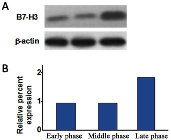
5 B7-H3 in osteosarcoma Figure 1. Relative B3-H7 mrna expression levels. Tumor volumes were 10, 15, and 40 cm 3 in the early, middle, and late phases, respectively. Figure 2. B3-H7 protein expression levels. Tumor volumes were 10, 15, and 40 cm 3 in the early, middle, and late phases, respectively. A. B7-H3 protein Western blot result. B. The semi-quantitative results of B7-H3 protein.

 (Figure 4). Figure 4.")
6 S.J. Yin et al Figure 3. B3-H7 localization in osteosarcoma tissue. B7-H3 s position in osteosarcoma. Green fluorescence indicated osteosarcoma cells, blue fluorescent indicated B7-H3. CD3+ T cell expression in osteosarcoma tissue CD3+ T cell numbers in osteosarcoma tissue in the early, middle, and late phases were 93 ± 13, 92 ± 12, and 46 ± 15, respectively. The cell number decreased significantly during the late stage (P < 0.05) (Figure 4). Figure 4. CD3+ T cell quantification results.expression of CD3+ t cells in bone sarcomas. Columns indicated the number of CD3+T cells.
7 B7-H3 in osteosarcoma DISCUSSION Lymphocytes play a central role in the process of tumor immunity, which is regulated by major histocompatibility antigens and costimulatory molecules; the latter is mainly composed of members of the B7/CD28 family including the newly discovered B7-H3 protein (Chapoval et al., 2001; Yi and Chen, 2009; Loos et al., 2010). Previous studies have shown that B7-H3 is expressed in numerous organs at the level of transcription, whereas it exhibits limited expressed in normal osteoblasts, fibroblasts, a fraction of epithelial cells, and in activated lymphocytes. Its expression, however, has been shown to be increased in tumor tissues. Our study provides the first demonstration that both B7-H3 mrna and protein are overexpressed in an osteosarcoma mouse model. In addition, our results also revealed that the expression levels of B7-H3 fluctuate during disease procession. B7-H3 mrna and protein are expressed at lower levels during the early and middle phases, whereas they are overexpressed during the late stage. In recent years, B7-H3 expression has been found to be correlated with survival time and high recurrence rates in different cancers, such as renal clear cell carcinoma, prostate cancer, and cervical cancer. Its expression level has also been associated with the severity of clinical pathology (Sun et al., 2006; Roth et al., 2007; Zang et al., 2007; Zhang et al., 2008; Yamato et al., 2009; Zhang et al., 2009; Sun et al., 2010). Our results also suggested that B7-H3 expression levels are associated with tumor volumes, and might be related to prognosis in the clinic; however, this suggestion will require confirmation through large-scale clinical research. T cell activation and proliferation depend on a dual signal; that is, the major histocompatibility complex antigen peptide complex on antigen presenting cells specifically combined with T cell receptors (TCR) to transmit the initial signal. The second signal is transmitted by members of the B7/CD28 family to determine whether T cell will be in a state to enhance, inhibit, weaken, or have no response. Costimulatory molecules of the B7/CD28 family, including B7-H3, play a very important role in determining the T cell immune response. In recent years, a large number of in vitro and in vivo experiments have confirmed that B7-H3 might be an inhibitory molecule that can be characterized by negative synergetic stimulation to inhibit activated T cell proliferation and cytokine synthesis (Chapoval et al., 2001; Yi and Chen, 2009; Loos et al., 2010). In addition, overexpressed B7-H3 has been negatively correlated with multiple malignant tumor prognoses (Zang et al., 2007; Zhang et al., 2008; Yamato et al., 2009; Sun et al., 2010). For example, a recent clinical study investigated the relationship of B7-H3 expression with the prognosis of patients with lung cancer and the degree of infiltration by CD3+ T lymphocytes. In that study, B7-H3 expression and CD3+ T lymphocyte infiltration degree were detected by immunohistochemistry, which found that B7-H3 was overexpressed and that B7-H3 expression was negatively correlated with patient survival and T cell invasion. Thus, B7-H3 exhibited a potential value in clinical application for lung cancer diagnosis and prognosis evaluation (Zhang et al., 2009). In addition, it has also been suggested that increased B7-H3 content might inhibit T cell activity to facilitate tumor growth (Chapoval et al., 2001; Sun et al., 2002; Yi and Chen, 2009; Loos et al., 2010). Whereas our results did not include immunologic analyses, they suggested that B7-H3 was overexpressed in osteosarcoma tissue, which in turn promoted tumor cell proliferation. Thus, osteosarcoma tissue growth might be associated with B7-H3 expression level. Tumor cell proliferation, as a basic feature, is known to be influenced by many factors. We believe that overexpressed B7-H3 is likely to facilitate its binding with corresponding ligands, and thus activate cell proliferation related genes directly, or stimulate secretion of certain cytokines to
8 S.J. Yin et al influence cell proliferation indirectly. Future research could be focused on the signaling pathways activated by B7-H3. In addition, as a transmembrane glycoprotein, B7-H3 contains an extracellular domain, a transmembrane region, and intracellular domains. Our study found that B7-H3 primarily exists in the cell nuclei within osteosarcoma tissue, indicating it might play a role in protein transcription, and might also activate downstream signaling pathways in the cell nucleus. Other studies have found that microrna29 participated in the B7-H3 posttranscriptional regulatory mechanism. In normal tissue, the expression level of microrna29 is high, which was shown to reduce the half-life of B7-H3 and also to inhibit the B7-H3 translational process. MicroRNA29 expression was shown to be markedly decreased in colon cancer tissue, resulting in insufficient levels of mirna29 to negatively regulate the B7-H3 posttranscriptional mechanism (Chapoval et al., 2001; Sun et al., 2002; Steinberger et al., 2004; Yi and Chen, 2009; Loos et al., 2010). Recently, tumor biological therapy, which mainly includes gene therapy and immune therapy, has increasingly become a hot clinical topic. The latter is closely related to the T cellmediated immune response (Chapoval et al., 2001; Luetke et al., 2014), in which costimulatory molecules play an important role. Our study confirmed that upregulated B7-H3 inhibited the activity and quantity of infiltrated CD3+ T cells, which alters the tumor microenvironment and promotes tumor proliferation. Furthermore, inhibition of B7-H3 on T cells also can down-regulate a variety of cytokines secreted by T cells such as IFN-α, IL-2, IL-17, and IL-10. Thus, B7-H3 might play an important role in tumor immunity. Our study also suggested that osteosarcoma cells might escape immune surveillance through B7-H3; for example, overexpressed B7-H3 might down-regulate T cell mediated antitumor immunity (Chapoval et al., 2001; Sun et al., 2002; Steinberger et al., 2004; Yi and Chen, 2009; Loos et al., 2010; Wang, Kang et al., 2014). Therefore, if we could reduce the increased B7-H3 level through molecular biology methods such as sirna or gene knockout to maintain the antitumor immune function of T cells, it would provide a new strategy for osteosarcoma treatment. Our study indicated that B7-H3 plays the role of a negative synergetic stimulating molecule in the process of tumor occurrence and development. It has a central role in the tumor cell immune escape mechanism by inhibiting the activation and proliferation of T cells to negatively regulate the immune response. Furthermore, B7-H3 expression is also closely related to tumor proliferation, invasion, and metastasis. Numerous tumors escape immune surveillance by downregulating positive synergetic stimulators and up-regulating negative synergetic stimulating signals. Blocking the negative synergetic stimulating pathway can enhance anti-tumor immunity, and can be effected with traditional cancer treatments in clinical or preclinical studies (Chapoval et al., 2001; Sun et al., 2002; Steinberger et al., 2004; Yi and Chen, 2009; Loos et al., 2010; Wang et al., 2014). Our experiments had several limitations, as our observations were limited to B7-H3 expression in a mouse model but not in clinical patients. In addition, the question of whether the overexpressed B7-H3 in osteosarcoma tissue is or functions the same as the normal B7-H3 still needs further verification. In summary, this study demonstrated the presence of B7-H3 mrna and protein expression in an osteosarcoma mouse model for the first time. Our results also showed the fluctuation of B7- H3 expression during the disease process. B7-H3 was expressed at lower levels in the early and middle phases, while it was overexpressed in the late stage. Accordingly, T cell numbers were decreased in the osteosarcoma tissue. Our research also revealed that the B7-H3 expression
9 B7-H3 in osteosarcoma level is associated with tumor volume. Thus, the level of B7-H3 expression might serve as an osteosarcoma marker, a proposition that needs further in-depth research. Conflicts of interest The authors declare no conflict of interest. REFERENCES Botter SM, Neri D and Fuchs B (2014). Recent advances in osteosarcoma. Curr. Opin. Pharmacol. 16: Chapoval AI, Ni J, Lau JS, Wilcox RA, et al. (2001). B7-H3: a costimulatory molecule for T cell activation and IFN-gamma production. Nat. Immunol. 2: Crispen PL, Sheinin Y, Roth TJ, Lohse CM, et al. (2008). Tumor cell and tumor vasculature expression of B7-H3 predict survival in clear cell renal cell carcinoma. Clin. Cancer Res. 14: Fleuren ED, Versleijen-Jonkers YM, Boerman OC and van der Graaf WT (2014). Targeting receptor tyrosine kinases in osteosarcoma and Ewing sarcoma: current hurdles and future perspectives. Biochim. Biophys. Acta 1845: Li Y, Flores R, Yu A, Okcu MF, et al. (2011). Elevated expression of CXC chemokines in pediatric osteosarcoma patients. Cancer 117: Loeb DM (2009). Is there a role for immunotherapy in osteosarcoma? Cancer Treat. Res. 152: Loos M, Hedderich DM, Friess H and Kleef J (2010). B7-h3 and its role in antitumor immunity. Clin. Dev. Immunol. 2010: Luetke A, Meyers PA, Lewis I and Juergens H (2014). Osteosarcoma treatment - where do we stand? A state of the art review. Cancer Treat. Rev. 40: Merchant MS, Melchionda F, Sinha M, Khanna C, et al. (2007). Immune reconstitution prevents metastatic recurrence of murine osteosarcoma. Cancer Immunol. Immunother. 56: Moore C, Eslin D, Levy A, Roberson J, et al. (2010). Prognostic significance of early lymphocyte recovery in pediatric osteosarcoma. Pediatr. Blood Cancer 55: Roth TJ, Sheinin Y, Lohse CM, Kuntz SM, et al. (2007). B7-H3 ligand expression by prostate cancer: a novel marker of prognosis and potential target for therapy. Cancer Res. 67: Ségaliny AI, Mohamadi A, Dizier B, Lokajczyk A, et al. (2014). Interleukin-34 promotes tumor progression and metastatic process in osteosarcoma through induction of angiogenesis and macrophage recruitment. Int. J. Cancer 137: Steinberger P, Majdic O, Derdak SV, Pfistershammer K, et al. (2004). Molecular characterization of human 4Ig-B7-H3, a member of the B7 family with four Ig-like domains. J. Immunol. 172: Sun J, Chen LJ, Zhang GB, Jiang JT, et al. (2010). Clinical significance and regulation of the costimulatory molecule B7-H3 in human colorectal carcinoma. Cancer Immunol. Immunother. 59: Sun M, Richards S, Prasad DV, Mai XM, et al. (2002). Characterization of mouse and human B7-H3 genes. J. Immunol. 168: Sun Y, Wang Y, Zhao J, Gu M, et al. (2006). B7-H3 and B7-H4 expression in non-small-cell lung cancer. Lung Cancer 53: Wang L, Kang FB and Shan BE (2014). B7-H3-mediated tumor immunology: Friend or foe? Int. J. Cancer 134: Yamato I, Sho M, Nomi T, Akahori T, et al. (2009). Clinical importance of B7-H3 expression in human pancreatic cancer. Br. J. Cancer 101: Yi KH and Chen L (2009). Fine tuning the immune response through B7-H3 and B7-H4. Immunol. Rev. 229: Zang X, Thompson RH, Al-Ahmadie HA, Serio AM, et al. (2007). B7-H3 and B7x are highly expressed in human prostate cancer and associated with disease spread and poor outcome. Proc. Natl. Acad. Sci. U. S. A. 104: Zhang G, Hou J, Shi J, Yu G, et al. (2008). Soluble CD276 (B7-H3) is released from monocytes, dendritic cells and activated T cells and is detectable in normal human serum. Immunology 123: Zhang G, Xu Y, Lu X, Huang H, et al. (2009). Diagnosis value of serum B7-H3 expression in non-small cell lung cancer. Lung Cancer 66: Zheng W, Xiao H, Liu H and Zhou Y (2014). Expression of programmed death 1 is correlated with progression of osteosarcoma. APMIS 123:
Advances in Computer Science Research, volume 59 7th International Conference on Education, Management, Computer and Medicine (EMCM 2016)
") 7th International Conference on Education, Management, Computer and Medicine (EMCM 2016) Expression of Beta-Adrenergic Receptor in Glioma LN229 Cells and Its Effect on Cell Proliferation Ping Wang1, Qingluan
7th International Conference on Education, Management, Computer and Medicine (EMCM 2016) Expression of Beta-Adrenergic Receptor in Glioma LN229 Cells and Its Effect on Cell Proliferation Ping Wang1, Qingluan
Plasmids Western blot analysis and immunostaining Flow Cytometry Cell surface biotinylation RNA isolation and cdna synthesis
 Plasmids psuper-retro-s100a10 shrna1 was constructed by cloning the dsdna oligo 5 -GAT CCC CGT GGG CTT CCA GAG CTT CTT TCA AGA GAA GAA GCT CTG GAA GCC CAC TTT TTA-3 and 5 -AGC TTA AAA AGT GGG CTT CCA GAG
(A) PCR primers (arrows) designed to distinguish wild type (P1+P2), targeted (P1+P2) and excised (P1+P3)14-
 PCR primers (arrows) designed to distinguish wild type (P1+P2), targeted (P1+P2) and excised (P1+P3)14-") 1 Supplemental Figure Legends Figure S1. Mammary tumors of ErbB2 KI mice with 14-3-3σ ablation have elevated ErbB2 transcript levels and cell proliferation (A) PCR primers (arrows) designed to distinguish
1 Supplemental Figure Legends Figure S1. Mammary tumors of ErbB2 KI mice with 14-3-3σ ablation have elevated ErbB2 transcript levels and cell proliferation (A) PCR primers (arrows) designed to distinguish
Expression and clinical significance of ADAM17 protein in esophageal squamous cell carcinoma
 Expression and clinical significance of ADAM17 protein in esophageal squamous cell carcinoma H.B. Liu, Y. Zhu, Q.C. Yang, Y. Shen, X.J. Zhang and H. Chen Department of Pathology First People s Hospital
SUPPLEMENTARY INFORMATION
 SUPPLEMENTARY INFORMATION FOR Liver X Receptor α mediates hepatic triglyceride accumulation through upregulation of G0/G1 Switch Gene 2 (G0S2) expression I: SUPPLEMENTARY METHODS II: SUPPLEMENTARY FIGURES
Profiles of gene expression & diagnosis/prognosis of cancer. MCs in Advanced Genetics Ainoa Planas Riverola
 Profiles of gene expression & diagnosis/prognosis of cancer MCs in Advanced Genetics Ainoa Planas Riverola Gene expression profiles Gene expression profiling Used in molecular biology, it measures the
Analysis of regulatory T cell subsets in the peripheral blood of immunoglobulin A nephropathy (IgAN) patients
 patients") Analysis of regulatory T cell subsets in the peripheral blood of immunoglobulin A nephropathy (IgAN) patients S. Yang, B. Chen, J. Shi, F. Chen, J. Zhang and Z. Sun Department of Nephrology, Huaihe Hospital
Analysis of regulatory T cell subsets in the peripheral blood of immunoglobulin A nephropathy (IgAN) patients S. Yang, B. Chen, J. Shi, F. Chen, J. Zhang and Z. Sun Department of Nephrology, Huaihe Hospital
Protein MultiColor Stable, Low Range
 Product Name: DynaMarker Protein MultiColor Stable, Low Range Code No: DM670L Lot No: ******* Size: 200 μl x 3 (DM670 x 3) (120 mini-gel lanes) Storage: 4 C Stability: 12 months at 4 C Storage Buffer:
Characterization and significance of MUC1 and c-myc expression in elderly patients with papillary thyroid carcinoma
 Characterization and significance of MUC1 and c-myc expression in elderly patients with papillary thyroid carcinoma Y.-J. Hu 1, X.-Y. Luo 2, Y. Yang 3, C.-Y. Chen 1, Z.-Y. Zhang 4 and X. Guo 1 1 Department
Supplementary data Supplementary Figure 1 Supplementary Figure 2
 Supplementary data Supplementary Figure 1 SPHK1 sirna increases RANKL-induced osteoclastogenesis in RAW264.7 cell culture. (A) RAW264.7 cells were transfected with oligocassettes containing SPHK1 sirna
Correlation between estrogen receptor β expression and the curative effect of endocrine therapy in breast cancer patients
 1568 Correlation between estrogen receptor β expression and the curative effect of endocrine therapy in breast cancer patients LIYING GUO 1, YU ZHANG 2, WEI ZHANG 3 and DILIMINA YILAMU 1 1 Department of
VENTANA PD-L1 (SP142) Assay Guiding immunotherapy in NSCLC
 Assay Guiding immunotherapy in NSCLC") VENTANA (SP142) Assay Guiding immunotherapy in NSCLC Hiker s path: VENTANA (SP142) Assay on non-small cell lung cancer tissue Location: Point Conception, CA VENTANA (SP142) Assay Assess NSCLC patient benefit
VENTANA (SP142) Assay Guiding immunotherapy in NSCLC Hiker s path: VENTANA (SP142) Assay on non-small cell lung cancer tissue Location: Point Conception, CA VENTANA (SP142) Assay Assess NSCLC patient benefit
Serum Amyloid A3 Gene Expression in Adipocytes is an Indicator. of the Interaction with Macrophages
 Serum Amyloid A3 Gene Expression in Adipocytes is an Indicator of the Interaction with Macrophages Yohei Sanada, Takafumi Yamamoto, Rika Satake, Akiko Yamashita, Sumire Kanai, Norihisa Kato, Fons AJ van
Supplementary Information
 Supplementary Information Supplementary Figure 1. CD4 + T cell activation and lack of apoptosis after crosslinking with anti-cd3 + anti-cd28 + anti-cd160. (a) Flow cytometry of anti-cd160 (5D.10A11) binding
Clinical significance of CD44 expression in children with hepatoblastoma
 Clinical significance of CD44 expression in children with hepatoblastoma H.-Y. Cai 1 *, B. Yu 1 *, Z.-C. Feng 2, X. Qi 1 and X.-J. Wei 1 1 Department of General Surgery, General Hospital of Beijing Military
High expression of fibroblast activation protein is an adverse prognosticator in gastric cancer.
 Biomedical Research 2017; 28 (18): 7779-7783 ISSN 0970-938X www.biomedres.info High expression of fibroblast activation protein is an adverse prognosticator in gastric cancer. Hu Song 1, Qi-yu Liu 2, Zhi-wei
General Laboratory methods Plasma analysis: Gene Expression Analysis: Immunoblot analysis: Immunohistochemistry:
 General Laboratory methods Plasma analysis: Plasma insulin (Mercodia, Sweden), leptin (duoset, R&D Systems Europe, Abingdon, United Kingdom), IL-6, TNFα and adiponectin levels (Quantikine kits, R&D Systems
Effects of VEGF/VEGFR/K-ras signaling pathways on mirna21 levels in hepatocellular carcinoma tissues in rats
 Effects of VEGF/VEGFR/K-ras signaling pathways on mirna21 levels in hepatocellular carcinoma tissues in rats J.Z. Gao 1,2 *, Y.L. Wang 1 *, J. Li 2 and L.X. Wei 2 1 Medical College, Xinxiang Medical University,
Data Sheet TIGIT / NFAT Reporter - Jurkat Cell Line Catalog #60538
 Data Sheet TIGIT / NFAT Reporter - Jurkat Cell Line Catalog #60538 Background: TIGIT is a co-inhibitory receptor that is highly expressed in Natural Killer (NK) cells, activated CD4+, CD8+ and regulatory
a) Primary cultures derived from the pancreas of an 11-week-old Pdx1-Cre; K-MADM-p53
 Primary cultures derived from the pancreas of an 11-week-old Pdx1-Cre; K-MADM-p53") 1 2 3 4 5 6 7 8 9 10 Supplementary Figure 1. Induction of p53 LOH by MADM. a) Primary cultures derived from the pancreas of an 11-week-old Pdx1-Cre; K-MADM-p53 mouse revealed increased p53 KO/KO (green,
1 2 3 4 5 6 7 8 9 10 Supplementary Figure 1. Induction of p53 LOH by MADM. a) Primary cultures derived from the pancreas of an 11-week-old Pdx1-Cre; K-MADM-p53 mouse revealed increased p53 KO/KO (green,
Toluidin-Staining of mast cells Ear tissue was fixed with Carnoy (60% ethanol, 30% chloroform, 10% acetic acid) overnight at 4 C, afterwards
 overnight at 4 C, afterwards") Toluidin-Staining of mast cells Ear tissue was fixed with Carnoy (60% ethanol, 30% chloroform, 10% acetic acid) overnight at 4 C, afterwards incubated in 100 % ethanol overnight at 4 C and embedded in
Toluidin-Staining of mast cells Ear tissue was fixed with Carnoy (60% ethanol, 30% chloroform, 10% acetic acid) overnight at 4 C, afterwards incubated in 100 % ethanol overnight at 4 C and embedded in
Effects of AFP gene silencing on Survivin mrna expression inhibition in HepG2 cells
 mrna expression inhibition in HepG2 cells Z.L. Fang 1, N. Fang 2, X.N. Han 3, G. Huang 2, X.J. Fu 2, G.S. Xie 2, N.R. Wang 2 and J.P. Xiong 1 1 Department of Medical Oncology, The First Affiliated Hospital
CD31 5'-AGA GAC GGT CTT GTC GCA GT-3' 5 ' -TAC TGG GCT TCG AGA GCA GT-3'
 Table S1. The primer sets used for real-time RT-PCR analysis. Gene Forward Reverse VEGF PDGFB TGF-β MCP-1 5'-GTT GCA GCA TGA ATC TGA GG-3' 5'-GGA GAC TCT TCG AGG AGC ACT T-3' 5'-GAA TCA GGC ATC GAG AGA
Abbreviations: P- paraffin-embedded section; C, cryosection; Bio-SA, biotin-streptavidin-conjugated fluorescein amplification.
 Supplementary Table 1. Sequence of primers for real time PCR. Gene Forward primer Reverse primer S25 5 -GTG GTC CAC ACT ACT CTC TGA GTT TC-3 5 - GAC TTT CCG GCA TCC TTC TTC-3 Mafa cds 5 -CTT CAG CAA GGA
Supplementary Appendix
 Supplementary Appendix This appendix has been provided by the authors to give readers additional information about their work. Supplement to: Sherman SI, Wirth LJ, Droz J-P, et al. Motesanib diphosphate
c Tuj1(-) apoptotic live 1 DIV 2 DIV 1 DIV 2 DIV Tuj1(+) Tuj1/GFP/DAPI Tuj1 DAPI GFP
 apoptotic live 1 DIV 2 DIV 1 DIV 2 DIV Tuj1(+) Tuj1/GFP/DAPI Tuj1 DAPI GFP") Supplementary Figure 1 Establishment of the gain- and loss-of-function experiments and cell survival assays. a Relative expression of mature mir-484 30 20 10 0 **** **** NCP mir- 484P NCP mir- 484P b Relative
Supplementary Figure 1 Establishment of the gain- and loss-of-function experiments and cell survival assays. a Relative expression of mature mir-484 30 20 10 0 **** **** NCP mir- 484P NCP mir- 484P b Relative
Islet viability assay and Glucose Stimulated Insulin Secretion assay RT-PCR and Western Blot
 Islet viability assay and Glucose Stimulated Insulin Secretion assay Islet cell viability was determined by colorimetric (3-(4,5-dimethylthiazol-2-yl)-2,5- diphenyltetrazolium bromide assay using CellTiter
Supplementary Materials and Methods
 Supplementary Materials and Methods Immunoblotting Immunoblot analysis was performed as described previously (1). Due to high-molecular weight of MUC4 (~ 950 kda) and MUC1 (~ 250 kda) proteins, electrophoresis
(a) Significant biological processes (upper panel) and disease biomarkers (lower panel)
 Significant biological processes (upper panel) and disease biomarkers (lower panel)") Supplementary Figure 1. Functional enrichment analyses of secretomic proteins. (a) Significant biological processes (upper panel) and disease biomarkers (lower panel) 2 involved by hrab37-mediated secretory
Supplementary Figure 1. Functional enrichment analyses of secretomic proteins. (a) Significant biological processes (upper panel) and disease biomarkers (lower panel) 2 involved by hrab37-mediated secretory
SUPPLEMENTAL MATERIAL. Supplementary Methods
 SUPPLEMENTAL MATERIAL Supplementary Methods Culture of cardiomyocytes, fibroblasts and cardiac microvascular endothelial cells The isolation and culturing of neonatal rat ventricular cardiomyocytes was
Supplementary Table 3. 3 UTR primer sequences. Primer sequences used to amplify and clone the 3 UTR of each indicated gene are listed.
 Supplemental Figure 1. DLKI-DIO3 mirna/mrna complementarity. Complementarity between the indicated DLK1-DIO3 cluster mirnas and the UTR of SOX2, SOX9, HIF1A, ZEB1, ZEB2, STAT3 and CDH1with mirsvr and PhastCons
Sestrin2 and BNIP3 (Bcl-2/adenovirus E1B 19kDa-interacting. protein3) regulate autophagy and mitophagy in renal tubular cells in. acute kidney injury
 regulate autophagy and mitophagy in renal tubular cells in. acute kidney injury") Sestrin2 and BNIP3 (Bcl-2/adenovirus E1B 19kDa-interacting protein3) regulate autophagy and mitophagy in renal tubular cells in acute kidney injury by Masayuki Ishihara 1, Madoka Urushido 2, Kazu Hamada
Sestrin2 and BNIP3 (Bcl-2/adenovirus E1B 19kDa-interacting protein3) regulate autophagy and mitophagy in renal tubular cells in acute kidney injury by Masayuki Ishihara 1, Madoka Urushido 2, Kazu Hamada
Circulating PD-L1 in NSCLC patients and the correlation between the level of PD-L1 expression and the clinical characteristics
 Thoracic Cancer ISSN 1759-7706 ORIGINAL ARTICLE Circulating PD-L1 in NSCLC patients and the correlation between the level of PD-L1 expression and the clinical characteristics Jie Zhang, Jing Gao, Yanyan
Downregulation of serum mir-17 and mir-106b levels in gastric cancer and benign gastric diseases
 Brief Communication Downregulation of serum mir-17 and mir-106b levels in gastric cancer and benign gastric diseases Qinghai Zeng 1 *, Cuihong Jin 2 *, Wenhang Chen 2, Fang Xia 3, Qi Wang 3, Fan Fan 4,
An epithelial-to-mesenchymal transition-inducing potential of. granulocyte macrophage colony-stimulating factor in colon. cancer
 An epithelial-to-mesenchymal transition-inducing potential of granulocyte macrophage colony-stimulating factor in colon cancer Yaqiong Chen, Zhi Zhao, Yu Chen, Zhonglin Lv, Xin Ding, Renxi Wang, He Xiao,
CHAPTER 4 RESULTS. showed that all three replicates had similar growth trends (Figure 4.1) (p<0.05; p=0.0000)
 (p<0.05; p=0.0000)") CHAPTER 4 RESULTS 4.1 Growth Characterization of C. vulgaris 4.1.1 Optical Density Growth study of Chlorella vulgaris based on optical density at 620 nm (OD 620 ) showed that all three replicates had similar
CHAPTER 4 RESULTS 4.1 Growth Characterization of C. vulgaris 4.1.1 Optical Density Growth study of Chlorella vulgaris based on optical density at 620 nm (OD 620 ) showed that all three replicates had similar
Oncolytic Immunotherapy: A Local and Systemic Antitumor Approach
 Oncolytic Immunotherapy: A Local and Systemic Antitumor Approach Oncolytic immunotherapy Oncolytic immunotherapy the use of a genetically modified virus to attack tumors and induce a systemic immune response
Correlation between expression and significance of δ-catenin, CD31, and VEGF of non-small cell lung cancer
 Correlation between expression and significance of δ-catenin, CD31, and VEGF of non-small cell lung cancer X.L. Liu 1, L.D. Liu 2, S.G. Zhang 1, S.D. Dai 3, W.Y. Li 1 and L. Zhang 1 1 Thoracic Surgery,
(A) RT-PCR for components of the Shh/Gli pathway in normal fetus cell (MRC-5) and a
 RT-PCR for components of the Shh/Gli pathway in normal fetus cell (MRC-5) and a") Supplementary figure legends Supplementary Figure 1. Expression of Shh signaling components in a panel of gastric cancer. (A) RT-PCR for components of the Shh/Gli pathway in normal fetus cell (MRC-5) and
Supplementary figure legends Supplementary Figure 1. Expression of Shh signaling components in a panel of gastric cancer. (A) RT-PCR for components of the Shh/Gli pathway in normal fetus cell (MRC-5) and
Protocol for Gene Transfection & Western Blotting
 The schedule and the manual of basic techniques for cell culture Advanced Protocol for Gene Transfection & Western Blotting Schedule Day 1 26/07/2008 Transfection Day 3 28/07/2008 Cell lysis Immunoprecipitation
The Schedule and the Manual of Basic Techniques for Cell Culture
 The Schedule and the Manual of Basic Techniques for Cell Culture 1 Materials Calcium Phosphate Transfection Kit: Invitrogen Cat.No.K2780-01 Falcon tube (Cat No.35-2054:12 x 75 mm, 5 ml tube) Cell: 293
Supplementary Figures
 Supplementary Figures Supplementary Figure 1. H3F3B expression in lung cancer. a. Comparison of H3F3B expression in relapsed and non-relapsed lung cancer patients. b. Prognosis of two groups of lung cancer
Supplementary Figure 1
 Supplementary Figure 1 3 3 3 1 1 Bregma -1.6mm 3 : Bregma Ref) Http://www.mbl.org/atlas165/atlas165_start.html Bregma -.18mm Supplementary Figure 1 Schematic representation of the utilized brain slice
CircHIPK3 is upregulated and predicts a poor prognosis in epithelial ovarian cancer
 European Review for Medical and Pharmacological Sciences 2018; 22: 3713-3718 CircHIPK3 is upregulated and predicts a poor prognosis in epithelial ovarian cancer N. LIU 1, J. ZHANG 1, L.-Y. ZHANG 1, L.
T-cell activation T cells migrate to secondary lymphoid tissues where they interact with antigen, antigen-presenting cells, and other lymphocytes:
 Interactions between innate immunity & adaptive immunity What happens to T cells after they leave the thymus? Naïve T cells exit the thymus and enter the bloodstream. If they remain in the bloodstream,
Cytokine Arrays Reveal Black Ops Tactics of Tumor-induced Immunosuppression
 Cytokine Arrays Reveal Black Ops Tactics of Tumor-induced Immunosuppression Jarad J Wilson, Ph.D. Technical Support & Marketing Specialist Ruo-Pan Huang, MD, Ph.D. Founder and CEO What are Antibody Arrays?
BMP6 treatment compensates for the molecular defect and ameliorates hemochromatosis in Hfe knockout mice
 SUPPLEMENTARY MATERIALS BMP6 treatment compensates for the molecular defect and ameliorates hemochromatosis in Hfe knockout mice Elena Corradini, Paul J. Schmidt, Delphine Meynard, Cinzia Garuti, Giuliana
Formylpeptide receptor2 contributes to colon epithelial homeostasis, inflammation, and tumorigenesis
 Supplementary Data Formylpeptide receptor2 contributes to colon epithelial homeostasis, inflammation, and tumorigenesis Keqiang Chen, Mingyong Liu, Ying Liu, Teizo Yoshimura, Wei Shen, Yingying Le, Scott
The Adaptive Immune Responses
 The Adaptive Immune Responses The two arms of the immune responses are; 1) the cell mediated, and 2) the humoral responses. In this chapter we will discuss the two responses in detail and we will start
Study on the expression of MMP-9 and NF-κB proteins in epithelial ovarian cancer tissue and their clinical value
 Study on the expression of MMP-9 and NF-κB proteins in epithelial ovarian cancer tissue and their clinical value Shen Wei 1,a, Chen Juan 2, Li Xiurong 1 and Yin Jie 1 1 Department of Obstetrics and Gynecology,
Epithelial interleukin-25 is a key mediator in Th2-high, corticosteroid-responsive
 Online Data Supplement: Epithelial interleukin-25 is a key mediator in Th2-high, corticosteroid-responsive asthma Dan Cheng, Zheng Xue, Lingling Yi, Huimin Shi, Kan Zhang, Xiaorong Huo, Luke R. Bonser,
FGL2 A new biomarker for cancer in a simple blood test
 FGL2 A new biomarker for cancer in a simple blood test WHO IS FGL2 Human gene (chromosome 7) is 7 kb long, 2 exons, monomer protein 70 KD, tetramer in solution. Fibrinogen-like protein 2 (Fgl2), a member
The toll-like receptor 4 ligands Mrp8 and Mrp14 play a critical role in the development of autoreactive CD8 + T cells
 1 SUPPLEMENTARY INFORMATION The toll-like receptor 4 ligands Mrp8 and Mrp14 play a critical role in the development of autoreactive CD8 + T cells Karin Loser 1,2,6, Thomas Vogl 2,3, Maik Voskort 1, Aloys
A smart acid nanosystem for ultrasensitive. live cell mrna imaging by the target-triggered intracellular self-assembly
 Electronic Supplementary Material (ESI) for Chemical Science. This journal is The Royal Society of Chemistry 2017 A smart ZnO@polydopamine-nucleic acid nanosystem for ultrasensitive live cell mrna imaging
Exploring the PD-L1 Pathway
 Active Within the tumor microenvironment Steps 1-3: Initiating and propagating anticancer immunity 1 may inhibit T-cell activity in the tumor microenvironment Dendritic cells capture cancer and then prime
Novel RCC Targets from Immuno-Oncology and Antibody-Drug Conjugates
 Novel RCC Targets from Immuno-Oncology and Antibody-Drug Conjugates Christopher Turner, MD Vice President, Clinical Science 04 November 2016 Uveal Melanoma Celldex Pipeline CANDIDATE INDICATION Preclinical
microrna-200b and microrna-200c promote colorectal cancer cell proliferation via
 Supplementary Materials microrna-200b and microrna-200c promote colorectal cancer cell proliferation via targeting the reversion-inducing cysteine-rich protein with Kazal motifs Supplementary Table 1.
Expression of mir-146a-5p in patients with intracranial aneurysms and its association with prognosis
 European Review for Medical and Pharmacological Sciences 2018; 22: 726-730 Expression of mir-146a-5p in patients with intracranial aneurysms and its association with prognosis H.-L. ZHANG 1, L. LI 2, C.-J.
Supporting Information
 Supporting Information Pang et al. 10.1073/pnas.1322009111 SI Materials and Methods ELISAs. These assays were performed as previously described (1). ELISA plates (MaxiSorp Nunc; Thermo Fisher Scientific)
Original Article B7-H3 repression by mir-539 suppresses cell proliferation in human gliomas
 Int J Clin Exp Pathol 2017;10(4):4363-4369 www.ijcep.com /ISSN:1936-2625/IJCEP0043994 Original Article B7-H3 repression by mir-539 suppresses cell proliferation in human gliomas Rong-Gang Li 1, Zhuo Gao
Research on the inhibitory effect of metformin on human oral squamous cell carcinoma SCC-4 and CAL-27 cells and the relevant molecular mechanism.
 Biomedical Research 2017; 28 (14): 6350-6354 ISSN 0970-938X www.biomedres.info Research on the inhibitory effect of metformin on human oral squamous cell carcinoma SCC-4 and CAL-27 cells and the relevant
Synergistic combinations of targeted immunotherapy to combat cancer
 Synergistic combinations of targeted immunotherapy to combat cancer Myung Ah Lee, M.D., Ph. D Division of Medical Oncology, Hepato-biliary pancreatic cancer center Seoul St. Mary s hospital, The Catholic
Supplemental Figures: Supplemental Figure 1
 Supplemental Figures: Supplemental Figure 1 Suppl. Figure 1. BM-DC infection with H. pylori does not induce cytotoxicity and treatment of BM-DCs with H. pylori sonicate, but not heat-inactivated bacteria,
(14): DOI: /j.issn ORCID: ( )
: DOI: /j.issn ORCID: ( )") 20 14 2016 04 01 Chinese Journal of Tissue Engineering Research April 1, 2016 Vol.20, No.14 ( 473000). [J]. 2016 20(14):2033-2039. DOI: 10.3969/j.issn.2095-4344.2016.14.008 ORCID: 0000-0002-8672-7109()
20 14 2016 04 01 Chinese Journal of Tissue Engineering Research April 1, 2016 Vol.20, No.14 ( 473000). [J]. 2016 20(14):2033-2039. DOI: 10.3969/j.issn.2095-4344.2016.14.008 ORCID: 0000-0002-8672-7109()
Effective activity of cytokine-induced killer cells against autologous metastatic melanoma including cells with stemness features
 Effective activity of cytokine-induced killer cells against autologous metastatic melanoma including cells with stemness features Loretta Gammaitoni, Lidia Giraudo, Valeria Leuci, et al. Clin Cancer Res
Supplementary Materials
 Supplementary Materials 1 Supplementary Table 1. List of primers used for quantitative PCR analysis. Gene name Gene symbol Accession IDs Sequence range Product Primer sequences size (bp) β-actin Actb gi
Supplementary Figure 1. Normal T lymphocyte populations in Dapk -/- mice. (a) Normal thymic development in Dapk -/- mice. Thymocytes from WT and Dapk
 Normal thymic development in Dapk -/- mice. Thymocytes from WT and Dapk") Supplementary Figure 1. Normal T lymphocyte populations in Dapk -/- mice. (a) Normal thymic development in Dapk -/- mice. Thymocytes from WT and Dapk -/- mice were stained for expression of CD4 and CD8.
Supplementary Figure 1. Normal T lymphocyte populations in Dapk -/- mice. (a) Normal thymic development in Dapk -/- mice. Thymocytes from WT and Dapk -/- mice were stained for expression of CD4 and CD8.
Figure S1. Analysis of genomic and cdna sequences of the targeted regions in WT-KI and
 Figure S1. Analysis of genomic and sequences of the targeted regions in and indicated mutant KI cells, with WT and corresponding mutant sequences underlined. (A) cells; (B) K21E-KI cells; (C) D33A-KI cells;
Supplementary Figure 1 a
 Supplementary Figure a Normalized expression/tbp (A.U.).6... Trip-br transcripts Trans Trans Trans b..5. Trip-br Ctrl LPS Normalized expression/tbp (A.U.) c Trip-br transcripts. adipocytes.... Trans Trans
Supplementary Figure 1. ROS induces rapid Sod1 nuclear localization in a dosagedependent manner. WT yeast cells (SZy1051) were treated with 4NQO at
 were treated with 4NQO at") Supplementary Figure 1. ROS induces rapid Sod1 nuclear localization in a dosagedependent manner. WT yeast cells (SZy1051) were treated with 4NQO at different concentrations for 30 min and analyzed for
Supplementary Figure 1. ROS induces rapid Sod1 nuclear localization in a dosagedependent manner. WT yeast cells (SZy1051) were treated with 4NQO at different concentrations for 30 min and analyzed for
Supplemental Data. Shin et al. Plant Cell. (2012) /tpc YFP N
 /tpc YFP N") MYC YFP N PIF5 YFP C N-TIC TIC Supplemental Data. Shin et al. Plant Cell. ()..5/tpc..95 Supplemental Figure. TIC interacts with MYC in the nucleus. Bimolecular fluorescence complementation assay using
MYC YFP N PIF5 YFP C N-TIC TIC Supplemental Data. Shin et al. Plant Cell. ()..5/tpc..95 Supplemental Figure. TIC interacts with MYC in the nucleus. Bimolecular fluorescence complementation assay using
TSH Receptor Monoclonal Antibody (49) Catalog Number MA3-218 Product data sheet
 Catalog Number MA3-218 Product data sheet") Website: thermofisher.com Customer Service (US): 1 800 955 6288 ext. 1 Technical Support (US): 1 800 955 6288 ext. 441 TSH Receptor Monoclonal Antibody (49) Catalog Number MA3-218 Product data sheet Details
Website: thermofisher.com Customer Service (US): 1 800 955 6288 ext. 1 Technical Support (US): 1 800 955 6288 ext. 441 TSH Receptor Monoclonal Antibody (49) Catalog Number MA3-218 Product data sheet Details
ACTIVATION AND EFFECTOR FUNCTIONS OF CELL-MEDIATED IMMUNITY AND NK CELLS. Choompone Sakonwasun, MD (Hons), FRCPT
, FRCPT") ACTIVATION AND EFFECTOR FUNCTIONS OF CELL-MEDIATED IMMUNITY AND NK CELLS Choompone Sakonwasun, MD (Hons), FRCPT Types of Adaptive Immunity Types of T Cell-mediated Immune Reactions CTLs = cytotoxic T lymphocytes
ACTIVATION AND EFFECTOR FUNCTIONS OF CELL-MEDIATED IMMUNITY AND NK CELLS Choompone Sakonwasun, MD (Hons), FRCPT Types of Adaptive Immunity Types of T Cell-mediated Immune Reactions CTLs = cytotoxic T lymphocytes
Positive nin one binding protein expression predicts poor outcome in prostate cancer
 MOLECULAR MEDICINE REPORTS 11: 2671-2676, 2015 Positive nin one binding protein expression predicts poor outcome in prostate cancer JIE CHEN *, JUNKAI WANG *, XINGANG CUI, YUSHAN LIU, LEI YIN, YAO LI,
RNA extraction, RT-PCR and real-time PCR. Total RNA were extracted using
 Supplementary Information Materials and Methods RNA extraction, RT-PCR and real-time PCR. Total RNA were extracted using Trizol reagent (Invitrogen,Carlsbad, CA) according to the manufacturer's instructions.
Protection against doxorubicin-induced myocardial dysfunction in mice by cardiac-specific expression of carboxyl terminus of hsp70-interacting protein
 Protection against doxorubicin-induced myocardial dysfunction in mice by cardiac-specific expression of carboxyl terminus of hsp70-interacting protein Lei Wang 1, Tian-Peng Zhang 1, Yuan Zhang 2, Hai-Lian
Simultaneous blockade of PD-1 and VEGFR2 induces synergistic. Short title: Synergistic antitumour effect by dual blockade of PD-1 and VEGFR2
 carticle Simultaneous blockade of PD-1 and VEGFR2 induces synergistic antitumour effect in vivo 1 Short title: Synergistic antitumour effect by dual blockade of PD-1 and VEGFR2 S. Yasuda 1, M. Sho 1, I.
Anti-Tumor Efficacy of Gene Vaccine Expressing PSMA
 6 Clin Oncol Cancer Res () 7: 6-5 DOI.7/s85--5-7 Anti-Tumor Efficacy of Gene Vaccine Expressing PSMA Xiao-ling YANG Jing LV Yue-hong ZHANG Bo NIU, Department of Laboratory Medicine, Shanxi Bo ai Hospital,
IMMUNOTHERAPY FOR CANCER A NEW HORIZON. Ekaterini Boleti MD, PhD, FRCP Consultant in Medical Oncology Royal Free London NHS Foundation Trust
 IMMUNOTHERAPY FOR CANCER A NEW HORIZON Ekaterini Boleti MD, PhD, FRCP Consultant in Medical Oncology Royal Free London NHS Foundation Trust ASCO Names Advance of the Year: Cancer Immunotherapy No recent
Immune Checkpoint Inhibitors: The New Breakout Stars in Cancer Treatment
 Immune Checkpoint Inhibitors: The New Breakout Stars in Cancer Treatment 1 Introductions Peter Langecker, MD, PhD Executive Medical Director, Global Oncology Clinipace Worldwide Mark Shapiro Vice President
[Abstract] Objective: Investigate the effect of KLT on the expression of Fas/Apo-1, FasL and PCNA genes in renal carcinoma cell line (GRC-1).
![[Abstract] Objective: Investigate the effect of KLT on the expression of Fas/Apo-1, FasL and PCNA genes in renal carcinoma cell line (GRC-1).](/thumbs/90/104150076.jpg "[Abstract] Objective: Investigate the effect of KLT on the expression of Fas/Apo-1, FasL and PCNA genes in renal carcinoma cell line (GRC-1).") Study on Effect of Kanglaite Injection(KLT) on the Expression of Fas/Apo-1, FasL and PCNA in Renal Carcinoma Cell Lines Wang Junjie, Sun Xinchen, Sheng Wenjiang, Yu Lizhang [Abstract] Objective: Investigate
Study on Effect of Kanglaite Injection(KLT) on the Expression of Fas/Apo-1, FasL and PCNA in Renal Carcinoma Cell Lines Wang Junjie, Sun Xinchen, Sheng Wenjiang, Yu Lizhang [Abstract] Objective: Investigate
Amniotic fluid stem cells provide considerable advantages in epidermal. regeneration: B7H4 creates a moderate inflammation
 Amniotic fluid stem cells provide considerable advantages in epidermal regeneration: B7H4 creates a moderate inflammation microenvironment to promote wound repair Qing Sun 1, +, Fang Li 1, +, Hong Li 2,
Intracellular MHC class II molecules promote TLR-triggered innate. immune responses by maintaining Btk activation
 Intracellular MHC class II molecules promote TLR-triggered innate immune responses by maintaining Btk activation Xingguang Liu, Zhenzhen Zhan, Dong Li, Li Xu, Feng Ma, Peng Zhang, Hangping Yao and Xuetao
MTC-TT and TPC-1 cell lines were cultured in RPMI medium (Gibco, Breda, The Netherlands)
") Supplemental data Materials and Methods Cell culture MTC-TT and TPC-1 cell lines were cultured in RPMI medium (Gibco, Breda, The Netherlands) supplemented with 15% or 10% (for TPC-1) fetal bovine serum
Supplemental data Materials and Methods Cell culture MTC-TT and TPC-1 cell lines were cultured in RPMI medium (Gibco, Breda, The Netherlands) supplemented with 15% or 10% (for TPC-1) fetal bovine serum
VEGFR2-Mediated Vascular Dilation as a Mechanism of VEGF-Induced Anemia and Bone Marrow Cell Mobilization
 Cell Reports, Volume 9 Supplemental Information VEGFR2-Mediated Vascular Dilation as a Mechanism of VEGF-Induced Anemia and Bone Marrow Cell Mobilization Sharon Lim, Yin Zhang, Danfang Zhang, Fang Chen,
Low levels of serum mir-99a is a predictor of poor prognosis in breast cancer
 Low levels of serum mir-99a is a predictor of poor prognosis in breast cancer J. Li 1, Z.J. Song 2, Y.Y. Wang 1, Y. Yin 1, Y. Liu 1 and X. Nan 1 1 Tumor Research Department, Shaanxi Provincial Tumor Hospital,
Mir-595 is a significant indicator of poor patient prognosis in epithelial ovarian cancer
 European Review for Medical and Pharmacological Sciences 2017; 21: 4278-4282 Mir-595 is a significant indicator of poor patient prognosis in epithelial ovarian cancer Q.-H. ZHOU 1, Y.-M. ZHAO 2, L.-L.
TFEB-mediated increase in peripheral lysosomes regulates. Store Operated Calcium Entry
 TFEB-mediated increase in peripheral lysosomes regulates Store Operated Calcium Entry Luigi Sbano, Massimo Bonora, Saverio Marchi, Federica Baldassari, Diego L. Medina, Andrea Ballabio, Carlotta Giorgi
Supporting Information
 Supporting Information M1 macrophage-derived nanovesicles potentiate the anticancer efficacy of immune checkpoint inhibitors Yeon Woong Choo, 1, Mikyung Kang, 2, Han Young Kim, 1 Jin Han, 1 Seokyung Kang,
Tumor Associated Macrophages as a Novel Target for Cancer Therapy
 Tumor mass Tumor Associated Macrophage Tumor Associated Macrophages as a Novel Target for Cancer Therapy This booklet contains forward-looking statements that are based on Amgen s current expectations
Peli1 negatively regulates T-cell activation and prevents autoimmunity
 Peli1 negatively regulates T-cell activation and prevents autoimmunity Mikyoung Chang 1,*, Wei Jin 1,5,*, Jae-Hoon Chang 1, Yi-chuan Xiao 1, George Brittain 1, Jiayi Yu 1, Xiaofei Zhou 1, Yi-Hong Wang
Examples of questions for Cellular Immunology/Cellular Biology and Immunology
 Examples of questions for Cellular Immunology/Cellular Biology and Immunology Each student gets a set of 6 questions, so that each set contains different types of questions and that the set of questions
L1 on PyMT tumor cells but Py117 cells are more responsive to IFN-γ. (A) Flow
 Flow") A MHCI B PD-L1 Fold expression 8 6 4 2 Fold expression 3 2 1 No tx 1Gy 2Gy IFN Py117 Py117 Supplementary Figure 1. Radiation and IFN-γ enhance MHCI expression and PD- L1 on PyMT tumor cells but Py117 cells
A MHCI B PD-L1 Fold expression 8 6 4 2 Fold expression 3 2 1 No tx 1Gy 2Gy IFN Py117 Py117 Supplementary Figure 1. Radiation and IFN-γ enhance MHCI expression and PD- L1 on PyMT tumor cells but Py117 cells
Neutrophils contribute to fracture healing by synthesizing fibronectin+ extracellular matrix rapidly after injury
 Neutrophils contribute to fracture healing by synthesizing fibronectin+ extracellular matrix rapidly after injury Bastian OW, Koenderman L, Alblas J, Leenen LPH, Blokhuis TJ. Neutrophils contribute to
Bihong Zhao, M.D, Ph.D Department of Pathology
 Bihong Zhao, M.D, Ph.D Department of Pathology 04-28-2009 Is tumor self or non-self? How are tumor antigens generated? What are they? How does immune system respond? Introduction Tumor Antigens/Categories
Posters and Presentations
 Posters and Presentations June 2017: American Society of Clinical Oncology (ASCO) Annual - Preliminary Correlative Analysis of PD-L1 expression from the SUNRISE Study. View April 2017: American Association
Expression and significance of CD28, CTLA-4, CD80 and CD86 in gastric cancer.
 Biomedical Research 017; 8 (): 10159-10164 ISSN 0970-938X www.biomedres.info Expression and significance of CD8, CTLA-4, CD80 and CD86 in gastric cancer. Xichao Yang, Tingting Lv, Jianhong Qiang, Xueyi
Supplementary Table 2. Conserved regulatory elements in the promoters of CD36.
 Supplementary Table 1. RT-qPCR primers for CD3, PPARg and CEBP. Assay Forward Primer Reverse Primer 1A CAT TTG TGG CCT TGT GCT CTT TGA TGA GTC ACA GAA AGA ATC AAT TC 1B AGG AAA TGA ACT GAT GAG TCA CAG
SUPPLEMENTARY METHODS
 SUPPLEMENTARY METHODS Histological analysis. Colonic tissues were collected from 5 parts of the middle colon on day 7 after the start of DSS treatment, and then were cut into segments, fixed with 4% paraformaldehyde,