Nasal Cavity and Paranasal Sinuses
|
|
|
- Kelley Patrick
- 5 years ago
- Views:
Transcription

 is a squamous cell carcinoma (SCC) characterized by a")

1 2017 Head and Neck Tumors Selected Topics Lester D. R. Thompson 1 2 Inclusion Criteria Nasal Cavity and Paranasal Sinuses 2005 edition: 76 diagnoses 2017 edition: 39 diagnoses 3 1. The tumor occurs exclusively at this site; 2. The tumor also occurs at other head and neck sites but has a predilection for the sinonasal tract; or 3. The tumor is important for differential diagnostic reasons Specifically, salivary gland neoplasms, bone and cartilage tumors, and hematolymphoid tumors were only recorded once in the book rather than repeated in each anatomic site 4 Non-keratinizing Squamous Cell Carcinoma Non-keratinizing squamous cell carcinoma (NKSCC) is a squamous cell carcinoma (SCC) characterized by a distinctive ribbon-like growth pattern with absent to limited maturation Synonyms: Schneiderian carcinoma; transitional cell carcinoma; cylindrical cell carcinoma Epidemiology: ~20% of sinonasal SCC Age: 6 th 7 th decades Sex: M > F Pathology: smooth stromal interface with a pushing border; immature appearance with minimal or no keratinization; high N:C ratio; may have peripheral palisading; numerous mitoses; necrosis 5 6 Update on New ENT WHO 1
 deficient carcinoma Rhabdoid and poorly differentiated features NUT carcinoma Abrupt\" keratinization 9 HPV-related carcinoma with adenoid cystic-like features")
 Sex: Female >> Male (7:2) Age: 40 75 years Highly cellular proliferation Solid nests with frequently encountered cribriform")

2 7 8 Non-keratinizing Squamous Cell Carcinoma Differential Diagnosis Sinonasal papilloma with malignant transformation Sinonasal undifferentiated carcinoma Neuroendocrine carcinoma Solid variant of adenoid cystic carcinoma SMARCB1 (INI-1) deficient carcinoma Rhabdoid and poorly differentiated features NUT carcinoma Abrupt" keratinization 9 HPV-related carcinoma with adenoid cystic-like features A distinctive human papillomavirus (HPV)-related carcinoma of the sinonasal tract with histologic and immunophenotypic features of both surface-derived and salivary gland carcinoma, the latter showing the appearance of a high grade adenoid cystic carcinoma (ACC) Sex: Female >> Male (7:2) Age: years Highly cellular proliferation Solid nests with frequently encountered cribriform structures separated by thin collagenized fibrous bands Basaloid cells align around cylindromatous microcystic spaces Hyperchromatic and slightly angulated nuclei with a high nuclear-to-cytoplasmic ratio True ductal cells are present, often with peripheral myoepithelial cells 10 Sinonasal Undifferentiated Carcinoma Undifferentiated carcinoma of the sinonasal tract without glandular or squamous features and not otherwise classifiable Sinonasal undifferentiated carcinoma (SNUC) is rare 3-5% of sinonasal carcinomas Age: years Sex: 70% male Pathology: sheets, lobules, and trabeculae; moderately large, round nuclei, variable amount of cytoplasm, and well-defined cell borders; limited pleomorphism; nuclei vary from hyperchromatic to vesicular, with open chromatin with prominent nucleoli; apoptosis, mitoses, and necrosis are frequent By definition: no squamous and no glandular differentiation But, carcinoma in situ and surface dysplasia may be seen HPV-HR ISH Update on New ENT WHO 2


3 13 14 CK Sinonasal Undifferentiated Carcinoma Differential Diagnosis Lymphoma, nonkeratinizing squamous cell carcinoma (SCC), basaloid SCC, and neuroendocrine carcinoma SCC has areas of histologic squamous differentiation, consistently reactive with cytokeratin 5/6, p63, and p40 Neuroendocrine carcinomas have speckled salt-andpepper nuclear chromatin, with strong reactivity with neuroendocrine markers Poorly differentiated carcinomas with rhabdoid features may show a loss of SMARCB1 (INI-1) protein by immunohistochemistry, suggesting a different tumor category NUT carcinoma has evidence of squamous differentiation, and is consistently and diffusely positive for p63 and p40, with strong NUT protein by immunohistochemistry CK5/6 p Update on New ENT WHO 3

 20 NUT Carcinoma NUT carcinoma is a poorly differentiated carcinoma, often with evidence of abrupt squamous differentiation, defined by the presence of NUT (nuclear")
4 SMARCB1 (INI1) deficient carcinoma Inactivation of SMARCB1 (INI-1) defines a diverse family of neoplasms Age: Wide age range (28 78 years, mean: 54 years) Sex: Slight female predominance Pathology: Undifferentiated basaloid nests High mitotic rates, frequent necrosis Relatively monotonous basaloid cells Rhabdoid or plasmacytoid features Round to oval nuclei No keratinization Biallelic inactivation of SMARCB1 (INI1) Loss of nuclear SMARCB1 immunohistochemistry Clinically aggressive carcinomas that are frequently associated with local recurrence, regional and distant metastases, and patient death 19 SMARCB1 (INI1) 20 NUT Carcinoma NUT carcinoma is a poorly differentiated carcinoma, often with evidence of abrupt squamous differentiation, defined by the presence of NUT (nuclear protein in testis, NUTM1) gene rearrangement Rare in the upper aerodigestive tract Age: median: 22 years Sex: slight female predominance F > M (55:45) 21 The etiology is unknown NUT Carcinoma No association with human papillomavirus, Epstein Barr virus, other viral infection, smoking, or other environmental factors If head & neck involvement, sinonasal tract is common (65%) Generally, but not always midline Non-specific symptoms; rapidly growing mass Lymph node metastases seen in up to 50% of sinonasal tract cases 22 NUT Carcinoma Pathology Poorly differentiated carcinoma arranged in sheets and nests Intermediate, round to oval nuclei in monotonously similar tumor cells Chromatin: vesicular with distinct nucleoli Cytoplasm: scant to moderate; may be clear Brisk mitoses Tumor necrosis often present Abrupt foci of keratinization, keratin pearl formation or squamous differentiation Intratumoral acute inflammation may be seen Glandular and mesenchymal differentiation is infrequent Update on New ENT WHO 4


, BRD3 (6%), or NSD3, creating chimeric genes that")
 in some cases Fluorescent in situ hybridization (FISH),")

5 25 26 NUT Carcinoma Unequivocal diagnosis when diffuse (>50%) nuclear staining with the NUT monoclonal antibody Positive: p63, p40 and cytokeratins; CD34 (~55%), Variable: neuroendocrine markers, p16 overexpression, TTF-1 Genetically defined by rearrangements of the nuclear protein in testis (NUTM1) gene NUTM1 on chromosome 15q14 is fused with BRD4 (70%), BRD3 (6%), or NSD3, creating chimeric genes that encode NUT fusion proteins NUTM1 is fused to an unknown partner gene (NUT variant) in some cases Fluorescent in situ hybridization (FISH), reversetranscriptase PCR, conventional cytogenetics, and targeted next-generation sequencing may be used to confirm diagnosis NUT p Update on New ENT WHO 5
![Differential diagnosis: NUT Carcinoma Poorly differentiated squamous cell carcinoma (including SMARCB1 [INI-1] deficient carcinoma), Ewing sarcoma, sinonasal undifferentiated](/docs-images/89/97831136/images/6-0.jpg "carcinoma, leukemia, germ cell tumors, olfactory neuroblastoma, rhabdomyosarcoma Prognosis is poor: median survival of 9.8 mo.")
 is a high-grade carcinoma with morphologic and immunohistochemical features of neuroendocrine differentiation, that include small cell carcinoma (SmCC) and large")


6 Differential diagnosis: NUT Carcinoma Poorly differentiated squamous cell carcinoma (including SMARCB1 [INI-1] deficient carcinoma), Ewing sarcoma, sinonasal undifferentiated carcinoma, leukemia, germ cell tumors, olfactory neuroblastoma, rhabdomyosarcoma Prognosis is poor: median survival of 9.8 mo. Some evidence suggests that NUT-variant patients may have a longer survival than BRD patients Neuroendocrine Carcinoma Neuroendocrine Carcinoma Sinonasal neuroendocrine carcinoma (SNEC) is a high-grade carcinoma with morphologic and immunohistochemical features of neuroendocrine differentiation, that include small cell carcinoma (SmCC) and large cell neuroendocrine carcinoma (LCNEC) Rare: ~3% of sinonasal tumors Age: mid to older aged men Mean: years for SmCC Mean: years for LCNEC Site: Ethmoid > nasal cavity > maxillary & sphenoid Rare association with transcriptionally-active high-risk HPV, previous irradiation No significant association with smoking Non-specific symptoms, rarely paraneoplastic syndromes Advanced local disease with regional or distant metastases at presentation 33 SmCC is histologically identical to lung Small cells, nuclear molding, cannibalism, necrosis, limited nucleoli, high mitoses Highly infiltrative with frequent perineural and lymphovascular invasion LCNEC contains large cells that show light microscopic neuroendocrine features 34 Small cell Large cell Update on New ENT WHO 6
 and EMA, often perinuclear or dot-like pattern Neuroendocrine")


7 Neuroendocrine Carcinoma Positive: Strongly positive for cytokeratins (CAM5.2, AE1/AE3) and EMA, often perinuclear or dot-like pattern Neuroendocrine markers (synaptophysin better than chromogranin, NSE or CD56) S100 protein, when positive is diffuse rather than sustentacular p16 Variable: p63, calretinin Negative: CK5/6, EBER, CK20 37 CK-pan 38 p16 Synaptophysin TTF Non-intestinal-type Adenocarcinoma Adenocarcinoma of the sinonasal tract that cannot be best classified as salivary gland neoplasia and do not have an intestinal phenotype. While the tumors are morphologically heterogeneous, this category may include some specific entities that are morphologically unique (e.g., renal cell-like carcinoma) 41 Low grade 42 Update on New ENT WHO 7


8 Non-intestinal-type Adenocarcinoma Renal cell-like Adenocarcinoma Composed predominately of clear cells, reminiscent of renal cell carcinoma Tumors are composed of monomorphous cuboidal to columnar glycogen-rich clear cells that lack mucin production The cytoplasm may be clear or slightly eosinophilic Absent: perineural invasion, lymphovascular invasion, necrosis, and marked pleomorphism Positive: CAIX, CD10 Negative: PAX8, RCC High grade Sinonasal papillomas No eponyms Conrad Victor Schneider German anatomist at University of Wittenberg Published in 1660 about nasal mucous membrane as the source of nasal secretions (rather than pituitary in the brain!) CAIX PAX Update on New ENT WHO 8


 suggests a benign neoplasm rather than a hamartoma Age: Median: 6 th decade Sex:")
9 Sinonasal papillomas Sinonasal papilloma inverted type A papilloma derived from sinonasal tract surface mucosa that usually shows inverted growth and has multilayered epithelium with mucocytes and transmigrating neutrophils Sinonasal papilloma oncocytic type A papilloma derived from the sinonasal epithelium composed of both exophytic fronds and endophytic invaginations lined by multiple layers of columnar cells with oncocytic features. Intraepithelial microcysts containing mucin and neutrophils are characteristic Sinonasal papilloma exophytic type A papilloma derived from the sinonasal mucosa composed of papillary fronds with delicate fibrovascular cores covered by multilayered epithelium Respiratory Epithelial Adenomatoid Hamartoma Respiratory epithelial adenomatoid hamartoma (REAH) is a benign acquired overgrowth of indigenous glands of the sinonasal tract arising from the surface epithelium Increased fractional allelic loss (30%) suggests a benign neoplasm rather than a hamartoma Age: Median: 6 th decade Sex: Male predominance Pathology: Widely-spaced, small to medium glands separated by stromal tissue The glands arise in direct continuity with the surface epithelium, invaginated downward Glands composed of multilayered ciliated respiratory epithelium, admixed mucin secreting (goblet) cells Glandular dilatation distended with mucus can be seen Glands surrounded by a thickened, eosinophilic basement membrane is characteristic Seromucinous Hamartoma Seromucinous hamartoma (SH) is benign overgrowth of indigenous seromucinous glands of the nasal cavity and paranasal sinuses Extremely rare Age: Mean: 56 years Sex: Male > Female (3:2) Site: Posterior nasal septum or nasopharynx Update on New ENT WHO 9

: probably a spectrum of lesions Tubular glands may be encircled by thick basement membrane Bland oval")

![[basal] markers) 55 56 CK7 p63 57 58 Chondromesenchymal Hamartoma Benign, locally destructive, tumor-like](/docs-images/89/97831136/images/10-3.jpg "growth containing mixed mesenchymal elements Age: Infants Sex: Male > Female Pathology: Lobular proliferation")

10 Seromucinous Hamartoma Polypoid mass covered by respiratory epithelium Small to large glands and ducts, lined by a single layer of cuboidal or flattened epithelial cells Eosinophilic secretion can be seen in the lumen Intermingled with the pre-existing seromucinous acini similar to respiratory epithelial adenomatoid hamartoma (REAH): probably a spectrum of lesions Tubular glands may be encircled by thick basement membrane Bland oval to round nuclei and amphophilic to eosinophilic cytoplasm Goblet or clear cells may be seen Mitoses are absent Immunohistochemistry: Positive: Keratins, EMA and S100 protein Negative: p63, CK5/6 (myoepithelial [basal] markers) CK7 p Chondromesenchymal Hamartoma Benign, locally destructive, tumor-like growth containing mixed mesenchymal elements Age: Infants Sex: Male > Female Pathology: Lobular proliferation of mature and immature hyaline cartilage with variably cellular fibrous stroma; bony trabecular may be seen May be seen with pleuropulmonary blastoma associated DICER1 familial tumor susceptibility syndrome Update on New ENT WHO 10
")


11 Biphenotypic Sinonasal Sarcoma Biphenotypic sinonasal sarcoma (BSNS) is a low grade spindle cell sarcoma with distinctive histologic, immunohistochemical, and molecular features, most frequently characterized by a recurrent PAX3-MAML3 gene fusion Synonym: Low grade sinonasal sarcoma with neural and myogenic features Sex: Female > Male (2:1) Age: Site: Mean, 52 years (range: years) Multiple sites, especially superior aspect of the nasal cavity and ethmoid sinus, with extension to orbit or cribriform plate Symptoms: Nonspecific, but usually a mass Biphenotypic Sinonasal Sarcoma Pathology Cellular submucosal spindle cell proliferation Unencapsulated and infiltrative, including into bone Elongated spindle cells arranged in medium to long intersecting fascicles, sometimes "herringbone appearance Scant, delicate collagen matrix Nuclei are uniform and slender Few mitoses Striking proliferation of the epithelium, with invaginations intimately admixed with neoplastic cells Squamous or oncocytic metaplasia resembles sinonasal papilloma May show a prominent hemangiopericytoma-like vascular pattern Focal rhabdomyoblastic differentiation (11%) is associated with an alternate fusion partner Positive: S100 protein; SMA and/or MSA Staining may be focal, patchy, or diffuse Variable: CD34, desmin, myod1, myogenin, EMA, keratin S100 protein Update on New ENT WHO 11
 An in-frame fusion of exon 7 of transcription factor PAX3 to exon 2 of MAML3, a co-activator of the Notch signaling pathway PAX3-MAML3 is found in most")
 up to 9 years after initial treatment No metastatic disease or death from disease reported")
 Age: Peak 7 th decade Sex: Slight F > M Positive: Actins (SMA>MSA), nuclear β- catenin Negative: CD34, CD31, CD117, STAT6, EMA, keratin,")
 Activation of β-catenin with cyclin D1 over expression are important pathogenetic events 69 70 Solitary Fibrous Tumor Solitary fibrous tumor")

12 Biphenotypic Sinonasal Sarcoma Chromosomal translocation t(2;4)(q35;q31.1) An in-frame fusion of exon 7 of transcription factor PAX3 to exon 2 of MAML3, a co-activator of the Notch signaling pathway PAX3-MAML3 is found in most tumors (highly expressed) A subset harbor the alternate fusion genes, including PAX3-FOXO1 and PAX3-NCOA1 (similar to alveolar rhabdomyosarcoma) Slow progression with local destruction Local recurrences are common (50%) up to 9 years after initial treatment No metastatic disease or death from disease reported SMA Sinonasal Glomangiopericytoma A sinonasal tumor demonstrating perivascular myoid phenotype Rare tumors (<0.5% of SNT neoplasms) Age: Peak 7 th decade Sex: Slight F > M Positive: Actins (SMA>MSA), nuclear β- catenin Negative: CD34, CD31, CD117, STAT6, EMA, keratin, S100 protein, desmin Genetics: Somatic, single nucleotide substitution heterozygous mutations in CTNNB1 gene encoding β-catenin, specifically in GSK3β region (encoded by exon 3) Activation of β-catenin with cyclin D1 over expression are important pathogenetic events Solitary Fibrous Tumor Solitary fibrous tumor is a fusion gene-associated tumor of fibroblastic phenotype with a branching vasculature Solitary fibrous tumors (SFT) are rare Adults without gender predilection Pathology: Submucosal, pseudoencapsulated and variably cellular, with bland spindle-shaped cells Haphazard architecture Stellate to staghorn-like vessels Variable collagenous background Positive: STAT6 (nuclear), CD34, bcl-2 Genetics: NAB2-STAT6 fusion seems specific SMA ß-catenin71 72 Update on New ENT WHO 12


13 STAT Larynx 2005 edition: 44 diagnoses 2017 edition: 22 diagnoses Update on New ENT WHO 13

 Etiologically related to alcohol and tobacco smoking (synergistic),")


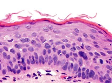
14 Dysplasia A spectrum of architectural and cytological epithelial changes caused by an accumulation of genetic changes that is associated with an increased likelihood of progression to squamous cell carcinoma Uncommon Age: Adults Sex: Male > Female (4.6:1) Etiologically related to alcohol and tobacco smoking (synergistic), gastroesophageal reflux with HPV a very minor role Dysplasia Most commonly along one vocal cord, although bilateral disease may occur Commissures are uncommonly involved Voice changes, hoarseness, sore throat, chronic cough Leukoplakia, erythroplakia or combination Diffuse, flat, exophytic to papillary Normal Update on New ENT WHO 14


15 Basal zone hyperplasia LG dysplasia LG dysplasia Update on New ENT WHO 15


16 LG dysplasia LG dysplasia HG dysplasia (moderate) Update on New ENT WHO 16


17 HG dysplasia (moderate) HG dysplasia HG dysplasia (severe) HG dysplasia (severe) HG dysplasia (severe) Update on New ENT WHO 17

 is a malignant epithelial neoplasm involving the oropharynx which includes: Soft")
18 HG dysplasia (severe) p Treatment and Outcome Biopsy, stripping, laser, cryotherapy and radiation variably employed Malignant progression: Low grade: 2% High grade: % (CIS is 40%) Anatomic site, multifocality, age, comorbidities (including alcohol and tobacco) contribute to progression Oropharyngeal Carcinoma Definition Oropharyngeal squamous cell carcinoma (OPSCC) is a malignant epithelial neoplasm involving the oropharynx which includes: Soft palate Tonsils and adenoids (Waldeyer ring) Uvula Base of tongue Oropharyngeal wall Update on New ENT WHO 18


19 Sagittal Axial Environmental Exposure Etiology/Pathogenesis Marijuana use is greater in HPV+ OPSCC Tobacco smoking and alcohol use greater in HPV- OPSCC, but still a major factor in HPV+ OPSCC Infectious Agents High-risk HPV associated with >80% of cases OPSCC HPV 16 is the predominant type Other HPV high-risk types are reported Epidemiology OPSCC increased 1-2% annually in USA males in past 20 years In USA, 225% increase in HPV+ OPSCC between ~13,000 new cases/year in US It is a sexually transmitted disease: Higher than average sex history Oral sex, multiple partners Epidemiology Sex: Male (~95%) Age: s Race: Caucasian History of smoking Mostly light or former Presentation Site: Anterior tonsillar pillar and fossa most common Tongue base Presentation Early lesions generally asymptomatic Tonsillar asymmetry Dysphagia Otalgia Trismus Enlarging cervical lymph node Often presenting symptom > 70% of patients present with stage III or IV disease Update on New ENT WHO 19
 &/or magnetic")


20 Imaging Findings Computed tomography (CT) &/or magnetic resonance imaging (MR) for preoperative tumor staging and planning Chest CT or plain film to rule out lung metastases Positron emission tomography (PET) useful particularly when dealing with unknown primary or in evaluating distant metastases Distant metastases uncommon in oral cavity cancer at presentation Update on New ENT WHO 20
 larger than 6 cm N N Criteria for Pathology Node Assessment NX Regional lymph nodes cannot be")

21 AJCC 8 th Edition Oropharynx N NX N0 N1 N2 N3 N Criteria for Clinical Node Assessment Regional lymph nodes cannot be assessed No regional lymph node metastasis One or more ipsilateral lymph nodes, none larger than 6 cm Contralateral or bilateral lymph nodes, none larger than 6 cm Lymph node(s) larger than 6 cm N N Criteria for Pathology Node Assessment NX Regional lymph nodes cannot be assessed pn0 No regional lymph node metastasis pn1 Metastasis in 4 or fewer lymph nodes pn2 Metastasis in more than 4 lymph nodes Squamous Cell Carcinoma There are 3 major carcinoma types in the upper aerodigestive tract that do not progress through dysplastic precursors Basaloid squamous cell carcinoma Nonkeratinizing squamous carcinoma Lymphoepithelial carcinoma Microscopic Features HPV positive types Oropharyngeal SCC, non-keratinizing Oropharyngeal SCC, keratinizing HPV negative types Oropharyngeal SCC, non-keratinizing Oropharyngeal SCC, keratinizing HPV Detection Methods Oncogenesis of HPV Show biologically or transcriptionally-active HPV Polymerase chain reaction RT-PCR for high risk E6/E7 mrna In situ hybridization Multiplexed (High risk vs. low risk) Type specific probes Other methods Hybrid capture (cytology samples) Other technologies p16 immunohistochemistry p16 Cyclin D1 p21 Apoptosis E7 Rb E2F ARF p53 E6 S-phase MDM2 125 HPV protein E7 degrades the retinoblastoma protein leading to aberrant overexpression of p Courtesy Dr. J. L. Hunt Update on New ENT WHO 21
 NP NP NP NP 2005 Weinberger, JCO 2006 18 17 (94%) NP NP NP NP Reimer, Int J Cancer 2007 29 25 (86%) NP NP NP NP Ang, NEJM")
 NP NP NP NP 147/148 (99%) Schlecht, Mod Pathol 2011 11 NP 6/10 (60%) NP 10 (90.9%) NP Thavaraj, J Clin Pathol 2011 90 NP 75 (83.")
 NP NP NP Totals 730 57/63 (90%) 555/675 (82%) 249/277 (89%) 10/11 (90%) 147/148 (99%) 127 HPV Detection methods Approximately 5%")
 gives best results Must be >70% nuclear &")
 Tumor often seen arising from epithelium of tonsillar crypts rather than overlying epithelium Basaloid oval to")

22 Study # of p16 Positive Patients* HPV DNA PCR HPV DNA ISH HPV DNA ISH & DNA PCR HPV RNA RTPCR HPV RNA ISH Dahlstrand, Anticancer Res (93%) NP NP NP NP 2005 Weinberger, JCO (94%) NP NP NP NP Reimer, Int J Cancer (86%) NP NP NP NP Ang, NEJM NP 192 (93%) NP NP NP Lewis, AJSP NP 139 (74%) Ukpo, AJSP NP 119/157 (75%) 161 (86.1%) NP NP NP NP 147/148 (99%) Schlecht, Mod Pathol NP 6/10 (60%) NP 10 (90.9%) NP Thavaraj, J Clin Pathol NP 75 (83.3%) 88 (97.8%) NP NP Doxtader, Hum Pathol NP 24 (96.0%) NP NP NP Totals /63 (90%) 555/675 (82%) 249/277 (89%) 10/11 (90%) 147/148 (99%) 127 HPV Detection methods Approximately 5% of p16 positive were HPV- However, p16 may be over-expressed by another mechanism Approximately 2% of p16 negative cases were HPV+ p16 is a sensitive marker for transcriptionally active HPV Clone E6H4 (MTM Laboratories) gives best results Must be >70% nuclear & cytoplasmic positive 128 WHO Classification Book-2017 p Microscopic Features HPV-positive OPSCC OPSCC, Nonkeratinizing type (75% of cases) Tumor often seen arising from epithelium of tonsillar crypts rather than overlying epithelium Basaloid oval to spindle-shaped cells with hyperchromatic nuclei and minimal cytoplasm forming trabeculae, sheets, or nests with sharply defined borders Comedo-necrosis frequently present Brisk mitotic rate and numerous scattered apoptotic cells Permeated by lymphocytes Squamous maturation and focal areas of keratinization can be seen but should comprise <10% Update on New ENT WHO 22


23 Microscopic Features HPV-positive OPSCC OPSCC, Lymphoepithelial-like Similar in histology to EBV-related nasopharyngeal carcinoma Syncytial-appearing large tumor cells with indistinct cell borders and vesicular nuclei intermingled with lymphocytes and plasma cells OPSCC, Papillary Exceedingly uncommon morphologic variant of SCC that can occur in oropharynx Finger-like projections of cytologically malignant epithelial cells with fibrovascular cores p Update on New ENT WHO 23


24 p16 p p OPSCC, Keratinizing Microscopic Features HPV-negative OPSCC Exhibits features of conventional-type SCC, including nests of epithelial cells with abundant eosinophilic cytoplasm and welldefined cell borders Frank keratinization present Basaloid morphology not seen Update on New ENT WHO 24
 p16 HPV 84% 16% 74% 26% Keratinizing")


25 p Reaction Positive Negative Positive Negative Nonkeratinizing (54%) 98% 2 % 88% 12% Tumor Type: p16 and HPV reactions Nonkeratinizing with maturation (21%) p16 HPV 84% 16% 74% 26% Keratinizing (25%) 19% 81% 21% Immunohistochemistry p16 strongly positive in HPV-associated OPSCC >70% both nuclear & cytoplasmic staining of tumor cells Normal epithelium is negative or shows minimal patchy staining p16 considered reliable surrogate marker for high risk BE AWARE: p16+ alone DOES NOT equal oropharyngeal carcinoma! There are many lesions that can be p16 positive 79% Treatment & Prognosis Approaches depend on clinical stage Tonsillectomy for small T1 tumors confined to tonsil Radiation therapy, specifically intensity-modulated radiation therapy (IMRT) (including brachytherapy) Concurrent radiotherapy with multiagent chemotherapy Targeted agents such as cetuximab Prognosis HPV-positive OPSCC associated with improved survival outcomes Tumor size and presence of metastases influence prognosis p Update on New ENT WHO 25


 Report p16 as part of the original report 155 Base of Tongue")
26 Prognosis Radiation: HPV+ HPV- 5 year 62% 26% Tobacco use: Decreased survival + Nodal status: Decreased survival Patients can be stratified into deescalation therapies E6 and E7 oncoprotein can be targeted, restoring p53 and retinoblastoma tumor suppressor pathways (degraded by E6/E7) Survival: Radiation vs. Chemotherapy Radiotherapy and Oncology, Volume 103, Issue 1, 2012, Pertinent Issues Do not diagnose OPSCC as in-situ Do not give tumor grades for OPSCC Report: Oropharyngeal squamous cell carcinoma Non-keratinizing; with maturation; or keratinizing types (for clarity) Report p16 as part of the original report 155 Base of Tongue p16156 Update on New ENT WHO 26


27 HPV-associated Neuroendocrine Oropharyngeal carcinoma HPV-associated Neuroendocrine Oropharyngeal carcinoma 157 Synaptophysin p16158 Branchial Cleft Cyst with p16 p Update on New ENT WHO 27
Salivary Glands 3/7/2017
 Salivary Glands 3/7/2017 Goals and objectives Focus on the entities unique to H&N Common board type facts Information for your future practice Salivary Glands Salivary Glands Major gland. Paratid. Submandibular.
Nasal Cavity and Paranasal Sinuses
 Chapter 2 Nasal Cavity and Paranasal Sinuses Introduction Included in this chapter are nasal cavities, frontal sinus, ethmoid complex, sphenoid sinus, and maxillary sinuses. These cavities and sinuses
Small (and large) Blue Cell Tumors of the Skull Base
 Blue Cell Tumors of the Skull Base") Small (and large) Blue Cell Tumors of the Skull Base Jennifer L. Hunt, MD, MEd Aubrey J. Hough Jr, MD, Endowed Professor of Pathology Chair of Pathology and Laboratory Medicine University of Arkansas for
Small (and large) Blue Cell Tumors of the Skull Base Jennifer L. Hunt, MD, MEd Aubrey J. Hough Jr, MD, Endowed Professor of Pathology Chair of Pathology and Laboratory Medicine University of Arkansas for
Lesions Mimicking Adenoid Cystic Carcinoma. Diagnostic Problems in Salivary Gland Pathology An Update 5/29/2009
 Diagnostic Problems in Salivary Gland Pathology An Update Lesions Mimicking Adenoid Cystic Carcinoma Stacey E. Mills, M.D. W.S. Royster Professor of Pathology Director of Surgical and Cytopathology University
Basaloid neoplasms of the head and neck. Basaloid SCC. Clinico-pathologic features 5/5/11. Basaloid Tumors Head and Neck
 Basaloid neoplasms of the head and neck Richard Jordan DDS PhD FRCPath Professor & Director UCSF Oral Pathology Laboratory University of California San Francisco Basaloid Tumors Head and Neck Basaloid
Human Papillomavirus and Head and Neck Cancer. Ed Stelow, MD
 Human Papillomavirus and Head and Neck Cancer Ed Stelow, MD No conflict of interest Declaration Cancer 1974 Lancet Oncol 2016; 17: e477-8 JAMA 1984; 252: 1857 JAMA 1988;259(13):1943-1944 Clin Cancer Res
Mody. AIS vs. Invasive Adenocarcinoma of the Cervix
 Common Problems in Gynecologic Pathology Michael T. Deavers, M.D. Houston Methodist Hospital, Houston, Texas Common Problems in Gynecologic Pathology Adenocarcinoma in-situ (AIS) of the Cervix vs. Invasive
Case 2. Dr. Sathima Natarajan M.D. Kaiser Permanente Medical Center Sunset
 Case 2 Dr. Sathima Natarajan M.D. Kaiser Permanente Medical Center Sunset History 24 year old male presented with a 3 day history of right flank pain, sharp in nature Denies fever, chills, hematuria or
Did President Grant die from too much Oral Sex?
 Case 6 Did President Grant die from too much Oral Sex? Lester D. R. Thompson www.lester-thompson.com 53 year old man Small nodule noted 2 months ago, just below the angle of the jaw during shaving Non-tender
Pitfalls in Sinonasal Pathology
 Pitfalls in Sinonasal Pathology Jennifer L. Hunt, MD, MEd Aubrey J. Hough Jr, MD, Endowed Professor of Pathology Chair of Pathology and Laboratory Medicine University of Arkansas for Medical Sciences jhunt2@uams.edu
Head and Neck Squamous Subtypes
 1 Head and Neck Squamous Subtypes Adel K. El-Naggar, M.D., Ph.D. The University of Texas MD Anderson Cancer Center, Houston, Texas HNSCC 5 th -6 th most common cancer 400,000/year 50% mortality Considerable
Note: The cause of testicular neoplasms remains unknown
 - In the 15- to 34-year-old age group, they are the most common tumors of men. - Tumors of the testis are a heterogeneous group of neoplasms that include: I. Germ cell tumors : 95%; all are malignant.
04/09/2018. Salivary Gland Pathology in the Molecular Era Old Friends, Old Foes, & New Acquaintances
 Salivary Gland Pathology in the Molecular Era Old Friends, Old Foes, & New Acquaintances Jennifer L. Hunt, MD, MEd Aubrey J. Hough Jr, MD, Endowed Professor of Pathology Chair of Pathology and Laboratory
Scotland and Northern Ireland EQA Scheme. Circulation 46
 Scotland and Northern Ireland EQA Scheme Circulation 46 Special Educational Cases E1 and E2 Presented by Dr K Robertson Case E1 Female 42 year old with heavy menstrual and intermenstrual bleeding. IUS
Synonyms. Nephrogenic metaplasia Mesonephric adenoma
 Nephrogenic Adenoma Synonyms Nephrogenic metaplasia Mesonephric adenoma Definition Benign epithelial lesion of urinary tract with tubular, glandular, papillary growth pattern Most frequently in the urinary
Papillary Lesions of the Breast A Practical Approach to Diagnosis. (Arch Pathol Lab Med. 2016;140: ; doi: /arpa.
 Papillary Lesions of the Breast A Practical Approach to Diagnosis (Arch Pathol Lab Med. 2016;140:1052 1059; doi: 10.5858/arpa.2016-0219-RA) Papillary lesions of the breast Span the spectrum of benign,
Salivary Gland Cytology
 Salivary Gland Cytology Diagnostic challenges and potential pitfalls Tarik M. Elsheikh, MD Professor and Medical Director Anatomic Pathology Cleveland Clinic FNA Salivary Gland Lesions Indications Distinguish
Molecular Diagnostics of Head and Neck Tumors Justin A. Bishop, M.D. Associate Professor of Pathology The Johns Hopkins University Baltimore, Maryland
 Molecular Diagnostics of Head and Neck Tumors Justin A. Bishop, M.D. Associate Professor of Pathology The Johns Hopkins University Baltimore, Maryland Two Main Topics Molecular insights in salivary gland
Case Presentation. Maha Akkawi, MD, Fatima Obeidat, MD, Tariq Aladily, MD. Department of Pathology Jordan University Hospital Amman, Jordan
 Case Presentation Maha Akkawi, MD, Fatima Obeidat, MD, Tariq Aladily, MD Department of Pathology Jordan University Hospital Amman, Jordan The 25th Annual Congress of the ADIAP The 8/11/2013 1 5th International
Respiratory Tract Cytology
 Respiratory Tract Cytology 40 th European Congress of Cytology Liverpool, UK Momin T. Siddiqui M.D. Professor of Pathology and Laboratory Medicine Director of Cytopathology Emory University Hospital, Atlanta,
Epithelial tumors. Dr. F.F. Khuzin, PhD Dr. M.O. Mavlikeev
 Epithelial tumors Dr. F.F. Khuzin, PhD Dr. M.O. Mavlikeev Epithelial tumors Tumors from the epithelium are the most frequent among tumors. There are 2 group features of these tumors: The presence in most
ARTHUR PURDY STOUT SOCIETY COMPANION MEETING: DIFFICULT NEW DIFFERENTIAL DIAGNOSES IN PROSTATE PATHOLOGY. Jonathan I. Epstein.
 1 ARTHUR PURDY STOUT SOCIETY COMPANION MEETING: DIFFICULT NEW DIFFERENTIAL DIAGNOSES IN PROSTATE PATHOLOGY Jonathan I. Epstein Professor Pathology, Urology, Oncology The Reinhard Professor of Urological
04/13/2017. Sinonasal Pathology. Sinonasal Tract Pathology Outline. 24 th Annual Seminar in Pathology Pittsburgh, PA April 27, 2017
 Sinonasal Pathology 24 th Annual Seminar in Pathology Pittsburgh, PA April 27, 2017 Bruce M. Wenig, MD Moffitt Cancer Center Tampa, FL Sinonasal Tract Pathology Outline Sinonasal (Schneiderian) papillomas
Difficult Diagnoses and Controversial Entities in Neoplastic Lung
 Difficult Diagnoses and Controversial Entities in Neoplastic Lung Lynette M. Sholl, M.D. Associate Pathologist, Brigham and Women s Hospital Chief, Pulmonary Pathology Service Associate Professor, Harvard
Diplomate of the American Board of Pathology in Anatomic and Clinical Pathology
 A 33-year-old male with a left lower leg mass. Contributed by Shaoxiong Chen, MD, PhD Assistant Professor Indiana University School of Medicine/ IU Health Partners Department of Pathology and Laboratory
THYMIC CARCINOMAS AN UPDATE
 THYMIC CARCINOMAS AN UPDATE Mark R. Wick, M.D. University of Virginia Medical Center Charlottesville, VA CARCINOMA OF THE THYMUS General Clinical Features No apparent gender predilection Age range of 35-75
Gross appearance of nodular hyperplasia in material obtained from suprapubic prostatectomy. Note the multinodular appearance and the admixture of
 Tiền liệt tuyến Tiền liệt tuyến Gross appearance of nodular hyperplasia in material obtained from suprapubic prostatectomy. Note the multinodular appearance and the admixture of solid and microcystic areas.
Case Report SMARCB1 (INI1)-deficient sinonasal carcinoma: a newly described entity
-deficient sinonasal carcinoma: a newly described entity") Int J Clin Exp Pathol 2016;9(3):3454-3458 www.ijcep.com /ISSN:1936-2625/IJCEP0024095 Case Report SMARCB1 (INI1)-deficient sinonasal carcinoma: a newly described entity Ming Zeng 1*, Changrong Chen 2*,
Int J Clin Exp Pathol 2016;9(3):3454-3458 www.ijcep.com /ISSN:1936-2625/IJCEP0024095 Case Report SMARCB1 (INI1)-deficient sinonasal carcinoma: a newly described entity Ming Zeng 1*, Changrong Chen 2*,
Enterprise Interest Nothing to declare
 Enterprise Interest Nothing to declare Diagnoses one would not like to miss in soft tissue pathology early in your career Marta Sbaraglia, MD Department of Pathology Hospital of Treviso University of Padua
HPV and Head and Neck Cancer: What it means for you and your patients
 HPV and Head and Neck Cancer: What it means for you and your patients Financial Disclosure: None November 8, 2013 Steven J. Wang, MD Associate Professor Department of Otolaryngology-Head and Neck Surgery
Differential Diagnosis of Oral Masses. Palatal Lesions
 Differential Diagnosis of Oral Masses Palatal Lesions Palatal Masses Periapical Abscess Torus Palatinus Mucocele Lymphoid Hyperplasia Adenomatous Hyperplasia Benign Salivary Neoplasms Malignant Salivary
Sarcomatoid (spindle cell) carcinoma of the cricopharynx presenting as dysphagia
 carcinoma of the cricopharynx presenting as dysphagia") Case Report Sarcomatoid (spindle cell) carcinoma of the cricopharynx presenting as dysphagia Jagtap Sunil V. 1, Shukla Dhirajkumar B. 2, Jagtap Swati S. 3, Havle Abhay D. 4 1 Associate Professor, Department
Case Report Sarcomatoid (spindle cell) carcinoma of the cricopharynx presenting as dysphagia Jagtap Sunil V. 1, Shukla Dhirajkumar B. 2, Jagtap Swati S. 3, Havle Abhay D. 4 1 Associate Professor, Department
4/12/2018. MUSC Pathology Symposium Kiawah Island April 18, Jesse K. McKenney, MD
 MUSC Pathology Symposium Kiawah Island April 18, 2018 Jesse K. McKenney, MD 1 Urothelial Carcinoma with Alternative Differentiation 2 Urothelial Carcinoma with Alternative Differentiation Recognition as
Oncocytic-Appearing Salivary Gland Tumors. Oncocytic, Cystic, Mucinous, and High Grade Salivary Gland Tumors SALIVARY GLAND FNA: PART II
 William C. Faquin, MD, PhD Professor of Pathology Harvard Medical School Director of Head and Neck Pathology Massachusetts Eye and Ear Massachusetts General Hospital SALIVARY GLAND FNA: PART II Oncocytic,
Presentation material is for education purposes only. All rights reserved URMC Radiology Page 1 of 98
 Presentation material is for education purposes only. All rights reserved. 2011 URMC Radiology Page 1 of 98 Radiology / Pathology Conference February 2011 Brooke Koltz, Cytopathology Resident Presentation
What is New in the 2015 WHO Lung Cancer Classification? Zhaolin Xu, MD, FRCPC, FCAP
 What is New in the 2015 WHO Lung Cancer Classification? Zhaolin Xu, MD, FRCPC, FCAP Professor, Dept of Pathology, Dalhousie University, Canada Pulmonary Pathologist and Cytopathologist, QEII HSC Senior
Nutn but a Small Biopsy How to approach small biopsy samples of the sinonasal tract
 California Society of Pathology Saturday Slide Seminar Nutn but a Small Biopsy How to approach small biopsy samples of the sinonasal tract Lester D. R. Thompson www.lester-thompson.com Learning Objectives
Pathology of Selected Head and Neck Lesions. Adel Assaad MD Department of Pathology
 Pathology of Selected Head and Neck Lesions Adel Assaad MD Department of Pathology 1 NOSE Infections 2 Zygomycosis (Mucormycosis) Opportunistic infection caused by "bread mold fungi," including Rhizopus,
04/10/2018. Intraductal Papillary Neoplasms Of Breast INTRADUCTAL PAPILLOMA
 Intraductal Papillary Neoplasms Of Breast Savitri Krishnamurthy MD Professor of Pathology Deputy Division Head The University of Texas MD Anderson Cancer Center 25 th Annual Seminar in Pathology Pittsburgh,
Diseases of the breast (1 of 2)
") Diseases of the breast (1 of 2) Introduction A histology introduction Normal ducts and lobules of the breast are lined by two layers of cells a layer of luminal cells overlying a second layer of myoepithelial
Diseases of the breast (1 of 2) Introduction A histology introduction Normal ducts and lobules of the breast are lined by two layers of cells a layer of luminal cells overlying a second layer of myoepithelial
Human Papillomavirus Testing in Head and Neck Carcinomas
 Human Papillomavirus Testing in Head and Neck Carcinomas Guideline from the College of American Pathologists Early Online Release Publication: Archives of Pathology & Laboratory Medicine 12/18/2017 Overview
Keratinizing Dysplasia and Select Variants of Head & Neck Squamous Cell Carcinoma
 Keratinizing Dysplasia and Select Variants of Head & Neck Squamous Cell Carcinoma Napa Valley Pathology Conference Silverado Resort & Spa May 18, 2018 Bruce M. Wenig, MD Moffitt Cancer Center Tampa, FL
57th Annual HSCP Spring Symposium 4/16/2016
 An Unusual Malignant Spindle Cell Lesion to Involve the Breast Erinn Downs-Kelly, D.O. Associate Professor of Pathology University of Utah & ARUP Laboratories No disclosures Case 39 y/o female with no
Case year old female presented with asymmetric enlargement of the left lobe of the thyroid
 Case 4 22 year old female presented with asymmetric enlargement of the left lobe of the thyroid gland. No information available relative to a prior fine needle aspiration biopsy. A left lobectomy was performed.
Case 4 Diagnosis 2/21/2011 TGB
 Case 4 22 year old female presented with asymmetric enlargement of the left lobe of the thyroid gland. No information available relative to a prior fine needle aspiration biopsy. A left lobectomy was performed.
Proliferative Epithelial lesions of the Breast. Sami Shousha, MD, FRCPath Charing Cross Hospital & Imperial College, London
 Proliferative Epithelial lesions of the Breast Sami Shousha, MD, FRCPath Charing Cross Hospital & Imperial College, London Amman, November2013 Proliferative Epithelial Lesions of the Breast Usual type
ACCME/Disclosures ALK FUSION-POSITIVE MESENCHYMAL TUMORS. Tumor types with ALK rearrangements. Anaplastic Lymphoma Kinase. Jason L.
 Companion Meeting of the International Society of Bone and Soft Tissue Pathology The Evolving Concept of Mesenchymal Tumors ALK FUSION-POSITIVE MESENCHYMAL TUMORS Jason L. Hornick, MD, PhD March 13, 2016
Basement membrane in lobule.
 Bahram Memar, MD Basement membrane in lobule. Normal lobule-luteal phase Normal lobule-follicular phase Lactating breast Greater than 95% are adenocarcinomas in situ carcinomas and invasive carcinomas.
Evening Specialty Conference Bone and Soft Tissue Pathology. Diagnostic pitfalls in bone and soft tissue pathology
 Evening Specialty Conference Bone and Soft Tissue Pathology. Case 1 Elizabeth G Demicco, MD, PhD Mount Sinai Hospital, New York Disclosure of Relevant Financial Relationships USCAP requires that all planners
Reporting HPV related carcinomas of the head and neck. dr. Nina Zidar Institute of Pathology Faculty of Medicine University of Ljubljana Slovenia
 Reporting HPV related carcinomas of the head and neck dr. Nina Zidar Institute of Pathology Faculty of Medicine University of Ljubljana Slovenia Conflict of interest/funding X None Company: Product royalties
CLINICAL SIGNIFICANCE OF BENIGN EPITHELIAL CHANGES
 Papillomas. Papillomas are composed of multiple branching fibrovascular cores, each having a connective tissue axis lined by luminal and myoepithelial cells ( Fig. 23-11 ). Growth occurs within a dilated
Update in Salivary Gland Pathology. Benjamin L. Witt University of Utah/ARUP Laboratories February 9, 2016
 Update in Salivary Gland Pathology Benjamin L. Witt University of Utah/ARUP Laboratories February 9, 2016 Objectives Review the different appearances of a selection of salivary gland tumor types Establish
Moffitt Weekends in Pathology Head & Neck, and Endocrine Pathology Course Outline
 Moffitt Weekends in Pathology Head & Neck, and Endocrine Pathology Course Outline Squamous Cell Lesions Lecture BMW: 8:30-9:15 Break: 9:15-9:30 Case Review LK: 9:30-10:15 Case Review JHP: 10:30-11:15 Break:
Cutaneous Mesenchymal Neoplasms with EWSR1 Rearrangement
 Cutaneous Mesenchymal Neoplasms with EWSR1 Rearrangement By Konstantinos Linos MD, FCAP, FASDP Bone, Soft Tissue and Dermatopathology Assistant Professor of Pathology Dartmouth-Hitchcock Medical Center
Objectives. Atypical Glandular Cells. Atypical Endocervical Cells. Reactive Endocervical Cells
 2013 California Society of Pathologists 66 th Annual Meeting San Francisco, CA Atypical Glandular Cells to Early Invasive Adenocarcinoma: Cervical Cytology and Histology Christina S. Kong, MD Associate
Pancreatitis: A Potential Pitfall in Endoscopic Ultrasound Guided Pancreatic FNA
 Pancreatitis: A Potential Pitfall in Endoscopic Ultrasound Guided Pancreatic FNA Jack Yang, MD Department of Pathology, Medical University of South Carolina Objectives Understand the indication of EUS
FNA OF SALIVARY GLANDS: A PRACTICAL APPROACH
 FNA OF SALIVARY GLANDS: A PRACTICAL APPROACH FNA of Salivary Glands: Challenges Wide range of neoplastic and non-neoplastic lesions Cytological overlap between the different benign and malignant tumors
CINtec p16 INK4a Staining Atlas
 CINtec p16 INK4a Staining Atlas Rating Rating Positive The rating positive will be assigned if the p16 INK4a -stained slide shows a continuous staining of cells of the basal and parabasal cell layers of
Papillary Lesions of the breast
 Papillary Lesions of the breast Emad Rakha Professor of Breast Pathology The University of Nottingham Papillary lesions of the breast are a heterogeneous group of disease, which are characterised by neoplastic
5/21/2018. Prostate Adenocarcinoma vs. Urothelial Carcinoma. Common Differential Diagnoses in Urological Pathology. Jonathan I.
 Common Differential Diagnoses in Urological Pathology Jonathan I. Epstein Prostate Adenocarcinoma vs. Urothelial Carcinoma 1 2 NKX3.1 NKX3.1 3 4 5 6 Proposed ISUP Recommendations Option to use PSA as a
Columnar Cell Lesions
 Columnar Cell Lesions Laura C. Collins, M.D. Department of Pathology Beth Israel Deaconess Medical Center and Harvard Medical School Boston, MA Question? Columnar cell lesions are: a) Annoying lesions
3/27/2017. Pulmonary Pathology Specialty Conference. Disclosure of Relevant Financial Relationships. Clinical History:
 Pulmonary Pathology Specialty Conference Saul Suster, M.D. Medical College of Wisconsin Disclosure of Relevant Financial Relationships USCAP requires that all planners (Education Committee) in a position
Notice of Faculty Disclosures
 William C. Faquin, MD, PhD Professor of Pathology Harvard Medical School Director of Head and Neck Pathology Massachusetts Eye and Ear Massachusetts General Hospital FNA OF SQUAMOUS CYSTS OF THE HEAD AND
4/17/2015. Case 1. A 37 year old man with a 2.2 cm solitary left thyroid mass.
 Case 1 A 37 year old man with a 2.2 cm solitary left thyroid mass. Case 1 Case 1 1 Case 1: Diagnosis? A. Benign B. Atypia of undetermined significance/follicular lesion of undetermined significance C.
Carcinoma of Unknown Primary site (CUP) in HEAD & NECK SURGERY
 in HEAD & NECK SURGERY") Carcinoma of Unknown Primary site (CUP) in HEAD & NECK SURGERY SEARCHING FOR THE PRIMARY? P r o f J P P r e t o r i u s H e a d : C l i n i c a l U n i t C r i t i c a l C a r e U n i v e r s i t y O f
Carcinoma of Unknown Primary site (CUP) in HEAD & NECK SURGERY SEARCHING FOR THE PRIMARY? P r o f J P P r e t o r i u s H e a d : C l i n i c a l U n i t C r i t i c a l C a r e U n i v e r s i t y O f
Objectives. Salivary Gland FNA: The Milan System. Role of Salivary Gland FNA 04/26/2018
 Salivary Gland FNA: The Milan System Dr. Jennifer Brainard Section Head Cytopathology Cleveland Clinic Objectives Introduce the Milan System for reporting salivary gland cytopathology Define cytologic
3/24/2017 DENDRITIC CELL NEOPLASMS: HISTOLOGY, IMMUNOHISTOCHEMISTRY, AND MOLECULAR GENETICS. Disclosure of Relevant Financial Relationships
 DENDRITIC CELL NEOPLASMS: HISTOLOGY, IMMUNOHISTOCHEMISTRY, AND MOLECULAR GENETICS Jason L. Hornick, M.D., Ph.D. Director of Surgical Pathology and Immunohistochemistry Brigham and Women s Hospital Professor
FINALIZED SEER SINQ QUESTIONS
 0076 Source 1: WHO Class CNS Tumors pgs: 33 MP/H Rules/Histology--Brain and CNS: What is the histology code for a tumor originating in the cerebellum and extending into the fourth ventricle described as
04/09/2018. Squamous Cell Neoplasia and Precursor Lesions. Agenda. Squamous Dysplasia. Squamo-proliferative lesions. Architectural features
 Squamous Cell Neoplasia and Precursor Lesions Jennifer L. Hunt, MD, MEd Aubrey J. Hough Jr, MD, Endowed Professor of Pathology Chair of Pathology and Laboratory Medicine University of Arkansas for Medical
PRELIMINARY CYTOLOGIC DIAGNOSIS: Suspicious for Acinic Cell Carcinoma. Cell Block: Immunohistochemical Studies CYTOLOGIC DIAGNOSIS:
 1 PRELIMINARY CYTOLOGIC DIAGNOSIS: Suspicious for Acinic Cell Carcinoma. Cell Block: Immunohistochemical Studies GCDFP-15 S-100 CYTOLOGIC DIAGNOSIS: Consistent with mammary analogue secretory carcinoma.
Polymorphous Low-Grade. December 5 th, 2008
 Polymorphous Low-Grade Adenocarcinoma December 5 th, 2008 Epidemiology Represents 2 nd or 3 rd most common minor salivary gland malignancy (17-26%) 1 st mucoepidermoid carcinoma Rare in reported Asian
Breast pathology. 2nd Department of Pathology Semmelweis University
 Breast pathology 2nd Department of Pathology Semmelweis University Breast pathology - Summary - Benign lesions - Acute mastitis - Plasma cell mastitis / duct ectasia - Fat necrosis - Fibrocystic change/
Benign and malignant epithelial lesions: Seborrheic keratosis: A common benign pigmented epidermal tumor occur in middle-aged or older persons more
 Benign and malignant epithelial lesions: Seborrheic keratosis: A common benign pigmented epidermal tumor occur in middle-aged or older persons more common on the trunk; but extremities, head and neck are
Disclosures. The Thin Red Line Between Neuropathology and Head & Neck Pathology. Introduction CASE 1. Current Issues Tihan
 Disclosures I have nothing to disclose The Thin Red Line Between Neuropathology and Head & Neck Pathology Tarik Tihan, MD, PhD UCSF, Department of Pathology Neuropathology Division Introduction Three cases
My Journey into the World of Salivary Gland Sebaceous Neoplasms
 My Journey into the World of Salivary Gland Sebaceous Neoplasms Douglas R. Gnepp Warren Alpert Medical School at Brown University Rhode Island Hospital Pathology Department Providence RI Asked to present
Differential diagnosis of hematolymphoid tumors composed of medium-sized cells. Brian Skinnider B.C. Cancer Agency, Vancouver General Hospital
 Differential diagnosis of hematolymphoid tumors composed of medium-sized cells Brian Skinnider B.C. Cancer Agency, Vancouver General Hospital Lymphoma classification Lymphoma diagnosis starts with morphologic
Neoplasia 2018 Lecture 2. Dr Heyam Awad MD, FRCPath
 Neoplasia 2018 Lecture 2 Dr Heyam Awad MD, FRCPath ILOS 1. List the differences between benign and malignant tumors. 2. Recognize the histological features of malignancy. 3. Define dysplasia and understand
LARYNGEAL DYSPLASIA. Tomas Fernandez M; 3 rd year ENT resident, Son Espases University Hospital
 LARYNGEAL DYSPLASIA Tomas Fernandez M; 3 rd year ENT resident, Son Espases University Hospital INTRODUCTION Laryngeal cancer constitutes 1-2% of all malignancies diagnosed worldwide Survival is related
Kidney Case 1 SURGICAL PATHOLOGY REPORT
 Kidney Case 1 Surgical Pathology Report February 9, 2007 Clinical History: This 45 year old woman was found to have a left renal mass. CT urography with reconstruction revealed a 2 cm medial mass which
Non Small Cell Lung Cancer Histopathology ד"ר יהודית זנדבנק
 Non Small Cell Lung Cancer Histopathology ד"ר יהודית זנדבנק 26.06.09 Lecture outlines WHO histological classification Macro/Micro assessment Early diagnosis Minimal pathology Main subtypes SCC, AdCa, LCLC
A 25 year old female with a palpable mass in the right lower quadrant of her abdomen
 May 2016 A 25 year old female with a palpable mass in the right lower quadrant of her abdomen Contributed by: Paul Ndekwe, MD, Resident Physician, Indiana University School of Department of Pathology and
Normal endometrium: A, proliferative. B, secretory.
 Normal endometrium: A, proliferative. B, secretory. Nội mạc tử cung Nội mạc tử cung Cyclic changes in endometrium.. Approximate relationship of useful microscopic changes. Arias-Stella reaction in endometrial
When Immunostains Can Get You in Trouble: Gynecologic Pathology p16: Panacea or Pandora s Box?
 When Immunostains Can Get You in Trouble: Gynecologic Pathology p16: Panacea or Pandora s Box? Teri A. Longacre, MD Stanford Medicine Stanford California pi6 in Gynecologic Pathology: Panacea or Pandora
2018 SEER Solid Tumor Manual 2018 KCR SPRING TRAINING
 2018 SEER Solid Tumor Manual 2018 KCR SPRING TRAINING Eight Groups are Revised for 2018 Head & Neck Colon (includes rectosigmoid and rectum for cases diagnosed 1/1/2018 forward) Lung (2018 Draft not yet
Biliary tract tumors
 Short Course 2010 Annual Fall Meeting of the Korean Society for Pathologists Biliary tract tumors Joon Hyuk Choi, M.D., Ph.D. Professor, Department of Pathology, Yeungnam Univ. College of Medicine, Daegu,
5/22/2018. Keratinizing Dysplasia and Select Variants of Head & Neck Squamous Cell Carcinoma
 Keratinizing Dysplasia and Select Variants of Head & Neck Squamous Cell Carcinoma Napa Valley Pathology Conference Silverado Resort & Spa May 18, 2018 Bruce M. Wenig, MD Moffitt Cancer Center Tampa, FL
DISCUSSION: PLGA accounts for about 2% of all salivary gland tumours and occurs almost exclusively in the minor salivary glands.
 SWELLING ON THE HARD PALATE PRESENTING AS POLYMORPHOUS LOW GRADE ADENOCARCINOMA: A AND REVIEW OF LITERATURE Swapnil D. Chandekar 1, Sunita S. Dantkale 2, Rahul R. Narkhede 3, Snehal V. Chavhan 4, Khushboo
Neuroendocrine Carcinoma. Lebanon Neuroendocrine Neoplasms of H&N Nov /7/2011. Broad Classification:
 H&N Neuroendocrine Neoplasms: Classification and Diagnostic Considerations Adel K. El-Naggar, M.D., Ph.D. The University of Texas MD Anderson Cancer Center, Houston, Texas Broad Classification: A. Epithelial:
Disclosure. Relevant Financial Relationship(s) None. Off Label Usage None MFMER slide-1
 None. Off Label Usage None MFMER slide-1") Disclosure Relevant Financial Relationship(s) None Off Label Usage None 2013 MFMER slide-1 Case Presentation A 43 year old male, with partial nephrectomy for a right kidney mass 2013 MFMER slide-2 2013
Disclosure Relevant Financial Relationship(s) None Off Label Usage None 2013 MFMER slide-1 Case Presentation A 43 year old male, with partial nephrectomy for a right kidney mass 2013 MFMER slide-2 2013
Diagnostically Challenging Cases in Gynecologic Pathology
 Diagnostically Challenging Cases in Gynecologic Pathology Eric C. Huang, M.D., Ph.D. Department of Pathology and Laboratory Medicine University of California, Davis Medical Center Case 1 Presentation 38
5/22/2017. An Aggressive Nasopharyngeal Tumor. Case History
 An Aggressive Nasopharyngeal Tumor Head & Neck/Endocrine Evening Specialty Conference Martin Bullock, MD, FRCPC Dalhousie University, Halifax, Nova Scotia Case History 52-year-old male, 6 month history
BREAST PATHOLOGY. Fibrocystic Changes
 BREAST PATHOLOGY Lesions of the breast are very common, and they present as palpable, sometimes painful, nodules or masses. Most of these lesions are benign. Breast cancer is the 2 nd most common cause
Macro- and microacinar proliferations of the prostate
 Macro- and microacinar proliferations of the prostate (with emphasis on cancer mimics) Rodolfo Montironi, MD (IT), FRCPath (UK), IFCAP (USA) Polytechnic University of Marche Region (Ancona) School of Medicine,
University Journal of Pre and Para Clinical Sciences
 ISSN 2455 2879 Volume 2 Issue 1 2016 Metaplastic carcinoma breast a rare case report Abstract : Metaplastic carcinoma of the breast is a rare malignancy with two distinct cell lines described as a breast
Dysplasia, Mimics and Other Controversies
 Dysplasia, Mimics and Other Controversies Mary S. Richardson, MD Dept. of Pathology Medical University of South Carolina Charleston, SC Notice of Faculty Disclosure In accordance with ACGME guidelines,
Management of Neck Metastasis from Unknown Primary
 Management of Neck Metastasis from Unknown Primary.. Definition Histologic evidence of malignancy in the cervical lymph node (s) with no apparent primary site of original tumour Diagnosis after a thorough
Urinary Bladder: WHO Classification and AJCC Staging Update 2017
 Urinary Bladder: WHO Classification and AJCC Staging Update 2017 Houston Society of Clinical Pathologists 58 th Annual Spring Symposium Houston, TX April 8, 2017 Jesse K. McKenney, MD Classification
Problem 1: Differential of Neuroendocrine Carcinoma 3/23/2017. Disclosure of Relevant Financial Relationships
 Differential of Neuroendocrine Carcinoma Alain C. Borczuk,MD Weill Cornell Medicine Disclosure of Relevant Financial Relationships USCAP requires that all faculty in a position to influence or control
Breast Pathology. Breast Development
 Breast Pathology Lecturer: Hanina Hibshoosh, M.D. Reading: Kumar, Cotran, Robbins, Basic Pathology, 6th Edition, pages 623-635 Breast Development 5th week - thickening of the epidermis - milk line 5th
Diseases of oral cavity
 Diseases of oral cavity Diseases of Teeth and Supporting Structures Inflammatory/Reactive Lesions Infections Oral Manifestations of Systemic Disease Precancerous and Cancerous Lesions Odontogenic Cysts