Microglia-derived extracellular vesicles regulate the proliferation and differentiation of oligodendrocyte precursor cells
|
|
|
- Solomon Haynes
- 5 years ago
- Views:
Transcription


1 University of Turin CNR Institute of Neuroscience Microglia-derived extracellular vesicles regulate the proliferation and differentiation of oligodendrocyte precursor cells Roberta Parolisi Turin, December 3, 2016

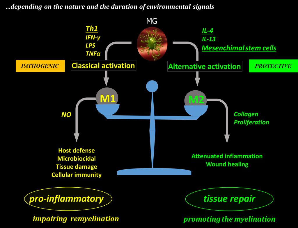
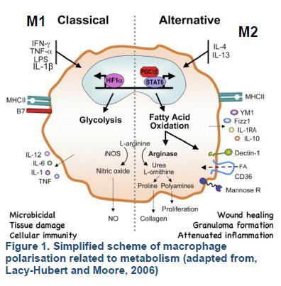
2 MICROGLIA & their roles Microglia (MG) are a type of small macrophage-like glial cells in the CNS. HOMEOSTASIS MG preserve homeostasis, and support neurons and glial cells M1 PATHOGENIC MG plays a role in disease progression, presenting antigens and secreting neurotoxic cytokines PROTECTIVE MG can suppress inflammation and cytodegeneration, and stimulate CNS repair, releasing trophic factors and anti-inflammatory cytokines Bogie et al., 2014
3


4 MICROGLIA-DERIVED EXTRACELLULAR VESICLES The phenotype of MG influences the production and the functions of EVs. EXTRACELLULAR VESICLES (EVs) EVs are small membranous vesicles. from plasma membrane The EVs have been related to several physiological and pathological conditions. exocytosis of multivesicular bodies Verderio et al., 2012 Microglia-derived EVs reflect disease activity in MS patients
5 WE STUDIED WHETHER EVS RELEASED FROM MICROGLIA CAN BOOST OR BLOCK THE PROLIFERATION AND THE TERMINAL DIFFERENTIATION OF OPCs. Oligodedrocytes precursor cells Oligodedrocytes cells Neuron OL OPC


6 METHODS IN VITRO and IN VIVO ANALYSIS PROLIFERATION AND DIFFERENTIATION OF OPC Lombardi M., Fumagalli M. and Verderio C.


7 1.EFFECTS OF MICROGLIA-DERIVED EVs ON OPC PROLIFERATION M0 M1 unstimulated pro-inflammatory+mscs pro-inflammatory anti-inflammatory M0 UNSTIMULATED METHODS / Th1 M0 M1 to evaluate the action of microglial Evs on OPC proliferation OPC culture Lombardi M., Fumagalli M. and Verderio C.


8 1.EFFECTS OF MICROGLIA-DERIVED EVs ON OPC PROLIFERATION M0 M1 unstimulated pro-inflammatory+mscs pro-inflammatory anti-inflammatory Ctrl M1 M0 M1 inhibit OPC proliferation M0 M1 Pre-conditioning with MSCs abrogates the anti-proliferative effects of M1 Lombardi M., Fumagalli M. and Verderio C.


9 2.EFFECTS OF MICROGLIA-DERIVED EVs ON OPC DIFFERENTIATION M0 M1 unstimulated pro-inflammatory anti-inflammatory M0 UNSTIMOLATED METHODS M0 M1 to evaluate the action of microglial EVs on OPC differentiation ATP OPC culture Lombardi M., Fumagalli M. and Verderio C.


10 MBP DAPI MBP positive OPCs/ DAPI normalized value 2.EFFECTS OF MICROGLIA-DERIVED EVs ON OPC DIFFENTIATION M0 M1 unstimulated pro-inflammatory anti-inflammatory MBP/HOECHST Ctrl M1-EVs -EVs M0 M1 M B P p o s itiv e O P C s /D A P I N o rm a liz e d v a lu e *** *** **** M1 and promote OPC differentiation M0 0.0 Ctrl C T R L M 0 -M V s M0 M1 M 1 - M V s M 2 -M V s Lombardi M., Fumagalli M. and Verderio C.


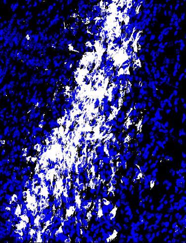
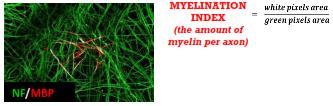
11 3.EFFECTS OF MICROGLIA-DERIVED EVs ON MYELIN DEPOSITION M0 M1 unstimulated pro-inflammatory+mscs pro-inflammatory anti-inflammatory Ctrl M1 ** *** promote myelin deposition M0 M1 Lombardi M., Fumagalli M. and Verderio C.


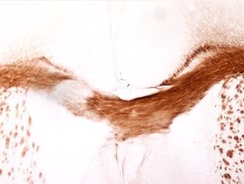
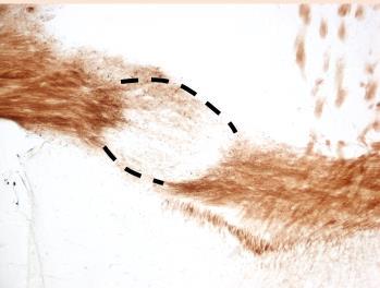

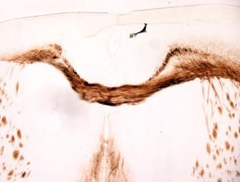

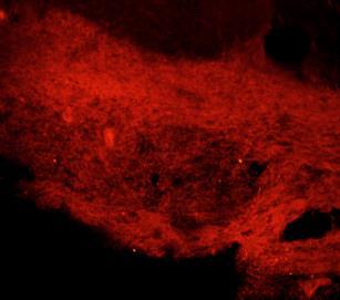
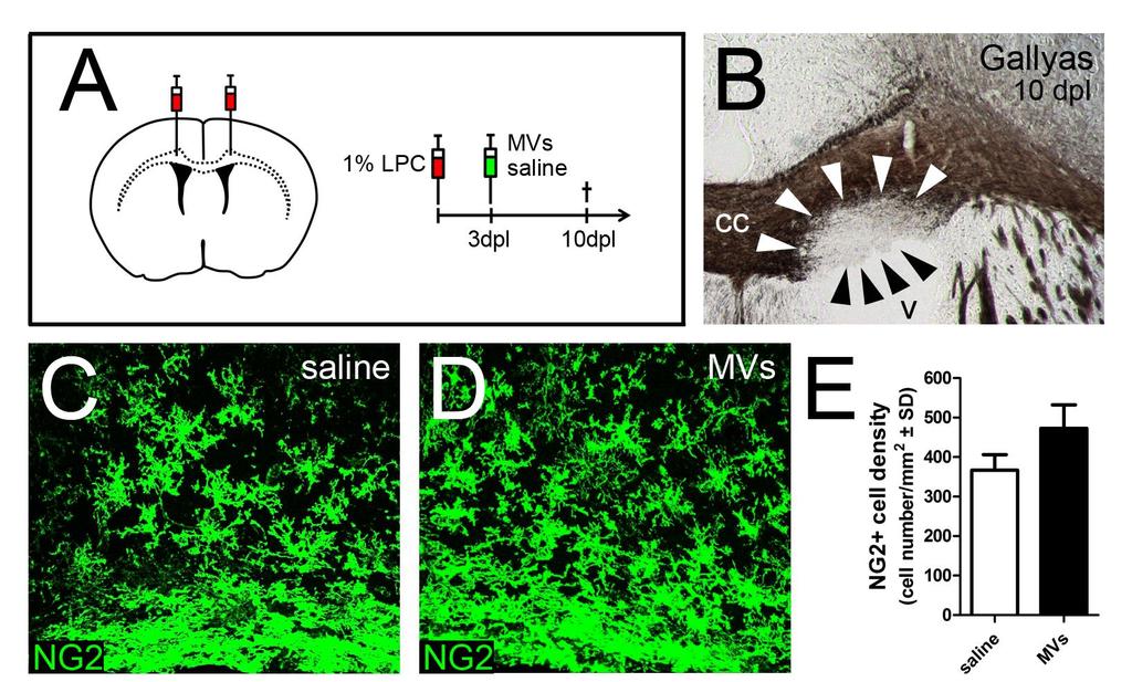
12 4.EFFECTS OF EVs ON OPC PROLIFERATION (and differentiation) IN VIVO LYSOLECITHIN MOUSE MODEL OF FOCAL DEMYELINATION 2%LPC 7dpi NG2DAPI 7dpi MBP Gallyas 14dpi
 7dpi Ctrl Ki67+ cells density")

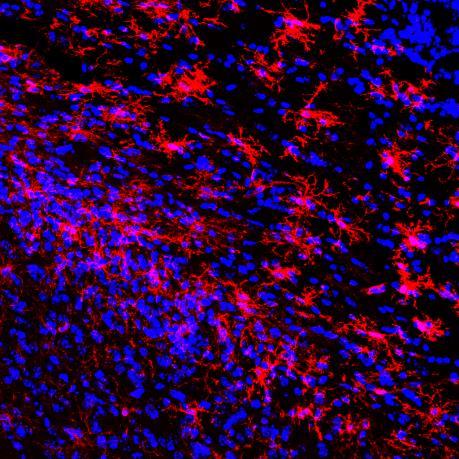
13 NG2DAPI anti-inflammatory pro-inflammatory+mscs Liposomes Ctrl Liposomes/IL-4/Th1+MSCs BrdU 2pulses (2h) 7dpi Ctrl Ki67+ cells density ** Ng2+ cells density 250 *** IL4 LIPO Ctrl 0 IL4 LIPO Ctrl induce cell proliferation and an increased recruitment of NG2+ OPCs.


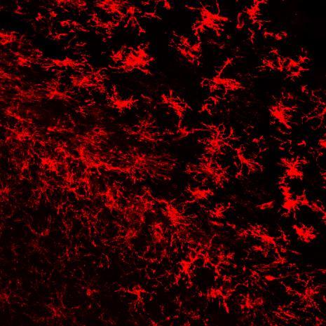
14 NG2BrdUDAPI anti-inflammatory pro-inflammatory+mscs Liposomes Ctrl Liposomes/IL-4/Th1+MSCs BrdU 2pulses (2h) 10dpi NG2BrdUDAPI NG2-BrdU+ NG2+BrdU+ Ng2 BrdU+ cells/ mm 2 Ng2 BrdU+ cells/ mm Ctrl With cronic administration of the new born NG2+ OPCs seem to be increased up to 10 days after lesion.


15 SUMMARY AMPLIFICATION DIFFERENTIATION M1 pro-inflammatory+mscs pro-inflammatory anti-inflammatory simplified system AMPLIFICATION DIFFERENTIATION +?? + multicellular environment EVs have a different effects in vivo and in vitro. In vivo, EVs could also affect other cell types, with secondary effects.
16 We are analysing the effects of EVs on differentiation to evaluate:? Hypothesis 1 this accumulation of OPCs pool is able to generate a greater number of mature cells (OLs) after differentiation, getting a massive myelination? Hypothesis 2 this accumulation of OPC pool is unable to differentiate and is blocked at this phase time


17 Laboratory of Neurobiology of brain plasticity Annalisa Buffo Enrica Boda CNR Institute of Neuroscience Claudia Verderio Marta Lombardi Martina Gabrielli University of Milan Maria Pia Abbracchio Marta Fumagalli Elisabetta Bonfanti
18

19 qrt-pcr analysis to confirm microglia polarization TNFalfa COX2 inos M1 M1 M1


20 Annex-PE MICROGLIA-DERIVED EXTRACELLULAR VESICLES Ectosomes 10 5 Flow cytometry 3% 32% % IB4-FICT Nanosight size plots
21 Concentration (particles/ml) Quantification and size of EVs from M0, M1 and microglia CTR M1 particles secreted by 1x10^6 MG 1.20E E E E E E E+000 CTR M1 Particle Diameter (nm) NO changes in the size distribution of M1-EVs or -EVs but increased EV production from M1 but not microglia as compared to unstimulated cells
22
23 IBA1DAPI

24 M0-EVs are able to act as chemoattractants for OPCs, similarly to -EVs overnight Evs are added to the culture medium of the outer well. OPCs are placed in the upper chamber Migratory cells pass through PET membrane. Non-migratory cells stay in the upper chamber % of migrated cells over control CTRL REST OPCs Free Medium PET membrane After removal of nonmigratory cells, migratory cells are stained and quantified Medium with EVs Staining solution Bar graph shows the percentage of migrated OPCs after an overnight exposure of EVs derived from REST or microglia with respect to control cells set to 100%. The number of migrated HOECHST + cells was counted in 60 optical fields at 20x magnification. Data are the mean±s.e.m. of cell count of 3 transwells/condition from one experiment.
25
26
27 MULTIPLE STRATEGIES TO PROMOTE ENDOGENOUS REMYELINATION VIA THE ACTION OF EVS RELEASED FROM ACTIVATED MG From Chandran et al., 2008
28 ACUTE ADMINISTRATION


![-Bis[4-(4,5-dihydro-1H-imidazol-2-yl)phenyl]-3,3](/docs-images/95/125847673/images/29-2.jpg "-p-phenylene-bis-acrylamide dihydrochloride)")
29 GW4869 inhibit nsmase2 in vivo. GW4869-treated mice produce reduced amounts of exosomes. GW4869 (N,N -Bis[4-(4,5-dihydro-1H-imidazol-2-yl)phenyl]-3,3 -p-phenylene-bis-acrylamide dihydrochloride) Nuclear Fas Red Luxol Fast Blue Intraperitoneal administration of GW4869 at 2 µg/g mouse body weight.
M2 microglia/ macrophages drive oligodendrocyte differentiation during CNS remyelination
 Supplemental Information Title: M2 microglia/ macrophages drive oligodendrocyte differentiation during CNS remyelination Authors: Veronique E. Miron, Amanda Boyd, Jing-Wei Zhao, Tracy J. Yuen, Julia M.
SUPPLEMENTARY FIG. S2. Representative counting fields used in quantification of the in vitro neural differentiation of pattern of dnscs.
 Supplementary Data SUPPLEMENTARY FIG. S1. Representative counting fields used in quantification of the in vitro neural differentiation of pattern of anpcs. A panel of lineage-specific markers were used
Supplementary Figure 1
 Supplementary Figure 1 AAV-GFP injection in the MEC of the mouse brain C57Bl/6 mice at 4 months of age were injected with AAV-GFP into the MEC and sacrificed at 7 days post injection (dpi). (a) Brains
GFP/Iba1/GFAP. Brain. Liver. Kidney. Lung. Hoechst/Iba1/TLR9!
 Supplementary information a +KA Relative expression d! Tlr9 5!! 5! NSC Neuron Astrocyte Microglia! 5! Tlr7!!!! NSC Neuron Astrocyte! GFP/Sβ/! Iba/Hoechst Microglia e Hoechst/Iba/TLR9! GFP/Iba/GFAP f Brain
Cord blood monocytes as a source of cell therapy products for treatment of brain injuries ISCT/CBA 2015 Cord Blood Workshop Wednesday, May 27, 2015
 Cord blood monocytes as a source of cell therapy products for treatment of brain injuries ISCT/CBA 2015 Cord Blood Workshop Wednesday, May 27, 2015 Andrew E. Balber, PhD Senior Scientific Advisor CT 2,
SUPPLEMENTARY INFORMATION
 DOI:.38/ncb3399 a b c d FSP DAPI 5mm mm 5mm 5mm e Correspond to melanoma in-situ Figure a DCT FSP- f MITF mm mm MlanaA melanoma in-situ DCT 5mm FSP- mm mm mm mm mm g melanoma in-situ MITF MlanaA mm mm
Nerve Cells and Behavior
 Nerve Cells and Behavior 27 th September, 2016 Touqeer Ahmed Ph.D. Atta-ur-Rahman School of Applied Biosciences National University of Sciences and Technology Nervous System and Behavior Nervous system
TITLE: Harnessing GPR17 Biology for Treating Demyelinating Disease
 AD Award Number: W81XWH-10-1-0721 TITLE: Harnessing GPR17 Biology for Treating Demyelinating Disease PRINCIPAL INVESTIGATOR: Nitin Karandikar, M.D., Ph.D. CONTRACTING ORGANIZATION: University of Texas
Supplemental Figures Supplemental Figure 1:
 Supplemental Figures Supplemental Figure 1: Representative FACS data showing Concurrent Brain cell type Acquisition using either Percoll PLUS (top row) or myelin removal beads (bottom two rows). Debris
The anti-inflammatory enzyme A20 in the neuropathology of Multiple Sclerosis
 More Than Neurons, 1-3 December, Turin The anti-inflammatory enzyme A20 in the neuropathology of Multiple Sclerosis Dr. Simona Perga, PhD Neuroscience Institute Cavalieri Ottolenghi (NICO) & Multiple Sclerosis
In vivo reprogramming reactive glia into ipscs to produce new neurons in the
 In vivo reprogramming reactive glia into ipscs to produce new neurons in the cortex following traumatic brain injury Xiang Gao 1, Xiaoting Wang 1, Wenhui Xiong 1, Jinhui Chen 1, * 1 Spinal Cord and Brain
Takayuki Hirai, Kenzo Uchida, Hideaki Nakajima, Tomoo Inukai, Naoto Takeura, Shuji Watanabe, Hisatoshi Baba
 Chronic progressive compression induces the phenotype changes of the activated microglia/macrophages in the spinal cord of spinal hyperostotic twy/twy mouse: implications in human cervical compressive
Contribution of microglia to tissue injury and repair in MS
 Contribution of microglia to tissue injury and repair in MS MS disease course histologic features Courtesy of Samuel Ludwin I ACUTE CHRONIC s ACTIVE CHRONIC Clinical Course Intra CNS Extra CNS Imaging
Supplementary Information
 Supplementary Information Distinct bone marrow-derived and tissue resident macrophage lineages proliferate at key stages during inflammation. 1 Luke C. Davies, 1 Marcela Rosas, 2 Stephen J. Jenkins, 1
Supplementary Information
 Supplementary Information Title Degeneration and impaired regeneration of gray matter oligodendrocytes in amyotrophic lateral sclerosis Authors Shin H. Kang, Ying Li, Masahiro Fukaya, Ileana Lorenzini,
Supplementary Figure 1
 Supplementary Figure 1 Genetic labeling of microglia Male and female 2-3 month-old CreERT2;R26-tdTomato mice or CreERT2;R26-tdTomato;Iba1-eGFP transgenic mice were treated with 1x, 2x (48 h apart), or
Supplementary Figure 1. Nature Neuroscience: doi: /nn.4547
 Supplementary Figure 1 Characterization of the Microfetti mouse model. (a) Gating strategy for 8-color flow analysis of peripheral Ly-6C + monocytes from Microfetti mice 5-7 days after TAM treatment. Living
Nature Neuroscience: doi: /nn Supplementary Figure 1
 Supplementary Figure 1 Quantification of myelin fragments in the aging brain (a) Electron microscopy on corpus callosum is shown for a 18-month-old wild type mice. Myelin fragments (arrows) were detected
Supplemental Table 1. Primer sequences for transcript analysis
 Supplemental Table 1. Primer sequences for transcript analysis Primer Sequence (5 3 ) Primer Sequence (5 3 ) Mmp2 Forward CCCGTGTGGCCCTC Mmp15 Forward CGGGGCTGGCT Reverse GCTCTCCCGGTTTC Reverse CCTGGTGTGCCTGCTC
TITLE: Harnessing GPR17 Biology for Treating Demyelinating Disease
 AD Award Number: W81XWH-10-1-0723 TITLE: Harnessing GPR17 Biology for Treating Demyelinating Disease PRINCIPAL INVESTIGATOR: Qing Lu, Ph.D. CONTRACTING ORGANIZATION: University of Texas Southwestern Medical
Supplementary Figure 1. Double-staining immunofluorescence analysis of invasive colon and breast cancers. Specimens from invasive ductal breast
 Supplementary Figure 1. Double-staining immunofluorescence analysis of invasive colon and breast cancers. Specimens from invasive ductal breast carcinoma (a) and colon adenocarcinoma (b) were staining
mm Distance (mm)
") b a Magnet Illumination Coverslips MPs Objective 2575 µm 1875 µm 1575 µm 1075 µm 875 µm 545 µm 20µm 2 3 0.5 0.3mm 1 1000 100 10 1 0.1 1000 100 10 1 0.1 Field Induction (Gauss) 1.5 0 5 10 15 20 Distance
b a Magnet Illumination Coverslips MPs Objective 2575 µm 1875 µm 1575 µm 1075 µm 875 µm 545 µm 20µm 2 3 0.5 0.3mm 1 1000 100 10 1 0.1 1000 100 10 1 0.1 Field Induction (Gauss) 1.5 0 5 10 15 20 Distance
(a) Significant biological processes (upper panel) and disease biomarkers (lower panel)
 Significant biological processes (upper panel) and disease biomarkers (lower panel)") Supplementary Figure 1. Functional enrichment analyses of secretomic proteins. (a) Significant biological processes (upper panel) and disease biomarkers (lower panel) 2 involved by hrab37-mediated secretory
Supplementary Figure 1. Functional enrichment analyses of secretomic proteins. (a) Significant biological processes (upper panel) and disease biomarkers (lower panel) 2 involved by hrab37-mediated secretory
Contact: Course outline: Contact for other times.
 Contact: kdelaney@uvic.ca Course outline: http://web.uvic.ca/~kdelaney/b367 Scheduled office hours: 1:00-3:00, M&Th Cunn. 259A Contact kdelaney@uvic.ca for other times. Quiz (0.5 hrs) midterm (1.4 hrs)
Modulation of TRP channels by resolvins in mouse and human
 July 9, 2015, Ion Channel Retreat, Vancouver Ion Channel and Pain Targets Modulation of TRP channels by resolvins in mouse and human Ru-Rong Ji, PhD Pain Research Division Department of Anesthesiology
Stewart et al. CD36 ligands promote sterile inflammation through assembly of a TLR 4 and 6 heterodimer
 NFκB (fold induction) Stewart et al. ligands promote sterile inflammation through assembly of a TLR 4 and 6 heterodimer a. mrna (fold induction) 5 4 3 2 1 LDL oxldl Gro1a MIP-2 RANTES mrna (fold induction)
Supplementary Table 1
 Supplementary Table 1 Flow Cytometry Antibodies Antibody Fluorochrome Clone Vendor CD45 PE-cyanine 7 30-F11 D ioscience CD3 Pacific lue 17A2 iolegend (San Diego, CA) CD11b APC M1/70 iolegend (San Diego,
Primary oligodendropathy is not a trigger of CNS autoimmunity
 Primary oligodendropathy is not a trigger of CNS autoimmunity Ari Waisman Institute for Molecular Medicine University Medical Center, JGU Mainz 1 How is an anti-myelin immune response initiated? Secondary
glial cells missing and gcm2 Cell-autonomously Regulate Both Glial and Neuronal
 glial cells missing and gcm2 Cell-autonomously Regulate Both Glial and Neuronal Development in the Visual System of Drosophila Carole Chotard, Wendy Leung and Iris Salecker Supplemental Data Supplemental
Supplemental Information. Menin Deficiency Leads to Depressive-like. Behaviors in Mice by Modulating. Astrocyte-Mediated Neuroinflammation
 Neuron, Volume 100 Supplemental Information Menin Deficiency Leads to Depressive-like Behaviors in Mice by Modulating Astrocyte-Mediated Neuroinflammation Lige Leng, Kai Zhuang, Zeyue Liu, Changquan Huang,
Neurodegeneration and macrophages; a beneficial or harmful role for macrophages and microglia in neuronal damage during multiple sclerosis
 Neurodegeneration and macrophages; a beneficial or harmful role for macrophages and microglia in neuronal damage during multiple sclerosis Marlijn van der Poel Writing assignment: literature review October
SUPPLEMENTARY FIGURES
 SUPPLEMENTARY FIGURES 1 Supplementary Figure 1, Adult hippocampal QNPs and TAPs uniformly express REST a-b) Confocal images of adult hippocampal mouse sections showing GFAP (green), Sox2 (red), and REST
Supplementary Figure 1 Expression of Crb3 in mouse sciatic nerve: biochemical analysis (a) Schematic of Crb3 isoforms, ERLI and CLPI, indicating the
 Schematic of Crb3 isoforms, ERLI and CLPI, indicating the") Supplementary Figure 1 Expression of Crb3 in mouse sciatic nerve: biochemical analysis (a) Schematic of Crb3 isoforms, ERLI and CLPI, indicating the location of the transmembrane (TM), FRM binding (FB)
Supplementary Figure 1 Expression of Crb3 in mouse sciatic nerve: biochemical analysis (a) Schematic of Crb3 isoforms, ERLI and CLPI, indicating the location of the transmembrane (TM), FRM binding (FB)
Supplementary Figure 1. Successful excision of genes from WBM lysates and
 Supplementary Information: Supplementary Figure 1. Successful excision of genes from WBM lysates and survival of mice with different genotypes. (a) The proper excision of Pten, p110α, p110α and p110δ was
Supplementary Figure 1.TRIM33 binds β-catenin in the nucleus. a & b, Co-IP of endogenous TRIM33 with β-catenin in HT-29 cells (a) and HEK 293T cells
 and HEK 293T cells") Supplementary Figure 1.TRIM33 binds β-catenin in the nucleus. a & b, Co-IP of endogenous TRIM33 with β-catenin in HT-29 cells (a) and HEK 293T cells (b). TRIM33 was immunoprecipitated, and the amount of
Supplementary Figure 1.TRIM33 binds β-catenin in the nucleus. a & b, Co-IP of endogenous TRIM33 with β-catenin in HT-29 cells (a) and HEK 293T cells (b). TRIM33 was immunoprecipitated, and the amount of
Supplemental Figure 1. Quantification of proliferation in thyroid of WT, Ctns -/- and grafted
 Supplemental Figure 1. Quantification of proliferation in thyroid of WT, Ctns -/- and grafted Ctns -/- mice. Cells immunolabeled for the proliferation marker (Ki-67) were counted in sections (n=3 WT, n=4
Electron micrograph of phosphotungstanic acid-stained exosomes derived from murine
 1 SUPPLEMENTARY INFORMATION SUPPLEMENTARY FIGURES Supplementary Figure 1. Physical properties of murine DC-derived exosomes. a, Electron micrograph of phosphotungstanic acid-stained exosomes derived from
Supplementary Figure 1
 Supplementary Figure 1 Global TeNT expression effectively impairs synaptic transmission. Injection of 100 pg tent mrna leads to a reduction of vesicle mediated synaptic transmission in the spinal cord
Demyelination arrest and remyelination induced by glatiramer acetate treatment of experimental autoimmune encephalomyelitis
 Demyelination arrest and remyelination induced by glatiramer acetate treatment of experimental autoimmune encephalomyelitis Rina Aharoni*, Avia Herschkovitz*, Raya Eilam, Michal Blumberg-Hazan, Michael
Supplementary Figures
 Supplementary Figures Supplementary Figure 1 Characterization of stable expression of GlucB and sshbira in the CT26 cell line (a) Live cell imaging of stable CT26 cells expressing green fluorescent protein
NG2-Glia (Polydendrocytes)
") NG2-Glia (Polydendrocytes) ii One liner Colloquium Chapter Title Digital Library of Life Sciences The Colloquium Digital Library of Life Sciences is an innovative information resource for researchers,
NG2-Glia (Polydendrocytes) ii One liner Colloquium Chapter Title Digital Library of Life Sciences The Colloquium Digital Library of Life Sciences is an innovative information resource for researchers,
Programmed necrosis, not apoptosis, is a key mediator of cell loss and DAMP-mediated inflammation in dsrna-induced retinal degeneration
 Programmed necrosis, not apoptosis, is a key mediator of cell loss and DAMP-mediated inflammation in dsrna-induced retinal degeneration The Harvard community has made this article openly available. Please
MOLECULAR AND CELLULAR NEUROSCIENCE
 MOLECULAR AND CELLULAR NEUROSCIENCE BMP-218 November 4, 2014 DIVISIONS OF THE NERVOUS SYSTEM The nervous system is composed of two primary divisions: 1. CNS - Central Nervous System (Brain + Spinal Cord)
Supplementary Figure 1 Induction of cellular senescence and isolation of exosome. a to c, Pre-senescent primary normal human diploid fibroblasts
 Supplementary Figure 1 Induction of cellular senescence and isolation of exosome. a to c, Pre-senescent primary normal human diploid fibroblasts (TIG-3 cells) were rendered senescent by either serial passage
Supplemental Figure 1. Intracranial transduction of a modified ptomo lentiviral vector in the mouse
 Supplemental figure legends Supplemental Figure 1. Intracranial transduction of a modified ptomo lentiviral vector in the mouse hippocampus targets GFAP-positive but not NeuN-positive cells. (A) Stereotaxic
Inhibition of DYRK1A stimulates human beta-cell proliferation
 Inhibition of DYRK1A stimulates human beta-cell proliferation Ercument Dirice 1,, Deepika Walpita 2,, Amedeo Vetere 2, Bennett C. Meier 2,5, Sevim Kahraman 1, Jiang Hu 1, Vlado Dančík 2, Sean M. Burns
SUPPLEMENTARY FIGURES
 SUPPLEMENTARY FIGURES 1 2 3 4 SUPPLEMENTARY TABLES Supplementary Table S1. Brain Tumors used in the study Code Tumor Classification Age Gender HuTuP51 Glioblastoma 57 Male HuTuP52 Glioblastoma 53 Male
Insight into cancer research from discovery to validation
 Insight into cancer research from discovery to validation 1 Exosomes in cancer research: from exosome isolation to biomarker discovery Zheng Songyue, PhD Technical Sales Specialists Life Technologies The
Supplementary Figure 1. SybII and Ceb are sorted to distinct vesicle populations in astrocytes. Nature Neuroscience: doi: /nn.
 Supplementary Figure 1 SybII and Ceb are sorted to distinct vesicle populations in astrocytes. (a) Exemplary images for cultured astrocytes co-immunolabeled with SybII and Ceb antibodies. SybII accumulates
Research Development: Bedside to Bench and Back
 Research Development: Bedside to Bench and Back Matt Bellizzi, MD PhD Department of Neurology University of Rochester School of Medicine and Dentistry Rochester, NY "I can walk down the hall just fine,
Supporting Information
 Supporting Information Valkenburg et al. 10.1073/pnas.1403684111 SI Materials and Methods ELISA and Microneutralization. Sera were treated with Receptor Destroying Enzyme II (RDE II, Accurate) before ELISA
Cells of the nervous system
 Neurobiology Cells of the nervous system Anthony Heape 2011 1 Cells of the nervous system Neuroglia : part 2 The non excitable cells of the nervous system that provide support to neuronal survival and
Progress Report for NJCSCR (Yu-Wen Chang)
") Progress Report for NJCSCR (Yu-Wen Chang) Overall Plan Summary: Traumatic injury to the spinal cord initiates a cascade of degenerative processes, known as secondary injury, which include various inflammatory
Progress Report for NJCSCR (Yu-Wen Chang) Overall Plan Summary: Traumatic injury to the spinal cord initiates a cascade of degenerative processes, known as secondary injury, which include various inflammatory
Microglia preconditioning (priming) in central nervous system pathologies
 in central nervous system pathologies") Microglia preconditioning (priming) in central nervous system pathologies Florence Perrin florence.perrin@umontpellier.fr Montpellier, October 2018 1 Spanish anatomists Glia = glue in Greek Santiago Ramon
Microglia preconditioning (priming) in central nervous system pathologies Florence Perrin florence.perrin@umontpellier.fr Montpellier, October 2018 1 Spanish anatomists Glia = glue in Greek Santiago Ramon
Nerve tissue & the Nervous System
 Nerve tissue & the Nervous System The human nervous system, by far the most complex system in the body, is formed by a network of many billion nerve cells (neurons), all assisted by many more supporting
activation with anti-cd3/cd28 beads and 3d following transduction. Supplemental Figure 2 shows
 Supplemental Data Supplemental Figure 1 compares CXCR4 expression in untreated CD8 + T cells, following activation with anti-cd3/cd28 beads and 3d following transduction. Supplemental Figure 2 shows the
Aggregated neutrophil extracellular traps limit inflammation by degrading cytokines and chemokines
 CORRECTION NOTICE Nat. Med. doi:10.1038/nm.3547; corrected online 25 August 2014 Aggregated neutrophil extracellular traps limit inflammation by degrading cytokines and chemokines Christine Schauer, Christina
Dietary cholesterol promotes repair of demyelinated lesions in the adult brain
 Received Apr 16 Accepted 1 Dec 16 Published Jan 17 DOI: 1.138/ncomms11 Dietary cholesterol promotes repair of demyelinated lesions in the adult brain OPEN Stefan A. Berghoff 1, Nina Gerndt 1, Jan Winchenbach
SUPPLEMENTARY INFORMATION
 doi:10.1038/nature10188 Supplementary Figure 1. Embryonic epicardial genes are down-regulated from midgestation stages and barely detectable post-natally. Real time qrt-pcr revealed a significant down-regulation
(A) PCR primers (arrows) designed to distinguish wild type (P1+P2), targeted (P1+P2) and excised (P1+P3)14-
 PCR primers (arrows) designed to distinguish wild type (P1+P2), targeted (P1+P2) and excised (P1+P3)14-") 1 Supplemental Figure Legends Figure S1. Mammary tumors of ErbB2 KI mice with 14-3-3σ ablation have elevated ErbB2 transcript levels and cell proliferation (A) PCR primers (arrows) designed to distinguish
1 Supplemental Figure Legends Figure S1. Mammary tumors of ErbB2 KI mice with 14-3-3σ ablation have elevated ErbB2 transcript levels and cell proliferation (A) PCR primers (arrows) designed to distinguish
Neurodevelopment II Structure Formation. Reading: BCP Chapter 23
 Neurodevelopment II Structure Formation Reading: BCP Chapter 23 Phases of Development Ovum + Sperm = Zygote Cell division (multiplication) Neurogenesis Induction of the neural plate Neural proliferation
Eosinophils are required. for the maintenance of plasma cells in the bone marrow
 Eosinophils are required for the maintenance of plasma cells in the bone marrow Van Trung Chu, Anja Fröhlich, Gudrun Steinhauser, Tobias Scheel, Toralf Roch, Simon Fillatreau, James J. Lee, Max Löhning
MII. Supplement Figure 1. CapZ β2. Merge. 250ng. 500ng DIC. Merge. Journal of Cell Science Supplementary Material. GFP-CapZ β2 DNA
 A GV GVBD MI DNA CapZ β2 CapZ β2 Merge B DIC GFP-CapZ β2 Merge CapZ β2-gfp 250ng 500ng Supplement Figure 1. MII A early MI late MI Control RNAi CapZαβ DNA Actin Tubulin B Phalloidin Intensity(A.U.) n=10
Elucidation of Viral Replication Mechanisms in an Animal Model for Multiple Sclerosis
 Elucidation of Viral Replication Mechanisms in an Animal Model for Multiple Sclerosis Introduction It is often difficult to research human diseases due to obvious ethical implications. However, when an
Supplementary Materials for
 www.sciencemag.org/content/348/6241/aaa825/suppl/dc1 Supplementary Materials for A mucosal vaccine against Chlamydia trachomatis generates two waves of protective memory T cells Georg Stary,* Andrew Olive,
Mary ET Boyle, Ph. D. Department of Cognitive Science UCSD
 ? Mary ET Boyle, Ph. D. Department of Cognitive Science UCSD Christian S Lobsiger & Don W Cleveland (2007) Nature Neuroscience 10, 1355-1360 Astrocytes: interlinked gatekeepers of glutamate astrocytes
Supplementary Figure 1.
 Supplementary Figure 1. Female Pro-ins2 -/- mice at 5-6 weeks of age were either inoculated i.p. with a single dose of CVB4 (1x10 5 PFU/mouse) or PBS and treated with αgalcer or control vehicle. On day
Supplementary Figure S1 Expression of mir-181b in EOC (A) Kaplan-Meier
 Kaplan-Meier") Supplementary Figure S1 Expression of mir-181b in EOC (A) Kaplan-Meier curves for progression-free survival (PFS) and overall survival (OS) in a cohort of patients (N=52) with stage III primary ovarian
Supplementary Figure S1 Expression of mir-181b in EOC (A) Kaplan-Meier curves for progression-free survival (PFS) and overall survival (OS) in a cohort of patients (N=52) with stage III primary ovarian
Supplemental Figure 1. Western blot analysis indicated that MIF was detected in the fractions of
 Supplemental Figure Legends Supplemental Figure 1. Western blot analysis indicated that was detected in the fractions of plasma membrane and cytosol but not in nuclear fraction isolated from Pkd1 null
Supplementary Information
 Nature Immunology doi:1.138/ni.2477 Supplementary Information Capillary and arteriolar pericytes attract innate leukocytes exiting through venules and instruct them with pattern recognition and motility
Generation of ST2-GFP reporter mice and characterization of ILC1 cells following infection
 Supplementary Figure 1 Generation of ST2-GFP reporter mice and characterization of ILC1 cells following infection with influenza virus. (a) ST2-GFP reporter mice were generated as described in Methods.
Microglia, Inflammation, and FTD
 FTD Minicourse April, 2009 Microglia, Inflammation, and FTD Li Gan, Ph.D Gladstone Institute of Neurological Disease University of California, San Francisco Outline Why study inflammation in neurodegeneration?
Liver-Resident Macrophage Necroptosis Orchestrates Type 1 Microbicidal Inflammation and Type-2- Mediated Tissue Repair during Bacterial Infection
 Liver-Resident Macrophage Necroptosis Orchestrates Type 1 Microbicidal Inflammation and Type-2- Mediated Tissue Repair during Bacterial Infection Camille Blériot, Théo Dupuis, Grégory Jouvion, Gérard Eberl,
EPIGENETIC RE-EXPRESSION OF HIF-2α SUPPRESSES SOFT TISSUE SARCOMA GROWTH
 EPIGENETIC RE-EXPRESSION OF HIF-2α SUPPRESSES SOFT TISSUE SARCOMA GROWTH Supplementary Figure 1. Supplementary Figure 1. Characterization of KP and KPH2 autochthonous UPS tumors. a) Genotyping of KPH2
Diseases of Immunity 2017 CL Davis General Pathology. Paul W. Snyder, DVM, PhD Experimental Pathology Laboratories, Inc.
 Diseases of Immunity 2017 CL Davis General Pathology Paul W. Snyder, DVM, PhD Experimental Pathology Laboratories, Inc. Autoimmunity Reflects a loss of immunologic tolerance Mechanisms Auto-antibodies
MagCapture Exosome Isolation Kit PS Q&A
 MagCapture Exosome Isolation Kit PS Q&A Specifications and performance P.1 Comparison of the conventional method P.2 Operation methods and composition P.4 Amount of starting sample P.5 Analysis after exosomes
effect on the upregulation of these cell surface markers. The mean peak fluorescence intensity
 SUPPLEMENTARY FIGURE 1 Supplementary Figure 1 ASIC1 disruption or blockade does not effect in vitro and in vivo antigen-presenting cell activation. (a) Flow cytometric analysis of cell surface molecules
Effector T Cells and
 1 Effector T Cells and Cytokines Andrew Lichtman, MD PhD Brigham and Women's Hospital Harvard Medical School 2 Lecture outline Cytokines Subsets of CD4+ T cells: definitions, functions, development New
Quantitative PPARγ expression affects the balance between tolerance and immunity
 Quantitative PPARγ expression affects the balance between tolerance and immunity Ya-Hui Liu 1, Yau-Sheng Tsai 1,2,3, Shih-Chieh Lin 4, Nan-Shih Liao 5, Ming-Shiou Jan 6, Chung-Tiang Liang 7, Shih-Wen Hsu
ROCK/Cdc42-mediated microglial motility and gliapse formation lead to phagocytosis of degenerating dopaminergic neurons in vivo
 Supplementary Information ROCK/Cdc42-mediated microglial motility and gliapse formation lead to phagocytosis of degenerating dopaminergic neurons in vivo Carlos Barcia* 1,2, Carmen M Ros 1,2, Valentina
Supplementary Information Titles Journal: Nature Medicine
 Supplementary Information Titles Journal: Nature Medicine Article Title: Corresponding Author: Supplementary Item & Number Supplementary Fig.1 Fig.2 Fig.3 Fig.4 Fig.5 Fig.6 Fig.7 Fig.8 Fig.9 Fig. Fig.11
Remyelination in the CNS: from biology to therapy
 NeuroN Glia interactions Remyelination in the CNS: from biology to therapy Robin J. M. Franklin* and Charles ffrench-constant Abstract Remyelination involves reinvesting demyelinated axons with new myelin
Supplementary Figure 1: Expression of NFAT proteins in Nfat2-deleted B cells (a+b) Protein expression of NFAT2 (a) and NFAT1 (b) in isolated splenic
 Protein expression of NFAT2 (a) and NFAT1 (b) in isolated splenic") Supplementary Figure 1: Expression of NFAT proteins in Nfat2-deleted B cells (a+b) Protein expression of NFAT2 (a) and NFAT1 (b) in isolated splenic B cells from WT Nfat2 +/+, TCL1 Nfat2 +/+ and TCL1 Nfat2
Supplementary Figure 1: Expression of NFAT proteins in Nfat2-deleted B cells (a+b) Protein expression of NFAT2 (a) and NFAT1 (b) in isolated splenic B cells from WT Nfat2 +/+, TCL1 Nfat2 +/+ and TCL1 Nfat2
stem cell products Basement Membrane Matrix Products Rat Mesenchymal Stem Cell Growth and Differentiation Products
 stem cell products Basement Membrane Matrix Products Rat Mesenchymal Stem Cell Growth and Differentiation Products Stem Cell Qualified Extracellular Matrix Proteins Stem cell research requires the finest
3rd International Conference on Neurology & Therapeutics.
 3rd International Conference on Neurology & Therapeutics www.neuroimmunology.ca Multiple sclerosis is a devastating disease The first description of the disease was mentioned in 14th century In 1838 Dr.
Exosomes secreted by Human Cardiac Progenitors contain mirna with cardioprotective and proangiogenic activities
 Exosomes secreted by Human Cardiac Progenitors contain mirna with cardioprotective and proangiogenic activities Elisabetta Cervio, PhD Molecular Cardiology Laboratory Cardiocentro Ticino, Lugano, CH International
SUPPLEMENTARY INFORMATION
 doi: 1.138/nature89 IFN- (ng ml ) 5 4 3 1 Splenocytes NS IFN- (ng ml ) 6 4 Lymph node cells NS Nfkbiz / Nfkbiz / Nfkbiz / Nfkbiz / IL- (ng ml ) 3 1 Splenocytes IL- (ng ml ) 1 8 6 4 *** ** Lymph node cells
! BIOL 2401! Week 5. Nervous System. Nervous System
 Collin County Community College! BIOL 2401! Week 5 Nervous System 1 Nervous System The process of homeostasis makes sure that the activities that occur in the body are maintained within normal physiological
Supplementary Figure 1. Characterization of basophils after reconstitution of SCID mice
 Supplementary figure legends Supplementary Figure 1. Characterization of after reconstitution of SCID mice with CD4 + CD62L + T cells. (A-C) SCID mice (n = 6 / group) were reconstituted with 2 x 1 6 CD4
Pathologic Stage. Lymph node Stage
 ASC ASC a c Patient ID BMI Age Gleason score Non-obese PBMC 1 22.1 81 6 (3+3) PBMC 2 21.9 6 6 (3+3) PBMC 3 22 84 8 (4+4) PBMC 4 24.6 68 7 (3+4) PBMC 24. 6 (3+3) PBMC 6 24.7 73 7 (3+4) PBMC 7 23. 67 7 (3+4)
Supplementary Figure 1 Chemokine and chemokine receptor expression during muscle regeneration (a) Analysis of CR3CR1 mrna expression by real time-pcr
 Analysis of CR3CR1 mrna expression by real time-pcr") Supplementary Figure 1 Chemokine and chemokine receptor expression during muscle regeneration (a) Analysis of CR3CR1 mrna expression by real time-pcr at day 0, 1, 4, 10 and 21 post- muscle injury. (b)
Supplementary Figure 1 Chemokine and chemokine receptor expression during muscle regeneration (a) Analysis of CR3CR1 mrna expression by real time-pcr at day 0, 1, 4, 10 and 21 post- muscle injury. (b)
Figure S1. Reduction in glomerular mir-146a levels correlate with progression to higher albuminuria in diabetic patients.
 Supplementary Materials Supplementary Figures Figure S1. Reduction in glomerular mir-146a levels correlate with progression to higher albuminuria in diabetic patients. Figure S2. Expression level of podocyte
regenerative medicine in the brain and the spinal cord spinal cord injuries
 regenerative medicine in the brain and the spinal cord spinal cord injuries primary and secondary events during SCI traumatic spinal cord injury (SCI) traumatic spinal cord injury (SCI) main goal is to
Figure S1. PMVs from THP-1 cells expose phosphatidylserine and carry actin. A) Flow
 Flow") SUPPLEMENTARY DATA Supplementary Figure Legends Figure S1. PMVs from THP-1 cells expose phosphatidylserine and carry actin. A) Flow cytometry analysis of PMVs labelled with annexin-v-pe (Guava technologies)
SUPPLEMENTARY DATA Supplementary Figure Legends Figure S1. PMVs from THP-1 cells expose phosphatidylserine and carry actin. A) Flow cytometry analysis of PMVs labelled with annexin-v-pe (Guava technologies)
NNZ-2566 in Rett Syndrome and Autism Spectrum Disorders Role and Update
 NNZ-2566 in Rett Syndrome and Autism Spectrum Disorders Role and Update 1 Overview The natural growth factor IGF-1 is broken down in the body to IGF-1[1-3] NNZ-2566 is an analogue of IGF-1[1-3] developed
April 29, Neurophysiology. Chul-Kyu Park, Ph.D. Assistant Professor Department of Physiology, Graduate School of Medicine, Gachon University,
 April 29, 2016 Neurophysiology Chul-Kyu Park, Ph.D. Assistant Professor Department of Physiology, Graduate School of Medicine, Gachon University, Cells in the brain Neurons glia 1. Astrocytes 2. Microglia
Nature Neuroscience: doi: /nn Supplementary Figure 1. Splenic atrophy and leucopenia caused by T3 SCI.
 Supplementary Figure 1 Splenic atrophy and leucopenia caused by T3 SCI. (a) Gross anatomy of representative spleens from control and T3 SCI mice at 28 days post-injury. (b and c) Hematoxylin and eosin
SUPPLEMENTARY INFORMATION
 doi:1.138/nature1554 a TNF-α + in CD4 + cells [%] 1 GF SPF 6 b IL-1 + in CD4 + cells [%] 5 4 3 2 1 Supplementary Figure 1. Effect of microbiota on cytokine profiles of T cells in GALT. Frequencies of TNF-α
T H E J O U R N A L O F C E L L B I O L O G Y
 T H E J O U R N A L O F C E L L B I O L O G Y Supplemental material Amelio et al., http://www.jcb.org/cgi/content/full/jcb.201203134/dc1 Figure S1. mir-24 regulates proliferation and by itself induces
The rationale to study EVs from the ES-2 cell line and use as control EVs from a 5th cell line, which has not been previously tested, is unclear.
 Reviewers' comments: Reviewer #1 (Remarks to the Author): This study attempts to demonstrate that extracellular vesicles (EVs) shed by aggressive ovarian cancer cells promote metastatic abilities of less