38 th Annual Gross Morbid Pathology of Laboratory
|
|
|
- Maude Ball
- 5 years ago
- Views:
Transcription


1 The Department of Veterinary Biosciences The OSU Comprehensive Cancer Center 38 th Annual Gross Morbid Pathology of Laboratory Rodents & Lagomorphs Krista M. D. La Perle, DVM, PhD, Dipl. ACVP Associate Professor Director, Comparative Pathology & Mouse Phenotyping Shared Resource ACVP GROSS EXAM 100 projected images Parts A & B 1-2 minutes/image shown once! 22 gross images and questions by each of 13 committee members (April) Section leader prepares p draft exam for review by entire committee (May/June) Exam finalized (July/August) Matrix Species Organ System Disease Process 2 ACVP GROSS EXAM Answers should represent the MOST LIKELY interpretation of lesions/diseases presented Be as specific as possible Questions Morphologic diagnosis Etiologic diagnosis Cause Name the disease or condition Histologic characteristics Histochemical or immunohistochemical characteristics Cytologic characteristics Pathogenesis Sequela(e) Associated lesions Associated clinical pathology findings ACVP GROSS EXAM Gross exam coversheet Sample of gross images, questions and answers from 2008 and 2009 exams reviewed at annual ACVP meeting 3 4 MICE MICE Nature 420, 2002 Albino BALB/c FVB Agouti 129 C3H Black C57BL/6 5 6 La Perle-2011 CL Davis Gross-Rodents & Lagomorphs 1


2 2


3 3


4 4


5 5


6 6


7 7


8 8


9 9


10 10


11 11


12 12


13 13


14 14


15 15


16 16


17 RATS 100 La Perle-2011 CL Davis Gross-Rodents & Lagomorphs 17


18 18


19 19


20 20


21 21


22 22


23 23


24 24


25 RABBITS 148 La Perle-2011 CL Davis Gross-Rodents & Lagomorphs 25


26 26


27 27


28 28


29 29


30 30


31 31


32 La Perle-2011 CL Davis Gross-Rodents & Lagomorphs 32


33 33


34 34


35 35


36 GUINEA PIGS 215 La Perle-2011 CL Davis Gross-Rodents & Lagomorphs 36


37 37


38 38


39 39


40 40


41 GERBILS 243 La Perle-2011 CL Davis Gross-Rodents & Lagomorphs 41


42 HAMSTERS 252 La Perle-2011 CL Davis Gross-Rodents & Lagomorphs 42


43 43


44 44


45 45

46 PHOTO CREDITS THANK YOU!...QUESTIONS? Sheree Beam Charlie Clifford Hai Nguyen Jo Lynne Raymond Teresa Southard Bruce Williams Cornell University College of Veterinary Medicine/Dr. John M. King s Necropsy Show & Tell Brian Caserto Michael Eckhaus Dean Percy Duncan Russell Paul Stromberg Uncited Individuals ACLAM Lab Animal Medicine and Science Series II University of Georgia College of Veterinary Medicine Noah s Arkive La Perle-2011 CL Davis Gross-Rodents & Lagomorphs 46
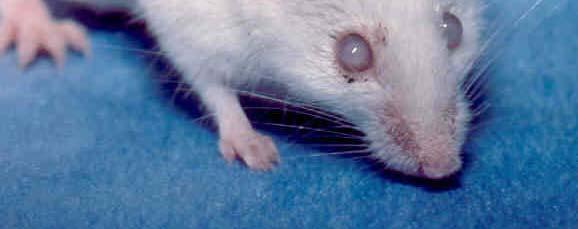
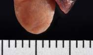
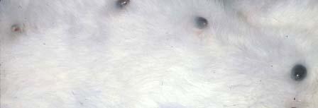
47 38 th Annual Gross Morbid Pathology of Laboratory Rodents & Lagomorphs Krista M. D. La Perle, DVM, PhD, Dipl. ACVP Associate Professor Director, Comparative Pathology & Mouse Phenotyping Shared Resource The Ohio State University, Department of Veterinary Biosciences 1925 Coffey Road Columbus, OH P: ; F: ; SLIDE # TISSUE/ORGAN MORPHOLOGIC DIAGNOSIS NOTES 1 TITLE SLIDE 2-4 ACVP GROSS EXAM INTRODUCTION 5 MICE (slides #6-99) 6 COAT COLORS AID IN IDENTIFICATION OF STRAIN/STRAIN-SPECIFIC LESIONS 7 Eye Bilateral cataracts Common in C57BL/6 (+/- microphthalmia) and B6C3F1 mice. 8 Eye Unilateral ulcerative and fibrinonecrotic keratitis Predisposing causes: Anesthesia (ketamine); environmental ammonia Eye, harderian gland Unilateral harderian gland adenoma with exophthalmia Fixed specimen. La Perle-2011 CL Davis Gross-Rodents & Lagomorphs Page 1
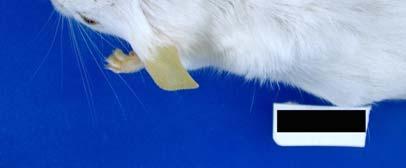
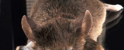
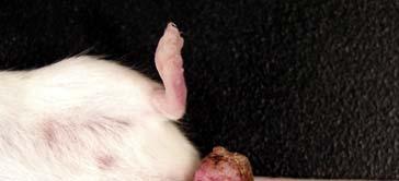
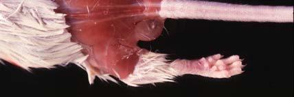
48 11 Eye Bilateral suppurative blepharoconjunctivitis or periocular dermatitis Etiology: Corynebacterium kutscheri; Staphylococcus spp.; Streptococcus spp.; Streptobacillus moniliformis; Pasteurella pneumotropica; Pseudomonas aeruginosa. 12 Eye Focal dorsal palpebral squamous papilloma 13 Ear Focal aural squamous papilloma 14 Ear Differential diagnoses: Auricular chondritis; pinnal squamous cell carcinoma; pinnal neural crest tumor (FVB). 15 Eye, ear, skin Icterus 16 Skin Name the condition: barbering, hair plucking, Dalia effect; trichotillomania Skin Unilateral pyogranulomatous perinasal dermatitis, furunculitis and cellulitis. Note barbering. Name the condition: Botryomycosis. Cause: Staphylococcus aureus (Pseudomonas aeruginosa). Histologic appearance: pyogranulomatous to necrosuppurative inflammation; gram positive cocci (gram negative rods); Splendora-Hoeppli material. 19 Skin Focal ulcerative nasal dermatitis Note barbering. Mechanical denudation from waters/cages. 20 Skin Multifocal ulcerative dermatitis of C57BL/6 mice Predisposing factor: Genetics (C57BL/6); ectoparasite sensitivity; secondary bacterial infection (Staphylococcus aureus or S. epidermidis); immune-mediated leukocytoclastic vasculitis. Exacerbated by scratching. La Perle-2011 CL Davis Gross-Rodents & Lagomorphs Page 2
49 21 Skin Focal dorsal cervical ulcerative dermatitis Cause: Fur mites (Myobia musculi; Myocoptes musculinus; Radfordia affinis). Differential diagnoses: Trauma; Staphylococcus aureus; S. xylosus; group G Streptococcus spp Skin Name the condition: Coryneform-associated hyperkeratosis. Etiology: Corynebacterium bovis. Predisposing factor: Immunodeficiency (not hairlessness). 24 Skin Multifocal cutaneous squamous papillomas. Cause: Carcinogen; mouse papillomavirus (Ingle, Veterinary Pathology, 48: , 2011); genotype (Tg.AC transgenic). 25 Skin, tail Name the condition: Ringtail. Predisposing factor: Low humidity; high temperature. Differential diagnoses: Strangulation by cotton nestlets in sucklings; frostbit; mousepox; Staphylococcal ulcerative/necrotizing dermatitis. 26 Skin, tail Multifocal ulcerative (nose, dorsum, tail) to proliferative (hind foot) dermatitis Etiologic diagnosis: Poxviral dermatitis; Ectromelial dermatitis. Name the condition: Mousepox. Cause: Ectromelia (mouse orthopox) virus. Histologic appearance: Epithelial ulceration and hyperplasia; epithelial ballooning degeneration; intracytoplasmic inclusion bodies. 27 Tail Coccygeal neural crest tumor FVB mice. Other common location is pinna of ear. 28 Mammary gland Widespread mammary gland hyperplasia. Predisposing factor: Pregnancy; pituitary pars distalis prolactinoma (FVB) Mammary gland Focal to multifocal mammary gland adenocarcinoma(s) Cause: Mouse mammary tumor retrovirus (MMTV); carcinogens; hormones (prolactin, progesterone, estrogen). Mammary tissue can be located anywhere on rodents! 32 Mammary gland/ Differential diagnoses: salivary gland myoepithelioma; mammary gland adenocarcinoma. La Perle-2011 CL Davis Gross-Rodents & Lagomorphs Page 3
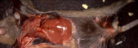
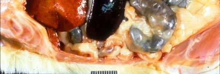
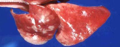
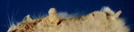
50 salivary gland 33 Preputial gland Unilateral preputial gland abscess or suppurative preputial adenitis Cause: Pasteurella pneumotropica; Klebsiella oxytoca; Staphylococcus spp.; Streptococcus spp. 34 Subcutis Widespread subcutaneous emphysema 35 Subcutis Widespread subcutaneous edema (anasarca) 36 Subcutis Multifocal subcutaneous hemangiosarcomas 37 Subcutis Focal subcutaneous angio-/lymphangiosarcoma Note the unilateral preputial gland abscess Subcutis Bilateral subcutaneous xenografts with unilateral ulceration Subcutis Focal lateral femoral hernia Comparative Medicine 58: , Heart Locally extensive epicardial mineralization Affected strains: BALB/c; C3H; DBA. Histochemical stain to aid in diagnosis: Von Kossa; Alizarin red. 44 Heart Left auricular thrombosis Pathogenesis: Amyloidosis proteinuria and hypoproteinemia loss of antithrombin III auricular thrombosis. 45 Lung Cause: Pneumocystis murina; acidophilic macrophage pneumonia; alveolar lipo-/proteinosis; pneumonia (paramyxo) virus of mice; Sendai (paramyxo) virus in immunodeficient mice; mouse norovirus-1 in immunodeficient mice. 46 Lung Focal pulmonary adenoma 47 Lung Multifocal pulmonary metastases (vertebral osteosarcoma) Various Lymphoma (lymph nodes = 48; thymus = 49; liver, spleen, peyer s patches and lymph nodes = 50) Cause: Moloney murine leukemia retrovirus (MuLV); radiation; chemicals; genotype (SCID and La Perle-2011 CL Davis Gross-Rodents & Lagomorphs Page 4
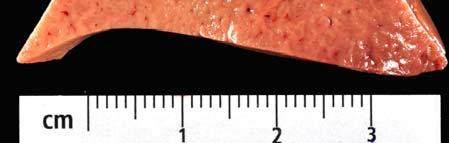
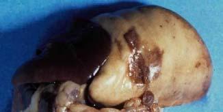
51 others) old age. 51 Liver and spleen Histiocytic sarcoma Associated findings: Renal tubular hyaline droplet formation; hepatic extramedullary hematopoiesis (erythroid); dysmyelopoiesis (decreased myeloid/increased erythroid) Liver Multifocal to coalescing suppurative to necrotizing hepatitis Cause: Ectromelia (mouse orthopox) virus; polytropic mouse hepatitis virus; Clostridium piliforme; salmonellosis; reovirus-3 (infants); cytomegalovirus (infants); adenovirus (infants). 54 Liver Multifocal to coalescing hepatic cysts Liver Focal hepatocellular adenoma (hepatoma; 55-56) or adenocarcinoma (57-58) Cause: Old age; Helicobacter hepaticus; carcinogen. 59 Liver Multifocal hepatic metastases (renal adenocarcinoma in TRAMP mice) 60 Liver Bilateral renal amyloidosis Pathogenesis: Chronic inflammation macrophages produce IL-1 and TNF serum precursor aposaa in liver degradation by macrophages AA amyloid deposition. Associated condition: Atrial thrombosis; nephrotic syndrome (proteinuria; hypoproteinemia; hypertriglyceridemia/hypercholesterolemia; edema/hydrothorax/ascites); uremia. 61 Kidney/ureter Unilateral hydronephrosis and hydroureter Cause: C3H, C57L, DDD strains; autosomal recessive mutation in C57BL/6; various genotypes; estrogen implantation in nude mice associated with urine retention. La Perle-2011 CL Davis Gross-Rodents & Lagomorphs Page 5
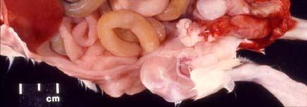
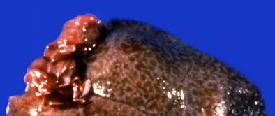
52 62 Kidney/urinary bladder Cystolithiasis; unilateral hydronephrosis; unilateral ascending pyelonephritis Cause: Proteus mirabilis; Enterococcus sp. and/or Klebsiella oxytoca in NOD-SCID-IL2Rγ (Foreman, Veterinary Pathology 48: , 2011) and C3H/HeJ (toll-like receptor 4 mutation). 63 Urinary bladder Cystolithiasis 64 Urinary bladder Name the condition: Mouse urologic syndrome. 65 Teeth Malocclusion Cause: Genetics (C57BL/6); trauma. 66 Teeth/subcutis Subcutaneous abscess extending from periodontitis, pulpitis and osteomyelitis 67 Salivary gland Salivary gland myoepithelioma Associated condition: Myeloid hyperplasia of bone marrow. 8/24 nonthymic tumors in SCID mice (Huang, Comparative Medicine 61: , 2011). 68 Esophagus Diffuse megaesophagus (lower right) Aging 129 mice +/- esophageal impaction and mortality. 69 Stomach Widespread gastric glandular hemorrhage and necrosis 70 Stomach Widespread gastric glandular hypertrophy and hyperplasia 71 Stomach Multifocal forestomach squamous papillomas Cause: Genotype (Tg.AC transgenic); carcinogen. 72 Body/GI Cause: Enterotropic mouse hepatitis virus; rotavirus A (epizootic diarrhea of infant mice-edim); salmonellosis; Clostridium piliforme; reovirus-3; mouse norovirus Intestine Multifocal intestinal adenomas Apc Min mouse. 74 Rectum Rectal prolapse Cause: Citrobacter rodentium (transmissible murine colonic hyperplasia [MAIDS, Fredrickson, La Perle-2011 CL Davis Gross-Rodents & Lagomorphs Page 6
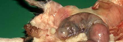
53 Veterinary Pathology 47: , 2010]); Escherichia coli (immunodeficient mice); Helicobacter spp. (bilis, typhlonius, mastomyrinus [telomerase deficient mice, Eaton, Veterinary Pathology 48: , 2011]); enterotropic mouse hepatitis virus (immunodeficient mice); pinworms. 75 Thyroid gland Bilateral papillary thyroid carcinomas ret/ptc1 transgenic mouse Pituitary gland (skull-76; brain-77) Pituitary gland adenoma (76) or adenocarcinoma with hemorrhage (77) 78 Ovary (whole body) Unilateral ovarian teratoma Histologic appearance: Presence of all 3 germ layers (endoderm, mesoderm, neuro-/ectoderm). 79 Ovaries and uterus Widespread cystic endometrial hyperplasia; Unilateral ovarian cyst 80 Uteri in vivo X 3 Muco-/hydrometra Predisposing factor: Imperforate vagina. 81 Uteri in vivo x 2 Multifocal segmental suppurative endometritis or pyometra Cause: Mycoplasma pulmonis; Klebsiella oxytoca; Pasteurella multocida; imperforate vagina. 82 Uterus in vivo Dystocia; vaginal prolapsed 83 Uterus in vivo Uterine histiocytic sarcoma 84 Penis Penile squamous papilloma 85 Testis (whole body) Unilateral testicular teratoma Most likely strain: Seminal vesicles Seminal vesicular dilatation (right) and contraction (left) La Perle-2011 CL Davis Gross-Rodents & Lagomorphs Page 7
54 87-90 Bulbourethral gland (whole body) Bilateral cystic bulbourethral glands (all) with unilateral hemorrhage and cholesterol granuloma (89-90) Note discoloration of left seminal vesicle in Reproductive tract (whole body-91) Brain (whole body- 93; skull-94) True hermaphrodite with: unilateral ovotestis and contralateral muco-hydrometra (91); unilateral testis, epididymis and seminal vesicle, and contralateral ovary and uterus Hydrocephalus (top-93; bottom-94) Common in C57BL/6. Fixed specimen (95). 96 Brain (head) Meningo-encephalocele 97 Skull (whole body) Unilateral pyogranulomatous mandibular osteomyelitis, cellulitis and dermatitis Name the condition: Botryomycosis. 98 Hindlimbs Bilateral tibiotarsal suppurative arthritis Cause: Corynebacterium kutscheri; Streptobacillus moniliformis; Mycoplasma pulmonis. 99 Vertebral column/ spinal cord Focal vertebral osteosarcoma with compression of subjacent spinal cord Fixed specimen. 100 RATS (slides # ) 101 Eyes (head) Chromodacryorrhea Cause: Rat sialodacryoadenitis coronavirus; sick rat! 102 Ear (head) Unilateral mild auricular chondritis Histologic appearance: Granulomatous inflammation with auricular chondrolysis. 103 Skin Keratoacanthoma Skin, tail Name the condition: Ringtail. Cause: Low humidity; genetics; high temperature; hydration status; nutritional status. Note gangrenous necrosis in 105. La Perle-2011 CL Davis Gross-Rodents & Lagomorphs Page 8
55 Mammary gland Focal subcutaneous fibroadenoma 108 Harderian gland Unilateral harderian gland necrosis with interstitial hemorrhage and edema Cause: Rat sialodacryoadenitis coronavirus. Fixed specimen. 109 Zymbal s gland Zymbal s gland adenocarcinoma 110 Preputial/clitoral gland Preputial/clitoral gland adenocarcinoma with ulceration 111 Scrotum Widespread scrotal dermal hemorrhage Cause: Kilham s rat parvovirus. 112 Lung Multifocal to coalescing necrosuppurative pneumonia Cause: Corynebacterium kutscheri; Name the condition: Pseudotuberculosis. Fixed specimen Lung Suppurative bronchopneumonia with bronchiectasis Cause: Mycoplasma pulmonis. Fixed specimen (114 and 116). 117 Lung Interstitial pneumonia Cause: Sendai (parainfluenza-1) virus; Pneumonia (paramyxo) virus of mice; Pneumocystis carinii ( rat respiratory virus ); rat coronavirus. 118 Lung Etiologic diagnosis: Pulmonary pneumocystosis. Cause: Pneumocystis carinii; P. wakefieldae. Histologic appearance: Intra-alveolar, foamy eosinophilic material with intralesional trophozoites and cysts OR lymphohistiocytic interstitial pneumonia ( rat respiratory virus ) [Clifford, Veterinary Pathology 46: , 2009; Livingston, Comparative Medicine 61: 45-59, 2011]. 119 Thorax Chylothorax 120 Liver Miliary necrotizing hepatitis Cause: Clostridium piliforme; Salmonella typhimurium; S. enteritidis. 121 Liver Differential diagnoses: Metastatic neoplasia (histiocytic sarcoma, lymphoma, fibrosarcoma); Hepatic abscess La Perle-2011 CL Davis Gross-Rodents & Lagomorphs Page 9
56 (Corynebacterium kutscheri, Streptococcus pneumoniae, Klebsiella pneumoniae, Pseudomonas aeruginosa). 122 Liver Cause: Cysticercus fasciolaris (larval stage of Taenia taeniaformis [cat tapeworm]). Associated lesion: Pulmonary arteriolar hypertrophy (Yi, Veterinary Pathology 47: , 2010); fibrosarcoma. 123 Kidney Name the condition: Chronic progressive nephropathy. Histologic appearance: Thickening of glomerular tufts by eosinophilic material, hyaline casts, dilated tubules with flattened epithelium, interstitial fibrosis, interstitial lymphoplasmacytic aggregates. Predisposing factor: Old age; male sex; Sprague Dawley strain; high protein diet; immune factors (IgM deposition); hormones (prolactin). 124 Kidney Unilateral nephroblastoma Precursor lesion in Sprague Dawleys is intralobar nephroblastematosis. 125 Urinary bladder Diffuse hemorrhagic cystitis Cause: Cystic calculi; Lewis/Brown Norway rats with hydronephrosis and renal papillary hyperplasia Urinary bladder and kidney (128) Cystolithiasis and unilateral nephrolithiasis with hydronephrosis (128) 129 Abdomen Large granular leukemia involving liver and spleen; icterus 130 Salivary gland Bilateral (left rat) salivary gland necrosis and edema Cause: Rat sialodacryoadenitis coronavirus. Sequela: Squamous metaplasia. 131 Stomach and duodenum 132 Intestines and mesentery Pancreaticoduodenal arterial aneurysm Mesenteric necrotizing polyarteritis with aneurysmal dilatation Cause: Polyarteritis nodosa. Sequela: Rupture with hemoabdomen. Name the condition: Polyarteritis nodosa. Medium-sized vessels in any tissue except lung. 133 Large intestine Etiologic diagnosis: Colonic nematodiasis or colonic oxyuriasis. Cause: Syphacia obvelata; S. muris; Aspiculuris tetraptera. La Perle-2011 CL Davis Gross-Rodents & Lagomorphs Page 10
57 134 Abdomen Hemoabdomen Cause: Polyarteritis nodosa with aneurysm and rupture Abdomen Peritoneal mesothelioma (epitheliomatous-136; sarcomatous-137) Fixed specimen. 137 Thyroid gland (pluck) Unilateral thyroid follicular adenoma 138 Adrenal glands Unilateral adrenomedullary pheochromocytoma (left); contralateral focal adrenocortical hyperplasia (right) 139 Pancreas (gastrointestinal tract) Focal pancreatic insulinoma Associated clinical pathologic finding: Hypoglycemia Pituitary (brain) Chromophobe adenoma of the pituitary pars distalis with compression of the overlying brain (141) Immunohistochemical finding: Immunoreactive for prolactin. 142 Testes (abdomen) Peritesticular, testicular, epididymal and scrotal hemorrhage Cause: Kilham s rat parvovirus. 143 Testis Testicular interstitial (Leydig) cell tumor Associated clinical pathologic finding: Hypercalcemia. 144 Testes Bilateral mesothelioma of the tunica vaginalis; unilateral (right) interstitial (Leydig) cell tumor; contralateral (left) testicular atrophy 145 Prostate gland (seminal vesicles, urinary bladder) Widespread suppurative prostatitis 146 Brain Multifocal cerebral and cerebellar hemorrhage and malacia Cause: Kilham s rat parvovirus. Sequela of neonatal infection: Cerebellar hypoplasia. Fixed specimen. La Perle-2011 CL Davis Gross-Rodents & Lagomorphs Page 11
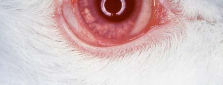
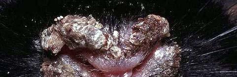
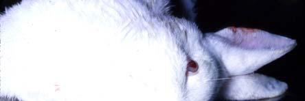
58 147 Brain Focal rostral cerebral meningeal granular cell tumor Histochemical appearance: PAS positive granules within neoplastic round cells. Ultrastructural appearance: Neoplastic round cells containing lysosomes. Note: Meninges of rat, uterus of mouse, bronchi of horse, tongue and meninges of dog. 148 RABBITS (slides # ) Eyes Unilateral (149-right) to bilateral buphthalmia Cause: Congenital (autosomal recessive). Pathogenesis: Absence or underdevelopment of outflow channel of iridocorneal angle intraocular pressure glaucoma. 151 Eye, nose Name the condition: Fibromatosis. Cause: Shope fibroma leporipoxvirus. Histologic appearance: Dense subcutaneous fibroblast proliferation with intracytoplasmic inclusion bodies and heterophilic inflammation. 152 Eye Name the condition: Myxomatosis. Cause: Myxomatosis leporipoxvirus. Histologic appearance: Proliferation of subcutaneous mesenchymal stellate cells interspersed within a mucinous/myxomatous matrix, degeneration to hyperplasia of overlying epidermis and follicular epithelium with intracytoplasmic inclusion bodies. 153 Head Torticollis Cause: Pasteurella multocida-induced unilateral. 154 Eye Lipid keratopathy Pathogenesis: LDL receptor deficient Watanabe rabbit hypercholesterolemia and hyperlipidemia lipid keratopathy. 155 Eye Episcleral abscess Cause: Pasteurella multocida. 156 Eye Cataract Cause: Encephalitozoon cuniculi. 157 Ears Unilateral (left) suppurative otitis media Cause: Pasteurella multocida. La Perle-2011 CL Davis Gross-Rodents & Lagomorphs Page 12
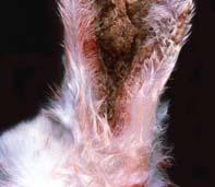
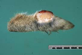
59 158 Ear Differential diagnoses: Shope fibroma; aural squamous papilloma Ear Proliferative otitis externa Etiologic diagnosis: Aural acariasis. Cause: Psoroptes cuniculi Skin, foot (161) Fibroma, carpal (161) or cutaneous/dermal (162) Cause: Shope fibroma leporipoxvirus. 163 Head Ventral cervical subcutaneous abscess Cause: Pasteurella multocida. 164 Skin (body) Exfoliative dermatitis Cause: Cheyletiella parasitovorax. OR Associated condition: Thymoma. 165 Perineum Ulcerative and exudative perineal dermatitis Cause: Treponema paraluiscuniculi. Name the condition: Venereal spirochetosis; vent disease; rabbit syphilis. Methods to confirm diagnosis: Warthin-Starry Histochemical stain; dark-field microscopy. 166 Feet Bilateral ulcerative pododermatitis Cause: Staphylococcus aureus. Predisposing factor: Poor sanitation; wire bottom cages; hereditary. Name the condition: Sore hocks. 167 Mammary gland Multifocal mammary gland dysplasia Associated condition: Prolactin-producing acidophil adenoma of the pituitary pars distalis. 168 Mammary gland Locally extensive necrohemorrhagic mastitis Cause: Pasteurella multocida; Staphylococcus aureus; Streptococcus pneumoniae. 169 Aorta Multifocal aortic atherosclerosis Pathogenesis: LDL receptor deficient Watanabe rabbit hypercholesterolemia and hyperlipidemia atherosclerosis. 170 Nose, front feet Mucopurulent rhinitis Cause: Pasteurella multocida. Name the La Perle-2011 CL Davis Gross-Rodents & Lagomorphs Page 13
60 condition: Snuffles. 171 Nose Nasal hyperkeratosis Cause: Treponema paraluiscuniculi. 172 Nose Name the condition: Myxomatosis. Differential diagnosis: Cyclosporine-induced gingival hyperplasia in New Zealand White causing ptyalism (Comparative Medicine 59: , 2009). 173 Nose Epistaxis Cause: Rabbit hemorrhagic disease calicivirus. 174 Nasal cavity Bilateral mucopurulent rhinosinusitis Cause: Pasteurella multocida. Name the condition: Snuffles. 175 Nasal cavity Bilateral turbinate atrophy Cause: Pasteurella multocida. Name the condition: Snuffles. 176 Lung Bilateral suppurative bronchopneumonia Cause: Pasteurella multocida; Bordatella bronchiseptica. 177 Lung Multifocal pulmonary abscesses Cause: Pasteurella multocida. 178 Lung Multifocal pulmonary hemorrhages Cause: Rabbit hemorrhagic disease calicivirus. 179 Lung Pulmonary metastases of uterine adenocarcinoma 180 Thymus (pluck) Thymoma Associated condition: Exfoliative dermatitis. 181 Teeth Malocclusion 182 Tongue Ventral lingual papillomatosis Cause: Rabbit oral papillomavirus. 183 Stomach Gastric trichobezoar Possible sequela: Gastric rupture with peritonitis. Predisposing factor: Excessive grooming; insufficient roughage; decreased gastric motility; La Perle-2011 CL Davis Gross-Rodents & Lagomorphs Page 14
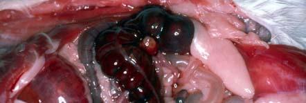
61 sedentary lifestyle. 184 Stomach Multifocal gastric ulcers 185 Stomach Multifocal gastric lymphoma 186 Intestines, mesentery Proliferative enteritis Cause: Lawsonia intracellularis. Histochemical stain: Warthin-Starry silver stain. 187 Intestinal tract Necrohemorrhagic enterotyphlocolitis Cause: Clostridium piliforme. Name the condition: Tyzzer s disease. 188 Intestinal tract Hemorrhagic typhlitis Cause: Clostridium piliforme; C. difficile; C. perfringens; C. piliforme; Escherichia coli; Klebsiella pneumoniae; Eimeria intestinalis; E. flavenscens. 189 Intestinal tract Necrohemorrhagic typhlitis Cause: Clostridium spiroforme. Pathogenesis: Disruption of gut flora bacterial colonization and proliferation spiroforme iota toxin production secretory and vascular changes necrosis and hemorrhage. 190 Large intestine Name the condition: Mucoid enteropathy; mucoid enteritis; bloat; hypoamylasemia. Fixed specimen. 191 Abdomen Miliary necrotizing hepatitis; necrohemorrhagic typhlocolitis Cause: Clostridium piliforme. Name the condition: Tyzzer s disease. 192 Thorax, abdomen Pulmonary congestion, hemorrhage and edema; renal hemorrhage; multifocal hepatic necrosis Cause: Rabbit hemorrhagic disease calicivirus. 193 Abdomen Lymphoma involving the liver, spleen and Peyer s patch 194 Abdomen Carcinomatosis (uterine origin) La Perle-2011 CL Davis Gross-Rodents & Lagomorphs Page 15
62 Liver Hepatic lipidosis Predisposing factor: Obesity; fasting; pregnancy/postpartum; hereditary (LDL receptor deficient Watanabe). 197 Liver Miliary to coalescing necrotizing hepatitis Cause: Clostridium piliforme; Francisella tularensis; Listeria monocytogenes; Yersinia pseudotuberculosis; Staphylococcus aureus Liver Multifocal biliary cystic ectasia ( ) to diffuse biliary papillary hyperplasia or proliferative cholangitis with cystic ectasia ( ) Cause: Eimeria stiedae. Name the condition: Hepatic coccidiosis. 202 Liver Multifocal biliary cysts 203 Kidney Granulomatous interstitial nephritis with cortical atrophy and fibrosis Cause: Encephalitozoon cuniculi. 204 Kidney Bilateral diffuse renal corticomedullary tubular lipidosis Pathogenesis: LDL receptor deficient Watanabe rabbit hypercholesterolemia and hyperlipidemia atherosclerosis. 205 Kidney Bilateral widespread renal lymphoma 206 Kidney Nephroblastoma 207 Pituitary, brain Acidophil adenocarcinoma of the pituitary pars distalis with compression of the overlying brain Immunohistochemical feature: Immunoreactive for prolactin. Associated condition: Mammary gland dysplasia. Fixed specimen Uterus Multifocal endometrial venous aneurysms Associated clinical sign: Vaginal hemorrhage; hematuria. Fixed specimen (209). La Perle-2011 CL Davis Gross-Rodents & Lagomorphs Page 16
63 210 Uterus Muco-/hydrometra Uterus Focal (211) to multicentric (212) uterine adenocarcinoma 213 Uterus, cervix, vagina, vulva Vaginal prolapsed Fixed specimen. 214 Prepuce, penis Chronic ulcerative and proliferative balanoposthitis Cause: Treponema paraluiscuniculi. 215 GUINEA PIGS (slides # ) 216 Eye Ocular dermoid 217 Eye Seropurulent conjunctivitis Cause: Chlamydophila caviae. 218 Ears, brain Bilateral suppurative otitis media Cause: Streptococcus pneumoniae; S. zooepidemicus; Bordatella bronchiseptica; Pseudomonas aeruginosa. Fixed specimen. 219 Skin (body) Etiologic diagnosis: Cutaneous dermatophytosis. Cause: Trichophyton mentagrophytes; Microsporum canis. 220 Skin (body) Etiologic diagnosis: Cutaneous demodecosis. Cause: Demodex caviae. 221 Skin (body) Name the condition: Scabies; sarcoptic mange. Cause: Trixacarus caviae. 222 Foot Multifocal ulcerative pododermatitis Cause: Staphylococcus aureus. Predisposing factor: Trauma; poor sanitation. Name the condition: Bumblefoot. 223 Mammary gland Focal necrohemorrhagic mastitis Cause: Escherichia coli; Klebsiella pneumoniae; Streptococcus zooepidemicus. 224 Heart Widespread myocardial rhabdomyomatosis Histologic appearance: Vacuolated cardiac La Perle-2011 CL Davis Gross-Rodents & Lagomorphs Page 17
64 myofibers containing granular to eosinophilic cytoplasm (glycogen). Histochemical stain: PAS. 225 Heart (pluck) Hemopericardium 226 Aorta Segmental aortic mineralization Name the condition: Metastatic mineralization. Cause: Diets low in calcium and/or magnesium, or high in phosphorus. 227 Lung Bilateral suppurative bronchopneumonia Cause: Bordatella bronchiseptica. 228 Lung Bilateral necrotizing bronchiolitis Cause: Guinea pig/caviae adenovirus. Rib impressions suggest concurrent interstitial disease due to parainfluenza-3 paramyxovirus infection. Fixed specimen Head, lymph nodes Multifocal suppurative cervical lymphadenitis Cause: Streptococcus zooepidemicus. 231 Lymph node Widespread suppurative lymphadenitis Cause: Streptococcus zooepidemicus. 232 Intestinal tract, lymph node Necrosuppurative or caseating mesenteric lymphadenitis Cause: Yersinia pseudotuberculosis. Name the condition: Pseudotuberculosis. 233 Head, lymph nodes Lymph nodes, lymphoma 234 Liver Hepatic lipidosis Name the condition: Pregnancy toxemia or ketosis; fasting ketosis or metabolic/nutritional form (no necrosis). Clinical pathological finding: Hypoglycemia; ketosis; hyperlipidemia. 235 Liver Multifocal hepatic abscesses Cause: Yersinia pseudotuberculosis. 236 Kidney Name the condition: Segmental nephrosclerosis. Fixed specimen. La Perle-2011 CL Davis Gross-Rodents & Lagomorphs Page 18
65 237 Teeth Malocclusion Name the condition: Slobbers. 238 Ribs, colon, kidney, stomach, spleen Intercostal myofiber mineralization; colonic mineralization; gastric mineralization Name the condition: Metastatic mineralization. Cause: Diets low in calcium and/or magnesium, or high in phosphorus Ovaries, uterus Bilateral cystic rete ovarii 241 Uterus, ovaries Multifocal suppurative endometritis Cause: Bordatella bronchiseptica; Streptococcus zooepidemicus; S. pneumoniae. 242 Body Multifocal intramuscular and periarticular hemorrhages Name the condition: Scurvy. Pathogenesis: dietary vitamin C lack of L-gulonolactone oxidase L-ascorbic acid lack of activation of prolyl and lysyl hydroxylases hydroxylation of procollagen inadequate collagen cross linkage impaired formation of collagenous support of blood vessels. Other species affected: Primates; fruit-eating bats; some birds; some fish; cetaceans (whales/dolphins). 243 GERBILS (slides # ) 244 Skin, nose Nasal ulcerative to exudative dermatitis Pathogenesis: Porphyrin-containing Harderian gland secretions nasolacrimal duct nares to no grooming chemical irritation dermatitis +/- Staphylococcus xylosus or S. aureus infection. Diagnostic test: UV fluorescence (+ culture). 245 Spleen Splenic amyloidosis (top) Majority of cases are secondary to chronic renal La Perle-2011 CL Davis Gross-Rodents & Lagomorphs Page 19
66 disease (glomerulonephropathy). 246 Liver (abdomen) Multifocal necrotizing hepatitis Cause: Clostridium piliforme. Name the disease: Tyzzer s disease. Differential diagnosis: Salmonella typhimurium. 247 Adrenal gland, kidneys, ovaries, uterus, bladder Unilateral adrenocortical adenoma; bilateral ovarian cysts 248 Ovaries (body) Bilateral ovarian cysts 249 Kidneys, ovaries, uterus Unilateral ovarian granulose cell tumor Body Conjoined twins Fixed specimen. 252 HAMSTERS (slides # ) 253 Skin (body) Etiologic diagnosis: Cutaneous demodecosis. Cause: Demodex criceti (epidermal pits); D. aurati (hair follicles and sebaceous ducts) Skin (body) Multifocal cutaneous/epidermotropic lymphoma Name the disease: Mycosis fungoides. Note: Hamster polyoma virus causes transmissible (visceral) lymphoma. 256 Skin (body) Multifocal cutaneous trichoepitheliomas Cause: Hamster polyoma virus. 257 Subcutis (body) Diffuse ventral subcutaneous edema (anasarca) Cause: Glomerulonephropathy; amyloidosis; atrial thrombosis; polycystic liver disease; cirrhosis; proliferative ileitis. 258 Heart Left auricular/atrial thrombosis Cause: Amyloidosis; glomerulonephropathy. La Perle-2011 CL Davis Gross-Rodents & Lagomorphs Page 20
67 Liver Multifocal to coalescing( 260) hepatic cysts with atrophy and loss (260) Name the condition: Polycystic disease. Color change due to autolysis (259). Fixed specimen (260). 261 Liver Hepatic amyloidosis 262 Kidneys Bilateral renal amyloidosis 263 Kidneys Bilateral renal glomerulonephropathy (right) Sequela: Nephrotic syndrome (anasarca); uremia; atrial thrombosis. 264 Kidneys Bilateral renal glomerulonephropathy (upper right); bilateral renal amyloidosis (below) 265 Perineum Name the condition: Wet tail. Cause: Clostridium piliforme; Escherichia coli; C. difficile; Salmonella enteritidis; S. typhimurium; Lawsonia intracellularis. 266 Intestinal tract (body) Intestinal tract (abomen) 269 Adrenal gland, kidneys 270 Male caudal abdomen Hemorrhagic enterocolitis Proliferative ileitis (267)/enteritis (268) Unilateral adrenocortical carcinoma with hemorrhage Peri-testicular/epididymal cysts Cause: Clostridium difficile. Cause: Lawsonia intracellularis. Histochemical stain: Warthin-Starry silver stain. Name the condition: Polycystic disease. 271 PHOTO CREDITS 272 THANK YOU! QUESTIONS? La Perle-2011 CL Davis Gross-Rodents & Lagomorphs Page 21
2015 Lab Animal 8/22/ Mouse. 2. Rat. 3. Mouse. Tissue from an Mouse: Morphologic Diagnosis: Rectal prolapse
 1. Mouse 2015 Lab Animal 2. Rat Tissue from an Mouse: Morphologic Diagnosis: Rectal prolapse Causes: 1. Syphacia obveluta 2. Citrobacter rodentum 3. Heicobacter sp 3. Mouse 2. Tissue from a Rat: Morphologic
AFIP MINIBOARD EXAMINATION MAY 2008 LAB ANIMAL PATHOLOGY
 AFIP MINIBOARD EXAMINATION MAY 2008 LAB ANIMAL PATHOLOGY 1. Hallmark lesions of infection with polytropic strains of mouse hepatitis virus include: A. Lymphocytic syncytia in mesenteric lymph nodes B.
AUSTRALIAN PHENOMICS FACILITY HEALTH REPORTS
 S Zone 2 - Level 3 High - High Barrier - No Researcher Access Room - 3.71, 3.72, 3.75, 3.76, 3.79, 3.80, 3.83, 3.84, 3.87, 3.88 Random Sampling and Sentinel screening by Cerberus Sciences Last Test Date
AUSTRALIAN PHENOMICS FACILITY HEALTH REPORTS
 AUSTRALIAN PHENOMICS FACILITY HEALTH REPORTS Zone 2 - Level 3 High - High Barrier - No Researcher Access Room - 3.71, 3.72, 3.75, 3.76, 3.79, 3.80, 3.83, 3.84, 3.87, 3.88 Random Sampling and Sentinel screening
2014 Descriptive Vet Path Course. Gross Exam #2 KEY
 2014 Descriptive Vet Path Course Gross Exam #2 KEY 2014 DESCRIPTIVE VETERINARY PATHOLOGY COURSE GROSS EXAM #2 1. Tissue from a chicken. Morphologic Diagnosis: Multifocal proliferative and ulcerative facial
Table of Contents. Preface xi. Acknowledgments xiii. Part I Overview of the Diagnostic Process 1. 1 Overview of Grading and Staging 3
 Table of Contents Preface xi Acknowledgments xiii Part I Overview of the Diagnostic Process 1 1 Overview of Grading and Staging 3 Identification of the process 3 Identification of tumor types 5 Grading
Pathology of the Alimentary Tract
 Pathology of the Alimentary Tract Lab 2: Lower alimentary tract SI, LI, cecum, and peritoneum GIST in the cecum of a dog Shannon Martinson: http://people.upei.ca/smartinson VPM 221: November, 2011 3 year
Vascular Related Torsion Venous compression Hemorrhagic infarct Young men At night Very painful Can be reduced Scrotal Masses Testicular Tumors (solid
 Pathology of the Male Reproductive System Testis and Epididymis Failure of Testis to Descend Testis are not always in scrotum at birth. Testes from in abdomen with kidneys Migrate to scrotum May get stuck
Theses for the Final Exam of Pathology
 Theses for the Final Exam of Pathology (Asterisks indicate the pool of theoretical questions for the semi-final practical exam) GENERAL PATHOLOGY I. POSTMORTEM SIGNS NECROSIS 1. Postmortem changes, causes
Pathology of the Liver and Biliary Tract 5 Diseases of the Biliary Tract. Shannon Martinson, March 2017
 Pathology of the Liver and Biliary Tract 5 Diseases of the Biliary Tract Shannon Martinson, March 2017 http://people.upei.ca/smartinson/ OUTLINE Normal anatomy & function Hepatobiliary injury and responses
IDEXX BioAnalytics Case # Received: 9/18/2018 Completed: 9/26/2018 Submitted By Justin Wilson
 FINAL REPORT OF LABORATORY EXAMINATION 4011 Discovery Drive, Columbia, MO 65201 1-800-669-0825 1-573-499-5700 idexxbioanalytics@idexx.com www.idexxbioanalytics.com IDEXX BioAnalytics Case # 26931-2018
Spontaneous Neoplasms and Survival in Wistar Han Rats: Compilation of Control Group Data. March, 2003
 Spontaneous Neoplasms and Survival in Wistar Han Rats: Compilation of Control Group Data March, 2003 Information Prepared by Mary L.A. Giknis Ph.D Charles B. Clifford D.V.M, Ph.D TABLE OF CONTENTS INTRODUCTION...1
-1- Pathology Department (code: 0605) Final Exam for Third year students Date: Time allowed: 2 hours. Paper II (75 marks).
 Final Exam for Third year students Date: Time allowed: 2 hours. Paper II (75 marks).") -1- BENHA UNIVERSITY FACULTY OF MEDICINE Pathology Department (code: 0605) Final Exam for Third year students Date: 28-5-2011 Time allowed: 2 hours. Paper II (75 marks). Please note that this question
-1- BENHA UNIVERSITY FACULTY OF MEDICINE Pathology Department (code: 0605) Final Exam for Third year students Date: 28-5-2011 Time allowed: 2 hours. Paper II (75 marks). Please note that this question
Respiratory Pathology Lab 2: Lung. Shannon Martinson,
 Respiratory Pathology Lab 2: Lung Shannon Martinson, 2017 http://people.upei.ca/smartinson/ Case 1 Signalment: 9 month old DSH cat History: Poor doer with stunted growth One month of lethargy one day the
Price List. Effective 1 October Effective 1 October SAHMRI 2126/B SAHMRI 2126/B
 Price List Effective 1 October 2013 Effective 1 October 2013 1 SEROLOGY EIA $14.50 PER INDIVIDUAL TEST*** IFA $25.00 PER INDIVIDUAL TEST*** ***A 20% discount is applied to each submission with 30 or more
Cellular Pathology Gross Pathology Laboratory 2 Cell Injury. VPM 152: General Pathology Instructor: Chelsea Martin Winter 2016
 Cellular Pathology Gross Pathology Laboratory 2 Cell Injury VPM 152: General Pathology Instructor: Chelsea Martin Winter 2016 Gross Specimens The following slides consist of images from the specimens presented
Learning Outcomes: The following list provides the learning objectives that will be covered in the lectures, and tutorials of each week:
 Course Code Course Title ECTS Credits MED-309 Pathology II 6 School Semester Prerequisites Medical School Spring (Semester 6) MED-304 Pathology I Type of Course Field Language of Instruction Required Medicine
IDEXX BioResearch Case # Received: 3/3/2014 Completed: 3/13/2014 Submitted By Sentinels
 FINAL REPORT OF LABORATORY EXAMINATION 4011 Discovery Drive, Columbia, MO 65201 1-800-669-0825 1-573-499-5700 idexx-radil@idexx.com www.idexxbioresearch.com IDEXX BioResearch Case # 8518-2014 Received:
2014 SEVPAC Case #63 (Slide ID: #1)
") 2014 SEVPAC Case #63 (Slide ID: #1) Tuskegee University College of Veterinary Medicine Dr. Ebony Gilbreath Tissues submitted to TUSVM diagnostic services for histopathology Puppies 4 weeks of age From
2014 SEVPAC Case #63 (Slide ID: #1) Tuskegee University College of Veterinary Medicine Dr. Ebony Gilbreath Tissues submitted to TUSVM diagnostic services for histopathology Puppies 4 weeks of age From
Inflammation Laboratory 3 Emphasis: Chronic inflammation and healing. Shannon Martinson: VPM 152: April 2013
 Inflammation Laboratory 3 Emphasis: Chronic inflammation and healing Shannon Martinson: http://people.upei.ca/smartinson VPM 152: April 2013 Example A Reproductive tract and colon/rectum from a sheep Previous
Price List Effective 1 September 2016
 Price List Effective 1 September 2016 SEROLOGY EIA $15.20 PER INDIVIDUAL TEST*** IFA $26.30 PER INDIVIDUAL TEST*** ***A 10% discount is applied to each submission with 30 or more serum samples for an
Pathology of the Respiratory System 4: Pneumonia
 Pathology of the Respiratory System 4: Pneumonia Shannon Martinson, March 2016 http://people.upei.ca/smartinson/ VPM 222 Systemic Pathology LUNG PNEUMONIA Review Classification of Pneumonia Diffuse LUNG
PATHOLOGY OF GUINEA PIGS, HAMSTERS and GERBILS
 PATHOLOGY OF GUINEA PIGS, HAMSTERS and GERBILS Instructor: Bruce H. Williams, DVM, DACVP Dept. of Veterinary Pathology, AFIP (202) 782-2650 Email: Williamsb@afip.osd.mil PURPOSE The purpose of this 1.5-hour
Overview of Anatomy & Physiology
 Overview of Anatomy & Physiology Anatomy the study of the structure of body parts and their relationships to one another Gross or macroscopic Microscopic Developmental Physiology the study of the function
Pathology of the Liver and Biliary Tract 5 Diseases of the Biliary Tract. Shannon Martinson, April 2016
 Pathology of the Liver and Biliary Tract 5 Diseases of the Biliary Tract Shannon Martinson, April 2016 http://people.upei.ca/smartinson/ OUTLINE Normal anatomy & function Hepatobiliary Injury and responses
DEGENERATION NECROSIS AND INFILTRATION
 DEGENERATION NECROSIS AND INFILTRATION Cellular Degenerations and Infiltrations 1. Cloudy swelling and hydropic degeneration Cloudy swelling and hydropic degeneration occur when the regulatory mechanisms
AFIP MINIBOARD EXAMINATION MAY 2007 DOG/CAT PATHOLOGY
 AFIP MINIBOARD EXAMINATION MAY 2007 DOG/CAT PATHOLOGY 1. is a specific and sensitive marker for canine transitional epithelial (urothelial) neoplasms, and is the marker of choice for diagnostic purposes.
Pathology of the Respiratory System 5: Lung and Thoracic Cavity
 Pathology of the Respiratory System 5: Lung and Thoracic Cavity Shannon Martinson, Jan 2017 http://people.upei.ca/smartinson/ VPM 222 Systemic Pathology DISORDERS OF THE LUNG Congenital Pigmentary deposition
INTRODUCTION TO ANIMALS
 AP BIOLOGY ANIMALS ACTIVITY #1 NAME DATE HOUR INTRODUCTION TO ANIMALS LEVELS OF ORGANIZATION Animals Activity #1 page 1 HOMEOSTASIS: DEFINITION IMPORTANCE MECHANISMS FOR MAINTAINING HOMEOSTASIS: Animals
SCOPE OF PRACTICE PGY-5
 Recognize normal cytomorphology of cells derived from the respiratory, gastrointestinal, and genitourinary tracts, and body fluid (Cerebrospinal fluid, pleural and peritoneal fluid) Recognize normal cytomorphology
Spontaneous Neoplastic Lesions in the Crl:CD-1 (ICR)BR Mouse. March, 2000
BR Mouse. March, 2000") Spontaneous Neoplastic Lesions in the Crl:CD-1 (ICR)BR Mouse March, 2000 Information Prepared by Mary L. A. Giknis, Ph.D. Charles B. Clifford, D.V.M., Ph.D. CHARLES RIVER LABORATORIES TABLE OF CONTENTS
Spontaneous Neoplastic Lesions in the Crl:CD-1 (ICR)BR Mouse March, 2000 Information Prepared by Mary L. A. Giknis, Ph.D. Charles B. Clifford, D.V.M., Ph.D. CHARLES RIVER LABORATORIES TABLE OF CONTENTS
8. POST- MORTEM PROTOCOL (John Trupkiewicz)
") 8. POST- MORTEM PROTOCOL (John Trupkiewicz) 8.1. GENERAL COMMENTS Those individuals who may be charged with performing a necropsy examination should formulate a plan of action in the case of the sudden,
8. POST- MORTEM PROTOCOL (John Trupkiewicz) 8.1. GENERAL COMMENTS Those individuals who may be charged with performing a necropsy examination should formulate a plan of action in the case of the sudden,
WSC , Conference 9, Case 1. Tissue from a nyala.
 WSC 2009-2010, Conference 9, Case 1. Tissue from a nyala. MICROSCOPIC DESCRIPTION: Heart, atrium (1 pt.): Approximately 40% of the atrial myocardium is replaced by areas of fibrous connective tissue (1
SHN-1 Human Digestive Panel Test results
 SHN-1 Human Digestive Panel Test results HN-30 tongue HN-24 salivary gland HN-12 larynx HN-28 esophagus HN-29 stomach HN-20 pancreas HN-13 liver HN-14 gall bladder HN-27-1 duodenum HN-27-2 ileum HN-27-3
Liver Lab #2. Bacterial Hepatitis
 Liver Lab #2 Bacterial Hepatitis Case: O12561-04. Adult ewe. Describe the lesion: Multifocal large nodules ranging in size from 1-3.5cm in greatest diameter are present within the liver and are filled
Diagnostic Cytology of Cancer Cases
 Diagnostic Cytology of Cancer Cases Somporn Techangamsuwan Companion Animal Cancer Research Unit (CAC-RU) Department of Pathology, Faculty of Veterinary Science, Chulalongkorn University 1 Tumor or Non-tumor
Cellular Pathology. Histopathology Lab #2 (web) Paul Hanna Jan 2018
 Paul Hanna Jan 2018") Cellular Pathology Histopathology Lab #2 (web) Paul Hanna Jan 2018 Slide #91 Clinical History: a necropsy was performed on an aged cat the gross pathological changes included: widespread subcutaneous edema
Cellular Pathology Histopathology Lab #2 (web) Paul Hanna Jan 2018 Slide #91 Clinical History: a necropsy was performed on an aged cat the gross pathological changes included: widespread subcutaneous edema
3 Circulatory Pathways
 40 Chapter 3 Circulatory Pathways Systemic Arteries -Arteries carry blood away from the heart to the various organs of the body. -The aorta is the longest artery in the body; it branches to give rise to
WSC , Conference 9. Case 1. Tissue from a rhesus macaque.
 Case 1. Tissue from a rhesus macaque. MICROSCOPIC DESCRIPTION: Esophagus: There is multifocal loss of the mucosal lining (1 pt). In these areas, the denuded subepithelial fibrous connective tissue is infiltrated
Post-mortem Examinations. Floron C. Faries, Jr. DVM, MS
 Post-mortem Examinations Floron C. Faries, Jr. DVM, MS Objectives Define necropsy Discuss the importance of post-mortem examinations to veterinarians Discuss the process of site selection for postmortems
Appendix 5. EFSUMB Newsletter. Gastroenterological Ultrasound
 EFSUMB Newsletter 87 Examinations should encompass the full range of pathological conditions listed below A log book listing the types of examinations undertaken should be kept Training should usually
Respiratory Pathology. Kristine Krafts, M.D.
 Respiratory Pathology Kristine Krafts, M.D. Normal lung: alveolar spaces Respiratory Pathology Outline Acute respiratory distress syndrome Obstructive lung diseases Restrictive lung diseases Vascular
Gastrointestinal Pathology of Pigs. Jerome C. Nietfeld, DVM, MS, PhD Kansas State Veterinary Diagnostic Lab Department DMP Kansas State University
 Gastrointestinal Pathology of Pigs Jerome C. Nietfeld, DVM, MS, PhD Kansas State Veterinary Diagnostic Lab Department DMP Kansas State University Neonatal Diarrhea Likely the number 1 killer of neonatal
2013 Descriptive Vet Path Course. Gross Exam #1 KEY
 2013 Descriptive Vet Path Course Gross Exam #1 KEY 2013 DESCRIPTIVE VETERINARY PATHOLOGY COURSE GROSS EXAM #1 1. Tissue from a horse. Morphologic Diagnosis: Diffuse macronodular hepatocellular regeneration,
Dean s Signature: Date Reviewed: / /
 Fall 2015 22TBio 142 22THuman Anatomy and Physiology II Faculty Name: Virginia Garden Program Head: Virginia Garden Dean s Review: Dean s Signature: Date Reviewed: / / Revised: Semester/Year 22TBio 142
C.L. Davis Foundation Descriptive Veterinary Pathology Course
 C.L. Davis Foundation 2015 Descriptive Veterinary Pathology Course IHC Resources IHC Identification Targets Antibodies Antibodies 1 Antibodies Specimens Antigen Retrieval Unmasks antigen epitopes Methods
Pneumonia Virus of Mice
 Pneumonia Virus of Mice Host species mouse, rat, hamster, guinea pig, (rabbit) Organotropism respiratory tract Clinical disease asymptomatic in euthymic animals (Smith, et al., 1984) chronic pneumonia
Figure 2: Lymph node Cortical follicular (F) and paracortical (PC) atrophy, with narrowing of the cortex relative to the medulla (M).
 and paracortical (PC) atrophy, with narrowing of the cortex relative to the medulla (M).") Figure 1: Lymph node Follicular hyperplasia, with expansion of the follicular germinal centres (F) by large blast cells. Paracortical hyperplasia, with expansion of the paracortex (PC) by small lymphocytes.
Figure 1: Lymph node Follicular hyperplasia, with expansion of the follicular germinal centres (F) by large blast cells. Paracortical hyperplasia, with expansion of the paracortex (PC) by small lymphocytes.
What s your diagnosis? Malori Marotz. Squirt, an 8month old mix breed puppy. History:
 What s your diagnosis? Malori Marotz Squirt, an 8month old mix breed puppy History: The owner obtained squirt at 12 weeks of age. The owner reported that Squirt was passing soft stools lately and he is
Pathology of the Hematopoietic System GROSS/HISTO LAB
 Pathology of the Hematopoietic System GROSS/HISTO LAB Paul Hanna (thanks to Dr s Aburto, Martinson & Fenton) Fall 2014 Slide 1 Spleen from a Beaver Give a morphologic diagnosis and possible etiology &
S2 File. Clinical Classifications Software (CCS). The CCS is a
. The CCS is a") S2 File. Clinical Classifications Software (CCS). The CCS is a diagnosis categorization scheme based on the ICD-9-CM that aggregates all diagnosis codes into 262 mutually exclusive, clinically homogeneous
S2 File. Clinical Classifications Software (CCS). The CCS is a diagnosis categorization scheme based on the ICD-9-CM that aggregates all diagnosis codes into 262 mutually exclusive, clinically homogeneous
Fig. A.1. Frontal. plane. Transverse. plane. Sagittal plane. Copyright McGraw-Hill Education. Permission required for reproduction or display.
 Fig. A.1 Frontal plane Transverse plane Sagittal plane McGraw-Hill Education/Joe DeGrandis Fig. A.2 (a) Sagittal section (b) Frontal section (c) Transverse section Table A.1 Fig. A.3 Cephalic r. (head)
like humans, have well-developed mediastinal separation between the left and right hemithorax, thus unilateral changes can occur. On the other hand,
 Tutorial Module 6 Thoracic Cavity and Tumors of Lung and Pleura Alfonso López Atlantic Veterinary College University of Prince Edward Island Canada 2009 Enero 3 Thoracic Cavity There are significant anatomical
Charles Halsey, DVM, PhD, DACVP Pfizer, Inc. IHC Resources
 Charles Halsey, DVM, PhD, DACVP Pfizer, Inc. IHC Resources 1 IHC Identification Targets Specimens Controls 2 Tissue controls Trouble Spots 3 The Key to Description IHC Description 4 Intermediate Filaments
Descriptive Histology
 Atlas of Descriptive Histology Michael H. Ross University of Florida College of Medicine Gainesville, Florida Wojciech Pawlina Mayo Medical School College of Medicine, Mayo Clinic Rochester, Minnesota
Extracellular degeneration
 Extracellular degeneration By Dr. Hemn Hassan Othman PhD, Pathology Fall 2016 1/17/2017 1 Extracellular Degenerations I / Hyaline Degeneration (Hyalinization): The ward hyaline is derived from the Latin
RADPrimer Curriculum Breast Topics Covered Basic Intermediate 225
 Breast Anatomy & Normal Variants 11 Breast Imaging Modalities 13 BI RADS Lexicon 3 Mammography: Masses 9 Mammography: Calcifications 17 Mammography: Additional Findings 8 Ultrasound Features 10 Ultrasound
Alveolar Histiocytosis. Non-infectious conditions of Rats. Alveolar Histiocytosis
 Non-infectious conditions of Rats Charles B Clifford, DVM, PhD, DACVP Charles River Alveolar Histiocytosis Synonym: Alveolar lipidosis Prevalence: Very high in aged rats, moderate in younger animals may
Epithelial tumors. Dr. F.F. Khuzin, PhD Dr. M.O. Mavlikeev
 Epithelial tumors Dr. F.F. Khuzin, PhD Dr. M.O. Mavlikeev Epithelial tumors Tumors from the epithelium are the most frequent among tumors. There are 2 group features of these tumors: The presence in most
Crosswalk File of ICD9 Diagnosis Codes to Risk Group Assignment 1-Apr-15
 1 1500 MALIGNANT NEOPLASM OF CERVICAL ESOPHAGUS 1 1501 MALIGNANT NEOPLASM OF THORACIC ESOPHAGUS 1 1502 MALIGNANT NEOPLASM OF ABDOMINAL ESOPHAGUS 1 1503 MALIGNANT NEOPLASM OF UPPER THIRD OF ESOPHAGUS 1
Premium Specialty: Pediatrics
 Premium Specialty: Pediatrics Credentialed Specialties include: Adolescent Medicine, Pediatric Adolescent, and Pediatrics This document is designed to be used in conjunction with the UnitedHealth Premium
Health status. Microbial impact on research. Mouse hepatitis in the nude mouse. Microbiology Impact of infections on research
 Health status Micriobial status Microbial impact on research Specific infections Normal flora Axel Kornerup Hansen, Professor, DVM, Dr.Med.Vet. Department of Veterinary Disease Biology Section of Biomedicine
Inflammation Laboratory 2. Shannon Martinson: VPM 152: March 2012
 Inflammation Laboratory 2 Shannon Martinson: http://people.upei.ca/smartinson VPM 152: March 2012 Reminder - Creating a Morphologic Diagnosis for Inflammatory Lesions Organ and Process Exudate Distribution
IDEXX BioResearch Case # Received: 12/10/2014 Completed: 12/22/2014 Submitted By Beata Muszynska-Furas
 FINAL REPORT OF LABORATORY EXAMINATION Mörikestr. 28/3, D 71636 Ludwigsburg, Germany 001-877-635-4036 (International toll free number) IdexxBioresearch-Europe@idexx.com www.idexxbioresearch.com/europe
Inflammation Laboratory 1
 Inflammation Laboratory 1 Lab1 Emphasis: The exudates of acute inflammation Descriptions Morphologic Diagnoses Shannon Martinson: http://people.upei.ca/smartinson VPM 152: February 2012 Describing Lesions
Spontaneous Neoplastic Lesions in the CrI:CD-1(ICR) Mouse in Control Groups from 18 Month to 2 year Studies. March, 2005
 Mouse in Control Groups from 18 Month to 2 year Studies. March, 2005") Spontaneous Neoplastic Lesions in the CrI:CD-1(ICR) Mouse in Control Groups from 18 Month to 2 year Studies March, 2005 Information Prepared by Mary L.A. Giknis Ph.D Charles B. Clifford D.V.M, Ph.D 1063
Spontaneous Neoplastic Lesions in the CrI:CD-1(ICR) Mouse in Control Groups from 18 Month to 2 year Studies March, 2005 Information Prepared by Mary L.A. Giknis Ph.D Charles B. Clifford D.V.M, Ph.D 1063
Epithelial Tissue. Functions include: 1. Protection 4. Absorption 2. Secretion 5. Filtration 3. Sensory reception
 Tissues There are 4 primary tissue types in the human body: 1. Epithelial (covering/lining) 2. Connective (support) 3. Muscle (movement) 4. Nervous (control) Epithelium Epithelial Tissue Covers the surface
DIABETES MELLITUS: COMPLICATION. Benyamin Makes Dept. of Anatomic Pathology FMUI - Jakarta
 DIABETES MELLITUS: COMPLICATION Benyamin Makes Dept. of Anatomic Pathology FMUI - Jakarta COMPLICATION OF DIABETES Susceptibility to infections including tuberculosis, pneumonia, pyelonephritis, and mucocutaneous
Diseases of the breast (1 of 2)
") Diseases of the breast (1 of 2) Introduction A histology introduction Normal ducts and lobules of the breast are lined by two layers of cells a layer of luminal cells overlying a second layer of myoepithelial
Diseases of the breast (1 of 2) Introduction A histology introduction Normal ducts and lobules of the breast are lined by two layers of cells a layer of luminal cells overlying a second layer of myoepithelial
DUSTURBANCES OF GROWTH. MLS Basic histological diagnosis MLS HIST 422 Semester 8- batch 7 L8 Uz: Musa
 DUSTURBANCES OF GROWTH MLS Basic histological diagnosis MLS HIST 422 Semester 8- batch 7 L8 Uz: Musa Agnesia: means complete absence of an organ (Kidney). Aplasia: s defined in general as "defective development
Lab #9: Kidney: Gross Anatomy & Histology
 Name Date Lab #9: Kidney: Gross Anatomy & Histology Lab #10: Male Reproductive System: Human Models & Histology Lab #11: Female Reproductive System: Human Models & Histology Stuff to Know Dr. L. Bacha
CPT Codes: The following ICD-10-CM codes support the medical necessity of CPT code 82306:
 CPT s: 82306 Vitamin D; 25 hydroxy, includes fraction(s), if performed 82652 Vitamin D; 1, 25 dihydroxy, includes fraction(s), if performed The following ICD-10-CM codes support the medical necessity of
List of Qualifying Conditions
 List of Qualifying Conditions Cancer Conditions 1) Adrenal cancer 2) Bladder cancer 3) Bone cancer all forms 4) Brain cancer 5) Breast cancer 6) Cervical cancer 7) Colon cancer 8) Colorectal cancer 9)
Vitamin D Assay Testing For services performed on or after
 2018 MEDICARE LOCAL COVERAGE DETERMINATION (LCD) - L36692 CPT CODES: 82306, 82652 Vitamin D Assay Testing For services performed on or after 2-3-2017 DLS TEST CODE AND NAME 49907 (1,25 DIHYDROXY) (CPT
Pathology of the Alimentary System. Lecture 3 Teeth, tonsils, salivary glands & tongue
 Systemic Pathology I - VPM 221 Pathology of the Alimentary System Lecture 3 Teeth, tonsils, salivary glands & tongue Enrique Aburto Fall 2014 II. Diseases of teeth & dental tissues Structure & function
FELASA profile in 7 days!
 Lab Animal Health Screening & Biological Sample Testing FELASA profile in 7 days! Your Lab Animal Health Surveillance Partner We believe our work adds value to scientific research, and that every facility
IMPC phenotyping SOPs in JMC
 IMPC phenotyping SOPs in JMC Tissue Embedding and Block Banking IMPC_BLK_001 Purpose Collect and fix a standard list of tissues from the complete necropsy (see IMPC Gross Pathology & Tissue Collection
- Group Group supervisor
 Mansoura University Faculty of Medicine Pathology Department 2013-2014 - Student s Name:- - Address:- - Phone Number - Home - E-mail: - Mobile - Serial Number: - Section Section supervisor - Group Group
Interventions for non-metastatic squamous cell carcinoma of the skin: a systematic review and pooled analysis of observational studies
 Web appendix 2: SEARCH STRATEGIES Interventions for non-metastatic squamous cell carcinoma of the skin: a systematic review and pooled analysis of observational studies MEDLINE 1. exp epidemiologic studies/
Mammary glands 8/15/2012. Photograph Credits. Coloring. Guinea Pigs. Pathology of Guinea Pigs, Hamsters and Gerbils
 Pathology of Guinea Pigs, Hamsters and Gerbils Bruce H. Williams, DVM, DACVP Email: williamsb@afip.osd.mil Dr. Paul Stromberg Dr. Dean Percy Dr. Charles Clifford Dr. John King Dr. Marti Hanes Dr. Michael
The effects of natural laboratory animal pathogens on research
 The effects of natural laboratory animal pathogens on research Chang-Wu Tsai, D.V.M., Ph.D. Laboratory animal center, college of medicine, NTU 2006/9/7 1 Historical struggle against pathogens of laboratory
STEPHEN P. NONN OFFICE OF THE CORONER MADISON COUNTY, ILLINOIS 157 MAIN STREET SUITE 354 EDWARDSVILLE, IL
 MAIN OFFICE: (618) 692-7478 MORGUE: (618) 296-4525 FAX: (618) 692-6042 FAX: (618) 692-9304 STEPHEN P. NONN OFFICE OF THE CORONER MADISON COUNTY, ILLINOIS 157 MAIN STREET SUITE 354 EDWARDSVILLE, IL. 62025-1962
Epithelium. Four primary tissue types:
 Epithelium Four primary tissue types: Epithelial (covering) Connective (support) Nervous (control) Muscular (movement) Smooth muscle Cardiac muscle Skeletal muscle 1 Epithelial Tissue Features Epithelial
Renal Disease. Please refer to the assignment page Three online modules TBLs
 Renal Disease Please refer to the assignment page Three online modules TBLs 1 Renal Embryology 2 Lab Tests UA CBC Enzymes Creatinine Creatinine clearance Ammonia Abs C Bx 3 BUN Creatinine Creatinine Clearance
GENERAL ABDOMINAL IMAGING PERITONEAL SPACE, PANCREAS, & SPLEEN. VMB 960 March 25, 2013
 GENERAL ABDOMINAL IMAGING PERITONEAL SPACE, PANCREAS, & SPLEEN VMB 960 March 25, 2013 REFERENCE Chapters 35-36 Pages 650-678 Chapter 37 Pages 694-701 Chapter 3 Pages 38-49 OBJECTIVES Radiography and Ultrasound
CYSTIC DISEASES of THE KIDNEY. Dr. Nisreen Abu Shahin
 CYSTIC DISEASES of THE KIDNEY Dr. Nisreen Abu Shahin 1 Types of cysts 1-Simple Cysts 2-Dialysis-associated acquired cysts 3-Autosomal Dominant (Adult) Polycystic Kidney Disease 4-Autosomal Recessive (Childhood)
FINAL ANIMAL HEALTH MONITORING REPORT
 101 Blacks Road Gilles Plains SA 5086 T +61 8 8128 4617 F +61 8 8261 2280 info@compath.com.au www.compath.com.au FINAL ANIMAL HEALTH MONITORING REPORT Requested By University of Adelaide - Medical School
CODING TUMOUR MORPHOLOGY. Otto Visser
 CODING TUMOUR MORPHOLOGY Otto Visser INTRODUCTION The morphology describes the tissue of the tumour closest to normal tissue Well differentiated tumours are closest to normal Undifferentiated tumours show
Neoplasia part I. Dr. Mohsen Dashti. Clinical Medicine & Pathology nd Lecture
 Neoplasia part I By Dr. Mohsen Dashti Clinical Medicine & Pathology 316 2 nd Lecture Lecture outline Review of structure & function. Basic definitions. Classification of neoplasms. Morphologic features.
Group B: Organ systems (digestive, respiratory, urinary, genital system, heart, glands and skin) green
 green") Group B: Organ systems (digestive, respiratory, urinary, genital system, heart, glands and skin) green Digestive system 1. Teeth Main points: external and internal structure of a tooth, fixation of a tooth
Group B: Organ systems (digestive, respiratory, urinary, genital system, heart, glands and skin) green Digestive system 1. Teeth Main points: external and internal structure of a tooth, fixation of a tooth
Anatomy of the biliary tract
 Harvard-MIT Division of Health Sciences and Technology HST.121: Gastroenterology, Fall 2005 Instructors: Dr. Jonathan Glickman Anatomy of the biliary tract Figure removed due to copyright reasons. Biliary
Disorders of Cell Growth & Neoplasia. Histopathology Lab
 Disorders of Cell Growth & Neoplasia Histopathology Lab Paul Hanna April 2010 Case #84 Clinical History: 5 yr-old, West Highland White terrier. skin mass from axillary region. has been present for the
Genito Urinary Pathology
 Duration: 5 Weeks (25 days) Genito Urinary Pathology Concepts Objectives Time T/L Activity Dept. Comments 3/SBM-5/01 Kidneys and the urinary tract a. Auto regulation of renal blood flow b. The basic functional
Pathology of the Hematopoietic System - Lab.
 Pathology of the Hematopoietic System - Lab http://people.upei.ca/smartinson/ Shannon Martinson, September 2015 Case #1 Signalment: 96 kg gilt History: Pig from minimal disease herd. Sudden death Case
HISTOPATHOLOGY. Shannon Martinson
 HISTOPATHOLOGY Shannon Martinson March 2013 Case #1 History: 8 year old beagle Neck pain for the past couple of weeks Paresis, followed by paralysis developed over the past few days Gross Description courtesy
Semester I 2013/2014. Clinical Medicine & Pathology 316
 Semester I 2013/2014 Clinical Medicine & Pathology 316 Course Content Outline/Lecture Details & Timetable: 1. Introduction to Pathology Part I: Sunday* 01/09/13 (MD)*** a. Course introduction. b. Pathology
Disclaimer. The Laboratory Guinea Pig. Not ACLAM-sanctioned No specific knowledge of material on the 2009 ACLAM exam
 The Laboratory Guinea Pig Stephen B. Harvey, DVM, MS, DACLAM University of Georgia May 2009 Disclaimer Not ACLAM-sanctioned No specific knowledge of material on the 2009 ACLAM exam Many thanks to these
GASTROINTESTINAL IMAGING STUDY GUIDE
 GASTROINTESTINAL IMAGING STUDY GUIDE Pharynx Diverticula Foreign bodies Trauma o Motility Disorders Esophagus Diverticula Trauma Esophagitis Barrett esophagus Rings, webs, and strictures Varices Benign
General Pathology VPM 152. Disorders of Cell Growth & Neoplasia. Lecture 6 Carcinogenic agents (contd), Local and Systemic Effects of Neoplasms
, Local and Systemic Effects of Neoplasms") General Pathology VPM 152 Disorders of Cell Growth & Neoplasia Lecture 6 Carcinogenic agents (contd), Local and Systemic Effects of Neoplasms Enrique Aburto Apr 2010 Radiation Carcinogenesis ionizing radiation,
General Pathology VPM 152 Disorders of Cell Growth & Neoplasia Lecture 6 Carcinogenic agents (contd), Local and Systemic Effects of Neoplasms Enrique Aburto Apr 2010 Radiation Carcinogenesis ionizing radiation,
E. Staging a WHO (World Health Organization) b TNM (Tumor Node Metastasis)
 b TNM (Tumor Node Metastasis)") I KNOWLEDGE LIST ONCOLOGY Knowledge of neoplasms in animals (dog, cat, pocket pets, horses, cows) A. Clinical signs, History, Physical Exam (PE) B. Work up 1. Diagnostic Imaging a Bone Scan b Thyroid Scan
I KNOWLEDGE LIST ONCOLOGY Knowledge of neoplasms in animals (dog, cat, pocket pets, horses, cows) A. Clinical signs, History, Physical Exam (PE) B. Work up 1. Diagnostic Imaging a Bone Scan b Thyroid Scan