Update on Sellar & Orbital Imaging
|
|
|
- Karin Sparks
- 5 years ago
- Views:
Transcription
1 Update on Sellar & Orbital Imaging Gabriella Szatmáry, MD, PhD Director of Neuro-Ophthalmology Neuroimager Hattiesburg Clinic, PA American Society of Neuroimaging 36 th Annual Meeting

2 01/19/2013 2

 10.")
3 Intra +/-Suprasellar 1. Adenoma 2. Abscess 3. Craniopharyngioma 4. Eosinophilic granuloma 5. Hyperplasia 6. Langerhans cell histio 7. Lymphocytic hypophysitis 8. Mets 9. RCC (pars intermedia) 10. Granular cell tumor or choristoma (post pit) 01/19/2013 3
 sagittal T1 images show bright")

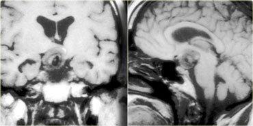
4 Ectopic Neurohypophysis A B Pre- (A) and post-gd (B) sagittal T1 images show bright spot in tuber cinereum of the hypothalamus. Post pit. hyperintesity may be lost in DI. 4


5 9/2008 2/2008 4/2008 Pituitary apoplexy 86 y/o VA, anorexia


6 Sellar lesions: Adenoma (15% of all IC tumors) enlarge bony sella (macro>10mm) 77 y/o diplopia PRL 28 x XRT Rx Dostinex, Steroids L-thyroxin Stalk effect Hook effect
7 Pituitary abscess 28-year-old man with febrile cephalalgia 7


8 Lymphocytic Hypophysitis Idiopathic inflammation of the pituitary gland Thickened, non tapering stalk (>2mm) Intense and homogeneous enhancement F:M = 8:1 8

9 Infundibular lesions 9


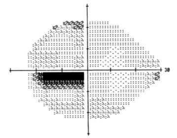

10 13 y/o VA SITA Standard 24-2 O.S. O.D.

11 Rathke s Cleft Cyst (RCC) RCC: intracystic nodule (mucin clump) with T1 & T2 Derived from remnants of Rathke s pouch. Byun et al. AJNR Am J Neuroradiol 21: , March

12 LANGERHANS CELL HISTIOCYTOSIS X 24 y/o with DI Bx of scalp and groin lesion T12 & pit mets Proliferative do. of Langerhan s cells

13 Granular cell tumour Sag T1WI C+ shows a well defined enhancing mass within the pituitary stalk. The pituitary gland is normal, as is the hypothalamus. Arise from stalk or neurohypophysis. Benign lesions, also called pituicytoma or choristoma. (Courtesy of L. Mechtler) 13
14 Pituitary protocol Image Plane Sequence Slice / Gap FOV Sagittal T1 3mm / 0mm 12 x 12 Coronal Coronal *Coronal (pituitary gland) T1 T2 Dynamic TSE/FSE T1+C 3mm / 0mm 12 x 12 3mm / 0mm 12 x 12 3mm / 0mm 12 x 12 Sagittal T1+C 3mm / 0mm 12 x 12 Coronal T1+C Fat Sat 3mm / 0mm 12 x 12 Axial whole brain T1+C Fat Sat x 12


15 Extrasellar: Suprasellar Craniopharyngioma Tuber cinereum hamartoma Arachnoid cyst Dermoid, Epidermoid Inflammation-infection Vascular 15


16 Craniopharyngioma epithelial: adamantinous or squamous-papillary Sartoretti-Schefer et al. Am J Neuroradiol 18:77 87, 16


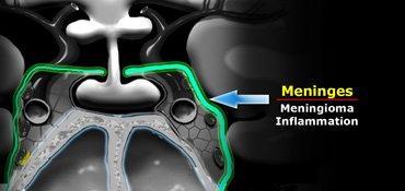
17 Germinoma ma 80-90% along IIIrd ventricle 5-10% basal ggl Bifocal germinoma: pineal + suprasellar DWI restriction: high cellularity Intense homogeneous Gad+ ± CSF seeding, ± brain invasion 17


18 Hypothalamic/Optic Chiasm Gliomas 15-30% associated with NF-1 18


19 Tuber Cinereum Hamartoma Post Gd T1, T2 sagittal, and post-gd T1 axial images. A mass is seen between the infundibulum and mammillary bodies, hyperintense to gray matter on T2. The mass appears similar to the brain parenchyma on T1 and shows no enhancement. This is a grey matter heterotopia. Large sessile causes gelastic seizures, and smaller pedunculated central precocious puberty. (courtesy of Dr. A. Osborn and Mechtler) 19


20 Arachnoid cyst in the suprasellar region markedly elevating the hypothalamus & stretching the pituitary stalk elevation of the right side of the chiasm by the cyst no cyst wall is evident pituitary tissue itself is nl. Same signal as CSF No enhancement No Ca++ 20


21 Colloid cysts are slow-growing lesions, close to foramen of Monro, rarely intra or suprasellar cistern, lined with epithelial cells and filled with mucus. Myriad of MRI signal: T1 T2. 21

22 Dermoid Cyst <0.5% if primary intracranial tumors Ectopic cyst containing dermal elements Less common than epidermoid Rupture causes significant morbidity and mortality 22

23 EPIDERMOID CYST -1% of all IC tumors -At CPA, parasellar, 4 th ventricular regions -Fast-FLAIR & DWI best sequences 23


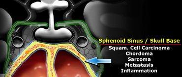
24 45 Y/O WOMAN WITH CONFUSION Neurosarcoidosis Solitary or multifocal CNS masses, dural plaque, parenchymal (non-caseating granulomas) 24


25 abnl. CXR in >90% of NS WM small vessel vasculitis/angiitis Coats CNs/fill IAC Leptomeningeal dz of the base of the brain similar to TB spreads along the Virchow- Robin spaces to form intraparenchymal masses 25

26 Extrasellar: Parasellar Meningioma Schwannoma THS CCF Cavernous hemang Thrombosis Lemierre syn 26


27 Patient presents with severe periorbital or retro-orbital pain of acute onset that is constant in nature; Diplopia due to opthalmoparesis usually follows the onset of the pain; The symptoms are usually unilateral 27
28 The International Headache Society criteria for THS One or more episodes of unilateral orbital pain lasting for an average of 8 weeks if left untreated Third, forth, and/or sixth cranial nerve palsy, which begins within two weeks of the onset of the orbital pain Symptoms that resolve within hours of initiation of steroid therapy Exclusion of other etiologies by appropriate investigation, including neuroimaging. 28

29 Dx? B 29 29


30 40 y/o galactorrhea, PRL:


31 Multiple Myeloma 73 y/o IgA MM 2 yrs s/p stem cell transplant; Confusion Headaches Intrasellar recurrent MM extension to left cavernous sinus PET/CT F18 FDG uptake 31
32 Extrasellar: Infrasellar 32
33 Chordoma Chordomas are the most common lesions of the clivus, also a favored location for metastases and chondrosarcomas. Hypointense intratumoral septations, Ca++, T2. 33

34 The CT shows some calcifications in this area. The differential diagnosis for this mass would be chordoma or chondrosarcoma Chordomas tend to occur in the midline, whereas chondrosarcomas tend to occur off the midline. 34

35 Chondrosarcoma 35

36 Patient with lung Ca presents with CN VIth palsy due to tumor metastasis. The normal fatty marrow has been replaced by abnl. tissue. Also lymphomas, myelomas or diffuse bone abnormalities can give this appearance. Therefore always take a moment to look at the clivus! 36


37 Squamous cell carcinoma Perineural spread 37 37

38 Hemangiopericytoma 38

39 ORBITAL ANATOMY 39
40 ORBITAL COMPARTMENTS The orbital space is divided into 4 compartments: 1. Intraconal (ON, orbital fat) 2. Conal (EOMs) 3. Extraconal (LG, preseptal) 4. Globe 1/27/

41 BONY ORBIT 1/27/
42 Intraconal: Inflammatory- Infectious Cellulitis IOD (Pseudotumor) Optic neuritis Sarcoid TB Tolosa-Hunt syndrome 42


43 49 Y WF pain on EM x1wk, VA: CF 1 OS DDx: ADEM, MS, NMO, sarcoid, idiopath Etiol: CMV, VZV, mycosis, toxo, syph, tb 43 43


44 34 y/o ON-itis OD 6 months later transverse myelitis

45 -Thickened & enhancing ONS -DDx: cat-scratch, Lyme, sarcoidosis, syphilis, tb, toxo 45 45
46 Intraconal: Neoplasms Cavernous hemangioma Hemangioblastoma (VHL) Lymphoma (most frequent)/leukemia ONG (child w/out NF-1, young adult, elderly) ONGG (ganglioglioma) may be a/w NF-1 Meningioma (ONSM) Mets Rhabdomyosarcoma Schwannoma (may be a/w NF-2) 46


47 10 year-old with incidentally found ON swelling 1/27/


48 12/23/2008 Intraneural & Perineural growth ONG Pilocytic astro 48 48


49 2/22/2008 4/25 6/18 75 WF subacute painless visual loss OS and gradual loss of vision OD 6 wks after OS; Labs: CRP 3.31 ESR 22 ACE 36; CSF: cell 0, pr 46, gl 63, MBP+, OCB- IgG index nl 49 49


50 75 y/o subacute painless sequential visual loss 50 50

51 Summary slide of the 3 cases 75 y/o 10 y/o 49 y/o ONG ONG ONitis 51 51

52 A B C D : fusiform axial ; eccentric extra axial proptosis; Ca

53 A B C 53 53
54 Intraconal 3. Metabolic: Krabbe s dz 4. Vascular: Cavernous hemangioma Dural malformations (ectasia) CCF, IIH Varix (thrombosis) 54

55 Krabbe dz: 6 month old with irritability, ON enlargement symmetric T2 of deep cerebral and peri-dentate nuclei white matter Shah S et al. Neurology 2012;78:e126-e by Lippincott Williams & Wilkins 55

56 IIH -Sx: TVO, puls. tinn -MRI/MRV -ONS diameter> 5mm -Correlate with ICP -Tortuous ONs -ONH indents sclera -Empty sella 56 56
57 Conal: EOM Size & Signal 1. Inflammatory/Infectious Grave s disease IOD (idiopathic orbital dz: orbital pseudotumor) 2. Tumor- direct extension mets (lymphoma, breast ca, lung ca, carcinoid) rhabdomyosarcoma 57

58 A Proptosis: from posterior lateral orbital rim to anterior globe > 21mm B 58 58


59 48 y/o with proptosis & diplopia Acute stage TED
 IOD 60")
60 A B Idiopathic orbital disorder Localized vs generalized -Violates compartments -Involves fat in 75% -Bone distruction -Relative T2 to muscle -DDx: orbital cellulitis (a/w skin inf or sinusitis) IOD 60 60

61 Localized IOD Myositis of the levator palpabrae superioris Almekhlafi, M. A. et al. Neurology 2008;71:


62 ORBITAL CELLULITIS 12 y/o OD pain & fever recurrent sinusitis WBC 62
 can be effected - Leukemia in children - T1 (to EOM), T2, marked enhancement - painless, extra-axial proptosis 63")
63 A B C -55% of malignant tumors of the orbit -Primary or Secondary -LG & conjuntiva most frequent sites but any tissue (EOM) can be effected - Leukemia in children - T1 (to EOM), T2, marked enhancement - painless, extra-axial proptosis 63 63


64 72 y/o painless diplopia on right gaze Bx: nl. muscle tissue Second opinion? 64


65 Invasive lobular breast carcinoma Anti-Estrogen-receptor
66 Atrophic & Dysplastic EOMs CN palsy : CN VI, III, IV NMJ disorder: MG, LEMS CPEO (chronic progressive external ophthalmoplegia) 66

67 67

68 68

69 CPEO Courtesy Prof. Demer 69
70 Extraconal 1. Bone lesions: fibrous dysplasia 2. Infection/inflam: granuloma, mucocele 3. Neoplastic: LCH, LG tumors, sinus Ca 4. Periosteal/mesenchymal lesions 5. Vascular: capillary/cavernous hemang., venous lymphatic malformation, varix 6. Trauma: blow-out fractures: inf, medial 70

71 A B cranio-orbital: sclerotic-ground glass, cystic degeneration 71 71

72 A B C D Frontoethmoidal: obstruction of the ostium CT: bone remodeling; MRI depends on H2O content 72

73 A B C D : encapsulated, solid/myxoid, Ca++, 73 mod enh. 73

74 Adenoid cystic carcinoma: erodes o. apex, lamina p.: bone erosion, infiltration,ca

75 A B C D : 80-85% intraconal, lat-to-on; proliferation followed 75 by involution 75


76 Orbital venous anomaly Intermittent proptosis Eye pain Thombosis: no signal void, if deoxy: T1, T2 if methgb: T1, T2 76
77 GLOBE COMPARTMENT 1. Coloboma, PHPV 2. staphy, detachments 3. CMV, Toxocara, VKHS, post. scleritis 4. Retinoblas, mets, melanoma, choroidal hemangioma (SWS), choroidal osteoma, astrocytic hamartoma (TS) 5. Retrolental fibroplasia 6. Coats dz, retinal angiomatosis (VHL) 1/27/

78 DDx of leukocoria: Retinoblastoma vs. Coats dz. vs. PHPV DDX of intraocular Ca++: Retinoblastoma vs. ONHD vs. larval granuloma Trilateral retinoblastoma : Retinoblastoma OU & pineal tumor 78

79 B C 79


80 Melanoma of the uveal tract: 85 % choroid, 15 % iris or ciliary body; T1, T2 80
81 MRI Orbit Protocol Noncontrast: Cor T1WI, Cor T2WI-FS, Ax DWI Contrast: axial & coronal T1WI-FS STIR paradoxical effect with IV contrast: suppresses tissues with a short T1 (fat, Gad, subacute blood) SPIR (selective partial IR) selectively inverts signal from fat without disturbing signal from water (SPIR/FLAIR) 81

82 Szatmáry G. Imaging of the orbit Neurol Clin Feb;27(1):

83 DISCLOSURES Nothing to disclose. Thank you for your attention! American Society of Neuroimaging 36 th Annual Meeting 83
PITUITARY PARASELLAR LESIONS. Kim Learned, MD
 PITUITARY PARASELLAR LESIONS Kim Learned, MD DIFFERENTIALS Pituitary Sella Clivus, Sphenoid Sinus Suprasellar Optic chiasm, Hypothalamus, Circle of Willis Parasellar Cavernous Sinus Case 1 17 YEAR-OLD
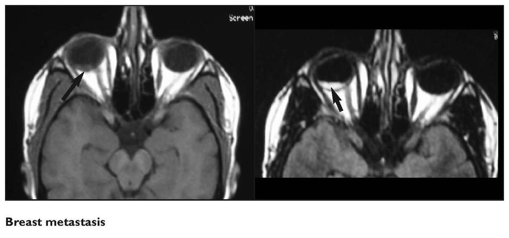
Metastasis. 57 year old with progressive Headache and Right Sided Visual Loss
 Metastasis 1% of sellar/parasellar masses Usually occurs with known primary Can involve third ventricle, hypothalamus, infundibular stalk May be both supra-, intrasellar 57 year old with progressive Headache
Laurie A. Loevner, MD
 Laurie A. Loevner, MD Chief, Division of Neuroradiology UPHS Professor of Radiology, Otorhinolaryngology: Head & Neck Surgery, Neurosurgery, and Ophthalmology University of Pennsylvania Health System Disclosures
EXPERT DIFFERENTIAL DIAGNOSIS:
 EXPERT DIFFERENTIAL DIAGNOSIS: Sellar Region Anne G. Osborn, M.D. DISCLOSURE: Published RSNA 2008 SELLA, PITUITARY: Normal Gross, 3T Anatomy SELLA, PITUITARY: Anatomically-Based Differential Diagnoses
Case Studies in Sella/Parasellar Region. Child thirsty, increased urination. Imaging. Suprasellar Germ Cell Tumor (Germinoma) No Disclosures
 No Disclosures") Case Studies in Sella/Parasellar Region No Disclosures 2018 Head and Neck Imaging Conference Child thirsty, increased urination Suprasellar Germ Cell Tumor (Germinoma) Midline Pineal >> Suprasellar > Other
Case Studies in Sella/Parasellar Region No Disclosures 2018 Head and Neck Imaging Conference Child thirsty, increased urination Suprasellar Germ Cell Tumor (Germinoma) Midline Pineal >> Suprasellar > Other
Where Has My Vision Gone? Evaluation of Sellar Lesions. Caleb Stowell,, HMS III Gillian Lieberman, MD November 2008
 Where Has My Vision Gone? Evaluation of Sellar Lesions Caleb Stowell,, HMS III Gillian Lieberman, MD November 2008 Objectives Present a case highlighting the clinical presentation and evaluation of a sellar
Case Studies in the Skull Base
 Case Studies in the Skull Base Amy C Tsai, MD Neuroradiology Fellow Department of Radiology and Imaging Sciences University of Utah Health Sciences Center Salt Lake City, Utah, USA No disclosures related
Imaging the Spinal Cord & Intradural Disease
 Department of Radiology University of California San Diego Imaging the Spinal Cord & Intradural Disease John R. Hesselink, M.D. Spinal Cord Diseases Tumors Syringohydromyelia Trauma Ischemia / Infarction
Imaging The Turkish Saddle. Russell Goodman, HMS III Dr. Gillian Lieberman
 Imaging The Turkish Saddle Russell Goodman, HMS III Dr. Gillian Lieberman Learning Objectives Review the anatomy of the sellar region Discuss the differential diagnosis of sellar masses Discuss typical
DISCLOSURES LEARNING OBJECTIVES WE WILL NOT DISCUSS. CSB: Birdseye View MESSAGE NAVIGATING THE SELLA AND CENTRAL SKULL BASE
 NAVIGATING THE SELLA AND CENTRAL SKULL BASE Christopher P. Hess, M.D., Ph.D. DISCLOSURES Research Support, General Electric SLIDES: http://www.radiology.ucsf.edu/research/meetings/rsna LEARNING OBJECTIVES
Cross sectional imaging of Intracranial cystic lesions Abdel Razek A
 Cross sectional imaging of Intracranial cystic lesions Abdel Razek A Department of Radiology. Mansoura Faculty of Medicine, Mansoura. Egypt. arazek@mans.edu.eg Introduction Intracranial cystic lesions
Neuro - imaging. Sella. ssregypt.com
 Neuro - imaging Sella ssregypt.com Bony Sella AP diameter Depth Contents 16mm 14mm Pituitary gland, part of infundibular stalk, CSF CT Technique 5 mm slices Axial and coronal Contrast injection Bone and
Orbital Tumor and Tumorlike. Thiparom Sananmuang, MD. Neuroradiologist Ramathibodi hospital, Mahidol University
 Orbital Tumor and Tumorlike lesion Thiparom Sananmuang, MD. Neuroradiologist Ramathibodi hospital, Mahidol University Outline Principle of orbital imaging Case-based approach tumor & tumor-like lesion
Diagnostic Neuroimaging in Neuro-Ophthalmology
 Diagnostic Neuroimaging in Neuro-Ophthalmology Gabriella Szatmáry, MD, PhD Director of Neuro-Ophthalmology Neuroimager Hattiesburg Clinic, PA DISCLOSURES Nothing to disclose! American Society of Neuroimaging
C. Douglas Phillips MD FACR Director of Head and Neck Imaging Weill Cornell Medical College/NewYork-Presbyterian Hospital
 C. Douglas Phillips MD FACR Director of Head and Neck Imaging Weill Cornell Medical College/NewYork-Presbyterian Hospital Disclosures Neither I nor any family members have any pertinent financial relations
RADIOLOGY TEACHING CONFERENCE
 RADIOLOGY TEACHING CONFERENCE John Athas, MD Monica Tadros, MD Columbia University, College of Physicians & Surgeons Department of Otolaryngology- Head & Neck Surgery September 27, 2007 CT SCAN IMAGING
Imaging of Hearing Loss
 Contemporary Imaging of Sensorineural Hearing Loss Imaging of Hearing Loss Discussion Outline (SNHL) Imaging Approaches Anatomic Relationships Lesions: SNHL KL Salzman, MD University of Utah School of
Brain Tumors. Medulloblastoma. Pilocytic astrocytoma: Ahmed Koriesh, MD. Pathological finding
 NeuroPathology Page 8 Brain Tumors Pathological finding Pseudorosette Rosenthal fibers Rosettes Wet Keratin Psammoma bodies Fried egg Tumor Ependymoma, SEGA Pilocytic astrocytoma Medulloblastoma Craniopharyngioma
Pediatric Ocular Sonography
 Pediatric Ocular Sonography Cicero J Torres A Silva, MD Associate Professor of Radiology 2016 SPR Pediatric Ultrasound Course Yale University School of Medicine None Disclosures Objectives of Presentation
CT and MRI Imaging Spectrum of Orbital Masses: A Pictorial Essay.
 CT and MRI Imaging Spectrum of Orbital Masses: A Pictorial Essay. Poster No.: C-1556 Congress: ECR 2014 Type: Educational Exhibit Authors: I. Chouchene, S. MAJDOUB, A. Achour, H. Zaghouani, M. 1 1 1 1
Case Studies In the Orbit
 Case Studies In the Orbit Ethan Neufeld MD Department of Radiology and Imaging Sciences University of Utah Health Sciences Center Salt Lake City, Utah, USA Case Studies In the Orbit Learning objectives:
Typical idiopathic intracranial hypertension Optic nerve appearance and brain MRI findings. Jonathan A. Micieli, MD Valérie Biousse, MD
 Typical idiopathic intracranial hypertension Optic nerve appearance and brain MRI findings Jonathan A. Micieli, MD Valérie Biousse, MD A 24 year old African American woman is referred for bilateral optic
Radiology of hypothalamic lesions: A pictorial essay depicting characteristic hypothalamic pathologies
 Radiology of hypothalamic lesions: A pictorial essay depicting characteristic hypothalamic pathologies Poster No.: C-2713 Congress: ECR 2010 Type: Scientific Exhibit Topic: Neuro Authors: A. J. B. Baxi,
Non-Traumatic Neuro Emergencies
 Department of Radiology University of California San Diego Non-Traumatic Neuro Emergencies John R. Hesselink, M.D. Nontraumatic Neuroemergencies 1. Acute focal neurological deficit 2. Worst headache of
13/02/1440 بسم ا هلل ا لرحمن ا لر حيم
 بسم ا هلل ا لرحمن ا لر حيم 1 Slowly progressive versus rapidly progressive proptosis by Ali M ISMAIL professor of ophthalmology @SOHAG U H Occuloplastic fellow @NNUH Occuloplastic fellow @Cambridge UH
RINGS N THINGS: Imaging Patterns in Differential Diagnosis. Anne G. Osborn, M.D.
 RINGS N THINGS: Imaging Patterns in Differential Diagnosis Anne G. Osborn, M.D. ExpDDxs: Intra-axial (Parenchymal) Lesions Ring-enhancing lesions, solitary 1 Ring-enhancing lesion crossing corpus callosum
What we will cover. Evaluation of the Child with Suspected Pituitary Disease. ituitary
 Evaluation of the Child with Suspected Pituitary Disease Craig Alter, MD University of Pennsylvania Children s Hospital of Philadelphia What we will cover * What laboratory tests to order * MRI: common
Adult Brain Tumours: an approach based on imaging findings
 Adult Brain Tumours: an approach based on imaging findings Robert J Sevick, MD, FRCPC, FACR Professor, Radiology and Clinical Neurosciences Cumming School of Medicine University of Calgary Learning objectives:
Imaging: When to get MRI, CT or PET-CT?
 Imaging: When to get MRI, CT or PET-CT? Alina Uzelac, D.O. Assistant Clinical Professor Neuroradiology UCSF Department of Radiology and Biomedical Imaging San Francisco General Hospital Overview CT MRI
Keep Imaging Simple: An Introduction To Neuroimaging
 Keep Imaging Simple: An Introduction To Neuroimaging Meghan Elkins, OD, FAAO Please silence all mobile devices and remove items from chairs so others can sit. Unauthorized recording of this session is
Part II - Revising the sellar and parasellar region: differential diagnosis of a sellar region mass
 Part II - Revising the sellar and parasellar region: differential diagnosis of a sellar region mass Poster No.: C-1390 Congress: ECR 2015 Type: Educational Exhibit Authors: I. Candelaria, C. Figueira,
Vertebral and Paravertebral Diseases
 Department of Radiology University of California San Diego Vertebral and Paravertebral Diseases John R. Hesselink, M.D. Vertebral / Paravertebral Disease (Extradural) Metastatic disease Primary bone tumors
Sellar and Parasellar Lesions: over and above adenomas.
 Sellar and Parasellar Lesions: over and above adenomas. Poster No.: C-2052 Congress: ECR 2013 Type: Educational Exhibit Authors: S. Paz Maya, P. Lemercier, I. lópez blasco, D. Soriano Mena, J. P. Ruiz
Neuro-imaging for the Ophthalmologist. Karl C. Golnik, MD, MEd University of Cincinnati & The Cincinnati Eye Institute
 Neuro-imaging for the Ophthalmologist Karl C. Golnik, MD, MEd University of Cincinnati & The Cincinnati Eye Institute Neuro-ophthalmology is that subspecialty where the diagnosis is made upon reinterpretation
RADIOANATOMY OF SELLA TURCICA
 RADIOANATOMY OF SELLA TURCICA O.BAKKACHA, H.MALAJATI, M.RHISSASSI, H. BENCHAABOUNE, N.CHAKIR, My R. EL HASSANI,M.JIDDANE Department of Neuroradiology specialties Hospital. Rabat Objective: New imaging
Pediatric Spine Tumors (and other masses)
") Pediatric Spine Tumors (and other masses) Francisco A Perez, MD, PhD Assistant Professor Neuroradiology and Pediatric Radiology Seattle Children s Hospital University of Washington, Seattle Commercial
Pediatric Spine Tumors (and other masses) Francisco A Perez, MD, PhD Assistant Professor Neuroradiology and Pediatric Radiology Seattle Children s Hospital University of Washington, Seattle Commercial
CNS TUMORS. D r. Ali Eltayb ( U. of Omdurman. I ). M. Path (U. of Alexandria)
. M. Path (U. of Alexandria)") CNS TUMORS D r. Ali Eltayb ( U. of Omdurman. I ). M. Path (U. of Alexandria) CNS TUMORS The annual incidence of intracranial tumors of the CNS ISmore than intraspinal tumors May be Primary or Secondary
CNS TUMORS D r. Ali Eltayb ( U. of Omdurman. I ). M. Path (U. of Alexandria) CNS TUMORS The annual incidence of intracranial tumors of the CNS ISmore than intraspinal tumors May be Primary or Secondary
Case Studies in CPA/IAC
 Outline Case Studies in CPA/IAC Atul K Mallik MD PhD Department of Radiology and Imaging Sciences University of Utah Health Sciences Center Salt Lake City, Utah, USA Case based review of cerebellopontine
Orbital Tumors - A Clinico Pathological Study
 Orbital Tumors - A Clinico Pathological Study Radha. J. DO, Ani Sreedhar. MS. Little Flower Hospital, Angamaly, Kerala ORIGINAL ARTICLES Abstract: Aim. To study the clinical and histopathological profiles
Supra- and infratentorial brain tumors from childhood to maternity
 Supra- and infratentorial brain tumors from childhood to maternity What to expect? I am going to show you the characteristic imaging findings of following tumors: Thierry A.G.M. Huisman, MD, FICIS, EQNR
Disclosures. Posterior Fossa Masses. I m from the Government. and I here to help! Differential Diagnosis
 Posterior Fossa Masses Differential Diagnosis James G. Smirniotopoulos, M.D. Radiology, Neurology, Biomedical Informatics Uniformed Services University Bethesda, Maryland http://rad.usuhs.edu http://medpix.usuhs.edu
MRI findings in childhood neurohypophyseal germinomas
 MRI findings in childhood neurohypophyseal germinomas Poster No.: C-1587 Congress: ECR 2015 Type: Scientific Exhibit Authors: C. Laganâ, S. I. Sirvent, M. A. Lopez-Pino, G. Albi, I. Solis Muniz, E. García
Traditional Approach. Pathways for Skull Base Pathology. Special Pathways Approach. 1. Traditional Approach. Central Skull Base. Anterior Skull Base
 Traditional Approach Pathways for Skull Base Pathology Anatomy Local Pathology Wade Wong DO FACR Professor of Radiology University of California, San Diego Special Pathways Approach Perineural Perivascular
Spinal Neoplasms. First Things First!! Localize the Lesion!! Ependymomas. Common Intramedullary Lesions
 Acta Radiológica Portuguesa, Vol.XXIII, nº 90, pág. 101-114, Abr.-Jun., 2011 Spinal Neoplasms Bruno A Policeni University of Iowa Hospitals and Clinics Assistant Professor of Radiology Disclosure of Commercial
Cholesteatoma and Non-cholesteatomatous Inflammatory Disease. Cholesteatoma. Disclosures. Overview EAC. Cholesteatoma. None
 Disclosures Cholesteatoma and Non-cholesteatomatous Inflammatory Disease None Amy F Juliano, MD Staff Radiologist, Massachusetts Eye and Ear Infirmary Assistant Professor of Radiology, Harvard Medical
Is there a role of CT in the evaluation of Proptosis
 International Journal of scientific research and management (IJSRM) Volume 3 Issue 4 Pages 2662-2666 2015 \ Website: www.ijsrm.in ISSN (e): 2321-3418 Is there a role of CT in the evaluation of Proptosis
The many faces of extranodal lymphoma
 The many faces of extranodal lymphoma Frank Pameijer Departments of Radiology and Radiation Oncology University Medical Center Utrecht Special thanks to Ilona M Schmalfuss, MD University of Florida Gainesville,
Pediatric CNS Tumors. Disclosures. Acknowledgements. Introduction. Introduction. Posterior Fossa Tumors. Whitney Finke, MD
 Pediatric CNS Tumors Disclosures Whitney Finke, MD Neuroradiology Fellow PGY-6 University of Utah Health Sciences Center Salt Lake City, Utah None Acknowledgements Introduction Nicholas A. Koontz, MD Luke
Contents. Basic Ultrasound Principles and Terminology. Ultrasound Nodule Characteristics
 Contents Basic Ultrasound Principles and Terminology Basic Ultrasound Principles... 1 Ultrasound System... 2 Linear Transducer for Superficial Images and Ultrasound-Guided FNA... 3 Scanning Planes... 4
Case 7391 Intraventricular Lesion
 Case 7391 Intraventricular Lesion Bastos Lima P1, Marques C1, Cabrita F2, Barbosa M2, Rebelo O3, Rio F1. 1Neuroradiology, 2Neurosurgery, 3Neuropathology, Coimbra University Hospitals, Portugal. University
Orbital facia. Periororbital facia Orbital septum Bulbar facia Muscular facia
 Anatomy Orbital facia Periororbital facia Orbital septum Bulbar facia Muscular facia Physiology of symptoms 1) Proptosis ( exophthalmos) Pseudoproptosis Axial Non axial Pulsating Positional Intermittent
ANATOMY AND IMAGING APPEARANCES OF COMMON PATHOLOGIES OF THE PITUITARY REGION: A PICTORIAL REVIEW
 ANATOMY AND IMAGING APPEARANCES OF COMMON PATHOLOGIES OF THE PITUITARY REGION: A PICTORIAL REVIEW Sitheeque F 1, Udupihille JJKH 2, Amarasinghe VGPS 1 1 Department of Radiology and Medical Imaging, Teaching
Neuro-Ocular Grand Rounds
 Neuro-Ocular Grand Rounds Anthony B. Litwak,OD, FAAO VA Medical Center Baltimore, Maryland Dr. Litwak is on the speaker and advisory boards for Alcon and Zeiss Meditek COMMON OPTIC NEUROPATHIES THAT CAN
Neuroimaging Core Curriculum
 Neuroimaging Core Curriculum Program Content The purpose of the training program is to prepare the physician for the independent practice of neuroimaging. Neuroimaging is the subspecialty of Neurology
Anatomy of the orbit. Lay-out. Imaging technique. 3 x 3. brief overview of the basic anatomy of the orbit and its structures
 Anatomy of the orbit Prof. Pia C Sundgren MD, PhD Department of Diagnostic Radiology, Clinical Sciences, Lund University, Sweden Lay-out brief overview of the basic anatomy of the orbit and its structures
Intrasphenoidal Rathke's Cleft Cyst: Case presentation and review of the literature
 Romanian Neurosurgery Volume XXX Number 4 2016 October - December Article Intrasphenoidal Rathke's Cleft Cyst: Case presentation and review of the literature Umit Kocaman, Muhammet Bahadir Yilmaz, Hakan
Imaging of Petrous Apex: Anatomy and Pathology
 University of Utah Head and Neck Conference 2018 Petrous apex Imaging of Petrous Apex: Anatomy and Pathology Philip Chapman MD University of Alabama, Birmingham Good News PAs tend to be symmetric A quick
Kathleen R. Fink, MD Virginia Mason Medical Center. 6 th Nordic Emergency Radiology Course 2017
 Kathleen R. Fink, MD Virginia Mason Medical Center 6 th Nordic Emergency Radiology Course 2017 Disclosure My spouse receives research salary support from: Guerbet Outline Acute neck and back pain Acute
Orbital Tumors And Pseudotumors
 Orbital Tumors And Pseudotumors Poster No.: C-2412 Congress: ECR 2015 Type: Educational Exhibit Authors: M. Limeme, N. Benzina, A. BelKhiria, H. Zaghouani, S. Majdoub, N. Mallat, H. Amara, D. Bakir, C.
Optic Pathway Gliomas, Germinomas, Spinal Cord Tumours. Colin Kennedy March 2015
 Optic Pathway Gliomas, Germinomas, Spinal Cord Tumours Colin Kennedy March 2015 Glioma of the optic chiasm. T1-weighted MRI with gadolinium enhancement, showing intense irregular uptake of contrast. The
Orbit and Sellar Region
 Orbit and Sellar Region 2 Francisco de Asís Bravo-Rodríguez and Rocío Díaz-Aguilera Contents Case 1 Choroidal Melanoma....................................... 24 Case 2 Grave s Ophthalmopathy....................................
Pathologic Analysis of CNS Surgical Specimens
 2015 Kenneth M. Earle Memorial Neuropathology Review Pathologic Analysis of CNS Surgical Specimens Peter C. Burger, MD Interdisciplinary Quality Control Familiarity with entities Use of diagnostic algorithm
Advances In Orbital Neuropathology
 Advances In Orbital Neuropathology Charles G. Eberhart, MD PhD Associate Professor of Pathology, Ophthalmology and Oncology Johns Hopkins University School of Medicine Overview Non-neoplastic lesions Microphthalmos/pseudoglioma
Posterior fossa tumors: clues to differential diagnosis with case-based review
 Posterior fossa tumors: clues to differential diagnosis with case-based review Poster No.: C-0323 Congress: ECR 2017 Type: Educational Exhibit Authors: H. A. Aboughalia, M. Abdelhady; Doha/QA Keywords:
Visual pathways in the chiasm
 Visual pathways in the chiasm Intracranial relationships of the optic nerve Fixation of the chiasm Chiasmatic pathologies The function of the optic chiasm may be altered by the presence of : 4) Artero
The Investigation of Proptosis in Paediatric Practice.
 The Investigation of Proptosis in Paediatric Practice. Ms Sayantani Ghosh 1, Mr Saugat Dey 1 1 Bankura Sammilani Medical College and Hospital, 136, Dr. Meghnad Saha Road, PRATYASHA APARTMENT Flat-4C, KOLKATA-
Dr. T. Venkat Kishan Asst. Prof Department of Radiodiagnosis
 Dr. T. Venkat Kishan Asst. Prof Department of Radiodiagnosis Schwannomas (also called neurinomas or neurilemmomas) constitute the most common primary cranial nerve tumors. They are benign slow-growing
Small lesions involving scalp and skull in pediatric age.
 Small lesions involving scalp and skull in pediatric age. Poster No.: C-1149 Congress: ECR 2013 Type: Educational Exhibit Authors: M. J. Yi, J. H. Yoo; Seoul/KR Keywords: Education and training, Education,
Magnetic Resonance Imaging. Basics of MRI in practice. Generation of MR signal. Generation of MR signal. Spin echo imaging. Generation of MR signal
 Magnetic Resonance Imaging Protons aligned with B0 magnetic filed Longitudinal magnetization - T1 relaxation Transverse magnetization - T2 relaxation Signal measured in the transverse plane Basics of MRI
Small lesions involving scalp and skull in pediatric age.
 Small lesions involving scalp and skull in pediatric age. Poster No.: C-1149 Congress: ECR 2013 Type: Educational Exhibit Authors: M. J. Yi, J. H. Yoo; Seoul/ Keywords: Education and training, Education,
Benign brain lesions
 Benign brain lesions Diagnostic and Interventional Radiology Hung-Wen Kao Department of Radiology, Tri-Service General Hospital, National Defense Medical Center Computed tomography Hounsfield unit (HU)
The central nervous system
 Sectc.qxd 29/06/99 09:42 Page 81 Section C The central nervous system CNS haemorrhage Subarachnoid haemorrhage Cerebral infarction Brain atrophy Ring enhancing lesions MRI of the pituitary Multiple sclerosis
Patients Treated with Leksell Gamma Knife
 Patients Treated with Leksell Gamma Knife 1968-2016 TREATMENTS REPORTED 2016 BY REGION AND INDICATION INDICATION Asia excl. Europe Latin Middle East & Africa North Grand Total Benign Tumors 12283 9778
Topical Diagnosis of Chiasmal and Retrochiasmal Disorders
 Topical Diagnosis of Chiasmal and Retrochiasmal Disorders Leonard A. Levin CHAPTER 12 TOPICAL DIAGNOSIS OF OPTIC CHIASMAL LESIONS Visual Field Defects Etiologies of the Optic Chiasmal Syndrome Masqueraders
Non-Functioning Tumours and Pituitary Hormone Testing. Miguel Debono Consultant in Endocrinology
 Non-Functioning Tumours and Pituitary Hormone Testing Miguel Debono Consultant in Endocrinology Agenda Pituitary masses Non functioning pituitary adenomas Testing pituitary function Pituitary Hormone Replacement
JMSCR Vol 3 Issue 11 Page November 2015
 www.jmscr.igmpublication.org Impact Factor 3.79 Index Copernicus Value: 5.88 ISSN (e)-2347-176x ISSN (p) 2455-0450 DOI: http://dx.doi.org/10.18535/jmscr/v3i11.37 Accuracy of CT in Diagnosis of Orbital
Unknown Cases from the Participants
 Unknown Cases from the Participants Case 1: 1 Case 1: Case 1: DDX? Answer on next slide Case 1: MS V5 Neuropathy Case 2: Case 2: 76 year old woman Ultrasound for multinodular goiter finds suspicious nodule
Slide 4. Slide 5. Slide 6
 Slide 1 Slide 4 Demographics El Paso Eye Care Border Healthcare-Based Grand Rounds Derek N. Cunningham, O.D. 80-90% Mexican-Americans Diabetes Hypertension Hyperlipidemia Obesity 70% uninsured High poverty
Evaluation of Neck Mass. Disclosure. Learning Objectives 3/24/2014. Karen T. Pitman MD, FACS Banner MDACC, Gilbert AZ. Nothing to disclose
 Evaluation of Neck Mass Karen T. Pitman MD, FACS Banner MDACC, Gilbert AZ Nothing to disclose Disclosure Learning Objectives 1. Describe a systematic method to evaluate a patient with a neck mass 2. Select
objectives Pitfalls and Pearls in PET/CT imaging Kevin Robinson, DO Assistant Professor Department of Radiology Michigan State University
 objectives Pitfalls and Pearls in PET/CT imaging Kevin Robinson, DO Assistant Professor Department of Radiology Michigan State University To determine the regions of physiologic activity To understand
Spinal cord tumours Luc van den Hauwe et al.
 overview spinal cord tumours L. van den Hauwe 1,2, D. Balériaux 3, J.W. Van Goethem 2, C. Venstermans 2, F. De Belder 2, P.M. Parizel 2 introduction imaging spinal tumour classification spinal cord tumours
Complicated Cataract to Intraocular Tumors, Beware of the unexpected
 Complicated Cataract to Intraocular Tumors, Beware of the unexpected Ihab Saad Othman, MD, FRCS Professor of Ophthalmology Cairo University In this part of the world: We Master Phakoemulsification 1 Intraoperative/Second
Neuro-Ocular Grand Rounds Anthony B. Litwak,OD, FAAO VA Medical Center Baltimore, Maryland
 Neuro-Ocular Grand Rounds Anthony B. Litwak,OD, FAAO VA Medical Center Baltimore, Maryland Dr. Litwak is on the speaker and advisory boards for Alcon and Zeiss Meditek COMMON OPTIC NEUROPATHIES THAT CAN
TABLES. Imaging Modalities Evidence Tables Table 1 Computed Tomography (CT) Imaging. Conclusions. Author (Year) Classification Process/Evid ence Class
 Imaging. Conclusions. Author (Year) Classification Process/Evid ence Class") TABLES Imaging Modalities Evidence Tables Table 1 Computed Tomography (CT) Imaging Author Clark (1986) 9 Reformatted sagittal images in the differential diagnosis meningiomas and adenomas with suprasellar
TABLES Imaging Modalities Evidence Tables Table 1 Computed Tomography (CT) Imaging Author Clark (1986) 9 Reformatted sagittal images in the differential diagnosis meningiomas and adenomas with suprasellar
MRI of the Pituitary Gland
 MRI of the Pituitary Gland Jean- François Bonneville Fabrice Bonneville Françoise Cattin Sonia Nagi MRI of the Pituitary Gland With a Foreword by A. Beckers Jean-François Bonneville, MD Department of
Patologie infiammatorie encefaliche e midollari
 Patologie infiammatorie encefaliche e midollari Maria Laura Stromillo Department of Medicine, Surgery and Neuroscience Inflammatory disorders of the CNS NMOSD ADEM Multiple Sclerosis Neuro-Myelitis Optica
Cystic Head and Neck Lesions
 Cystic Head and Neck Lesions Disclosures None Brad Wright, MD 19 March 2018 Key points Huge variety of cystic lesions in H&N May be cystic, necrotic, or solid but cystic-appearing Patient age, clinical
HEAD AND NECK IMAGING. James Chen (MS IV)
") HEAD AND NECK IMAGING James Chen (MS IV) Anatomy Course Johns Hopkins School of Medicine Sept. 27, 2011 OBJECTIVES Introduce cross sectional imaging of head and neck Computed tomography (CT) Review head
HEAD AND NECK IMAGING James Chen (MS IV) Anatomy Course Johns Hopkins School of Medicine Sept. 27, 2011 OBJECTIVES Introduce cross sectional imaging of head and neck Computed tomography (CT) Review head
CT & MRI Evaluation of Brain Tumour & Tumour like Conditions
 CT & MRI Evaluation of Brain Tumour & Tumour like Conditions Dr. Anjana Trivedi 1, Dr. Jay Thakkar 2, Dr. Maulik Jethva 3, Dr. Ishita Virda 4 1 M.D. Radiology, Professor and Head, P.D.U. Medical College
Disclosure. + Outline. Case-based approach to neurological emergencies that might present to the ED
 Kathleen R. Fink, MD University of Washington 5 th Nordic Emergency Radiology Course May 21, 2015 Disclosure My spouse receives research salary support from: Bracco BayerHealthcare Guerbet Outline Case-based
IMAGING OF INTRACRANIAL INFECTIONS
 IMAGING OF INTRACRANIAL INFECTIONS Dr Carolina Kachramanoglou LYSHOLM DEPARTMENT OF NEURORADIOLOGY NATIONAL HOSPITAL FOR NEUROLOGY AND NEUROSURGERY Plan Introduce MR sequences that are useful in the diagnosis
Diagnostic Imaging in Patients with Orbital Cellulitis and Inflammatory Pseudotumor. Fumiyuki Uehara, M.D. Norio Ohba, M.D.
 Diagnostic Imaging in Patients with Orbital Cellulitis and Inflammatory Pseudotumor Fumiyuki Uehara, M.D. Norio Ohba, M.D. We have had the opportunity, at an eye clinic, to examine and treat a patient
Transplanum Approach for Suprasellar pathology
 Transplanum Approach for Suprasellar pathology Omar A. El-Banhawy Prof. of otorhinolaryngology El Menoufyia University, Egypt Why Endoscopic Approach For Suprasellar Pathology Constant improvements in
Pictorial review of extraconal and osseous orbital pathology - what can be found 'around' the orbits?
 Pictorial review of extraconal and osseous orbital pathology - what can be found 'around' the orbits? Poster No.: C-2011 Congress: ECR 2013 Type: Educational Exhibit Authors: M. Meissnitzer, T. Meissnitzer,
Sellar and Parasellar pathologies: a comprehensive review on MRI
 Sellar and Parasellar pathologies: a comprehensive review on MRI Poster No.: C-1854 Congress: ECR 2016 Type: Educational Exhibit Authors: S. Sahni, K. Saggar, K. GUPTA, C. Kakkar, A. Banerjee ; 1 1 2 2
Imaging pituitary gland tumors
 November 2005 Imaging pituitary gland tumors Neel Varshney,, Harvard Medical School Year IV Two categories of presenting signs of a pituitary mass Functional tumors present with symptoms due to excess
The pituitary stalk is a funnel-like structure connecting the
 ORIGINAL RESEARCH N. Satogami Y. Miki T. Koyama M. Kataoka K. Togashi Normal Pituitary Stalk: High-Resolution MR Imaging at 3T BACKGROUND AND PURPOSE: Knowing the normal imaging appearance of the pituitary
Diagnostic Role of CT in the Evaluation of Proptosis
 IOSR Journal of Dental and Medical Sciences (IOSR-JDMS) e-issn: 2279-0853, p-issn: 2279-0861.Volume 14, Issue 4 Ver. IX (Apr. 2015), PP 25-31 www.iosrjournals.org Diagnostic Role of CT in the Evaluation
A Journey Down The Canal
 A Journey Down The Canal Radiological Assessment of Spinal Cord Masses John Berry-Candelario HMS III Gillian Lieberman, MD BIDMC Objectives Patient review Anatomy of the spine Imaging techniques Classification
DIFFERENTIAL DIAGNOSIS OF SELLAR MASSES
 ~~ ~~ ~ ADVANCES IN PITUITARY TUMOR THERAPY 0889-8529/99 $8.00 +.OO DIFFERENTIAL DIAGNOSIS OF SELLAR MASSES Pamela U. Freda, MD, and Kalmon D. Post, MD Pituitary adenomas are the most common cause of a