IN VITRO MODELS OF LYMPHATIC ENDOTHELIAL CELLS: CYTOKINE RECEPTOR EXPRESSION PROFILING AND INCORPORATION INTO ORGANOTYPIC CULTURE MODELS
|
|
|
- Raymond Daniels
- 5 years ago
- Views:
Transcription
1 IN VITRO MODELS OF LYMPHATIC ENDOTHELIAL CELLS: CYTOKINE RECEPTOR EXPRESSION PROFILING AND INCORPORATION INTO ORGANOTYPIC CULTURE MODELS by Fan Yang BS. Shandong University, China, 2014 Submitted to the Graduate Faculty of Graduate School of Public Health in partial fulfillment of the requirements for the degree of Master of Science University of Pittsburgh 2016
2 UNIVERSITY OF PITTSBURGH Graduate School of Public Health This thesis was presented by Fan Yang It was defended on April. 1 st, 2016 and approved by Thesis Director: Todd Reinhart, ScD Dean and Professor of Biology School of Sciences and Health Professions Saint Mary's University of Minnesota Committee Member: Frank Jenkins, PhD Associate Professor Infectious Diseases and Microbiology Graduate School of Public Health University of Pittsburgh Committee Member: Yue Chen, PhD Assistant Professor Infectious Diseases and Microbiology Graduate School of Public Health University of Pittsburgh ii
3 Copyright by Fan Yang 2016 iii
4 ABSTRACT Lymphatic endothelial cells (LECs) line the lymphatic vessels and lymph node sinuses. They function in balancing tissue interstitial fluid, trafficking of dendritic cells and lymphocyte movement into and out of lymph nodes, (lymph node-lecs), and can modulate self-tolerance. LECs also are important for viral pathogenesis and malignant cell migration. The discovery of LEC-specific markers has enabled the isolation and culture of LECs during the past decade, thereby increasing knowledge of LECs biology. Cytokines are small proteins secreted by cells that have pleiotropic effects on cell survival, growth, and functional activities. The expression of cytokine receptors on LECs, if present, could provide insight into the potential functions of LECs. The current study has defined the cytokine receptor expression profile, especially the IL6 family receptors, for three LEC populations: human dermal microvascular lymphatic endothelial cells (HMVEC-dLy), human lung lymphatic microvascular endothelial cells (HMVEC-LLy), and human telomerase reverse transcriptase transfected human dermal lymphatic endothelial cells (htert-hdlec), either with or without viral infection mimicry through the use of poly I:C treatment. Cytokine receptor expression was examined at the RNA, protein and function levels, as a strong reference for further LECs study. Todd Reinhart, ScD IN VITRO MODELS OF LYMPHATIC ENDOTHELIAL CELLS: CYTOKINE RECEPTOR EXPRESSION PROFILING AND INCORPORATION INTO ORGANOTYPIC CULTURE MODELS Fan Yang, M.S. University of Pittsburgh, 2016 In vitro 3-dimensional (3D) cell culture is an important substitute for in vivo experiments. It is also a meaningful improvement to 2-dimensional cell culture through the modification of iv
5 culture environments to better mimic in vivo situations. Using an extracellular matrix extraction called Matrigel TM as a simplified 3D model substrate, we demonstrated that five kinds of LECs (HMVEC-dLy, HMVEC-LLy, htert-hdlec, ferret lung isolated LECs and macaque jejunal LECs) can form a network-like structure on it (this process may be regarded as modeling the growth of lymphatic vessels or lymphangiogenesis). In addition, in this study we adapted an organotypic culture model containing lymphatic endothelial cells (LECs) to preliminary build a new powerful tool for in vitro study of LECs and also provide insights into the interactions between LECs and their environment. The work represented by this thesis holds public health significances lying mainly in enriching the knowledge of human LECs, considering the importance of LECs and lymphatic biology in cancer development and immunology. v
6 TABLE OF CONTENTS PREFACE... X 1.0 INTRODUCTION LYMPHATIC ENDOTHELIAL CELLS (LECS): FUNCTIONS, AND RELATED CYTOKINE/CYTOKINE RECEPTOR STUDY DIMENSIONAL TISSUE MODEL, AND 3-DIMENSIONAL CULTURE METHODS FOR LECS STATEMENT OF THE PROBLEM SPECIFIC AIM 1: DETERMINE LEC CYTOKINE RECEPTOR EXPRESSION PROFILE SPECIFIC AIM 2: DEVELOP 3D CULTURE METHODS OF LECS STATEMENT OF CONTRIBUTION RESULTS CYTOKINE RECEPTOR EXPRESSION PROFILES OF HUMAN LECS TRANSCRITIONAL AND CELL SURFACE EXPRESSION OF IL-6 FAMILY CYTOKINE RECEPTORS FUNCTIONAL RESPONSE OF LECS TO IL-6 FAMILY CYTOKINES DEVELOPMENT OF ORGANOTYPIC CULTURE MODELS CONTAINING LYMPHATIC ENDOTHELIAL CELLS Network formation by LECs on Matrigel TM vi
7 4.4.2 Develop of 3D mucosal tissue models containing LECs DISCUSSIONS CYTOKINE RECEPTOR EXPRESSION PROFILES OF LECS THE DEVELOPMENT OF 3D TISSUE MODELS CONTAINING LECS PUBLIC HEALTH SIGNIFICANCE MATERIALS AND METHODS APPENDIX: SUPPLEMENTARY FIGURES BIBLIOGRAPHY vii
8 LIST OF FIGURES Figure 1. HMVEC-dLy (Dly) or HMVEC-LLy (Lly) expression of cytokine receptors with or without the stimulation by poly I:C GeneCopoeia real-time RT-PCR assay Figure 2. HMVEC-dLy (DLY), HMVEC-LLy (LLY) and htert-hdlec (HTERT) expression of cytokine receptors with or without the stimulation by poly I:C Taqman assay Figure 3. Comparison between Taqman and GeneCopoeia data Figure 4. HMVEC-dLy (DLY), HMVEC-LLy (LLY) and htert-hdlec (HTERT) expression of IL6 family cytokine receptors with or without the stimulation by poly I:C flow cytometry assay Figure 5. Multiple IL6 family cytokines (IL6, LIF and OSM) induce the phosphorylation of STAT3 in Dly, Lly and htert-lec Figure 6. htert-hdlec formed a network-like structure on MatrigelTM coated plates Figure 7. Other kinds of LECs also form the network structure on MatrigelTM Figure 8. Human epithelial cells and lung fibroblasts do not form the networks on Matrigel TM. 28 Figure 9. CM-dil labeled htert-hdlecs (dil-htert-lecs) formed the networks on Matrigel TM Figure 10. Setup of the 3D tissue model Figure 11. 3D lung tissue model without LECs Figure 12. Growth process of the 3D lung mucosal tissue model containing LECs (between) cultured in DMEM media Figure 13. The 3D tissue models are able to express growth factors to support the survival of LECs viii
9 Figure 14. 3D lung tissue models containing LECs: Section views of fixed tissues and Top views of live tissues Figure 15. 3D lung tissue models containing LECs: 3D views of live tissues Figure 16. Representative gating strategy used for flow cytometric analyses of LEC cell surface cytokine receptor levels Figure 17. Representative gating strategy used for flow cytometric analyses of cell amount and survival status of cell isolates from tissue models Figure 18. Cell type specific staining on cell isolations from the 3D tissue models ix
10 PREFACE I would like to acknowledge my advisor Dr. Todd Reinhart for his patient guidance and professional direction not only on my research and study but also on my career development, my co-advisor Dr. Phalguni Gupta for his support, my thesis committee members Dr. Yue Chen and Dr. Frank Jenkins for their advice on this thesis, current and past Reinhart lab members Beth Juneckoa, Nicole Grant, and Dr. Stella Berendam for their kindly training and help on the lab techniques, Dr. Jayanth Venkatachari, Dr. Debjani Guha, Dr. Aki Hoji and Diana Campbell for their generous advises and help on my experiments and data analysis. Also I would like to take this chance to thank my girlfriend, Shuangping Zheng and my parents Mrs. Bing Liu and Prof. Jun Yang for their emotional support during the past two years. x
11 1.0 INTRODUCTION 1.1 LYMPHATIC ENDOTHELIAL CELLS (LECS): FUNCTIONS, AND RELATED CYTOKINE/CYTOKINE RECEPTOR STUDY Lymphatic vessels are part of a second, non-circulatory, vascular system with immune functions. Lymphatic vessels absorb the fluid that leaks from blood vessels into peripheral tissues and thereby contributes to tissue homeostasis and fluid balance. They are also are intimately involved in transporting antigen and antigen presenting cells (APCs) to lymph nodes (LNs) to initiate adaptive immune responses [1]. Lymphatic vessels and LN sinuses are lined by lymphatic endothelial cells (LECs). LECs were once difficult to isolate and culture, mainly because of a lack of efficient LEC-specific surface markers. The situation has been changed since 2000, when the specific expression of LYVE-1, vascular endothelial cell growth factor (VEGF)-C receptor, VEGFR-3, Podoplanin (PDPN) was found in LECs, but not in blood vascular endothelial cells (BECs) [2-4]. These discoveries have facilitated the enrichment and study of LECs and lymphatics. LECs function in the trafficking of dendritic cells (DCs) and lymphocytes into and out of lymph nodes (LNs) by expressing chemokines such as CCL21 and adhesion molecules such as ICAM-1. The LEC-related expression of CCL21 occurs in lymphatic vessels in multiple organs [5], which mediates the migration of antigen-loaded mature DCs from peripheral tissues to draining LNs via afferent lymphatic vessels [6]. The LECs lining the ceiling of the subcapsular 1
12 sinus are able to express chemokine receptor CCRL1 to scavenge CCL21 and maintain chemokine gradients across the sinus floor to enable the emigration of DCs [7]. After inflammatory cytokine treatment, human dermal LECs have increased expression of ICAM-1, VCAM-1, and E-selectin [8]. At the same time there also exists integrin-independent migration of DCs into LNs [9], which indicates the possible existence of other interactions between DCs and LECs such as the interaction between DC-expressed CLEC-2 and LEC-expressed podoplanin [10]. Lymphangiogenesis, which is the growth of new lymphatic vessels, is an important physiological activity of LECs. This process is extensive during embryonic or postnatal development. Lymphangiogenesis is also thought to be induced by most forms of human cancer cells leading to metastatic tumor spread through lymphatic vessels. In addition, lymphatic vessels are also observed to proliferate during inflammation [11]. The study of lymphangiogenesis can be very expansive and meaningful as it covers the field of cancer science, immunology and embryology, and the mechanisms of lymphangiogenesis have been studied for years. The most common view is that this process is mainly driven by the interaction between secreted vascular endothelial growth factors C and D (VEGF-C/D) and the cell-surface receptor VEGFR-3 on LECs [12]. Besides that, others have shown that podoplanin (a widely used LEC cell-surface marker) is necessary for lymphangiogenesis [13]. Interestingly, small interfering RNA (sirna)-induced TLR3 activation can inhibit blood and lymphatic vessel growth [14]. Also, IL-8 promoted LECs proliferation, tube formation, and migration without activating the VEGF signaling [15]. Cytokines are small proteins secreted by cells that have pleiotropic effects on cell survival, growth, and functional activities. The study of LEC-expressed cytokines and cellular receptors (including cytokine receptors) enrich the knowledge about LEC-associated activities especially about the immune potential of LEC, LECs that reside in LNs express MHC I molecules as well as 2
13 self-antigens expressed also in peripheral tissues but not co-stimulation molecules so they can work as tolerance-inducing antigen presenting cells (APC) to regulate T cells tolerance [16]. Besides expressing CCL family chemokines including CCL-1, 2, 5, 7, 8, 20, 21 and CXCL family chemokines CXCL-1, 3, 5, 6, 8, 9, 10, 11 to attract innate immune cells, LECs are also able to secrete different kinds of inflammatory cytokines including IL-1β, IL6, IL7, IL8, TGF-β, which directly take part in mediating immune response [17]. LECs also express several typical inflammatory factor receptors including IFNAR for IFN-α and IFNGR for IFN-γ [18] and toll-like receptors (TLRs) 1-6 and 9 [19], giving the cue that LECs have the ability not only to respond to the adaptive immune mediation signal created by other cells, but also to sense the environment and respond to infections diversely. In this thesis, to systematically profile cytokine receptor expression by LECs, we used GeneCopoeia and Taqman based methods to perform a RNA-level scan of 84 and 14 cytokine receptors mrnas, respectively, in untreated LECs or in a viral infection imitated environment (poly I:C treatment) in three LEC populations, human dermal microvascular lymphatic endothelial cells (HMVEC-dLy), human lung lymphatic microvascular endothelial cells (HMVEC-LLy) and human telomerase reverse transcriptase transfected human dermal lymphatic endothelial cells (htert-hdlec). Then the expression of several IL6 family receptors were followed on protein level and functional level. The cytokine receptor profile study in LECs will provide a strong reference for further LECs study. 3
14 1.2 3-DIMENSIONAL TISSUE MODEL, AND 3-DIMENSIONAL CULTURE METHODS FOR LECS Currently, cell based experiments are mainly performed at the 2-dimenssional (2D) level. Though 2D cell culture methods are convenient in their development, implementation and downstream analysis, the lack of cell-to-cell and cell-to-microenvironment interactions of 2D in vitro cell culture may set significant differences between the observations obtained from 2D-culture-based in vitro experiments and the observations obtained from in vivo experiments [20]. These differences may strongly limit the significance of observations obtained by 2D cell culture. It is thought that tissue homeostasis and the interaction among different cell types within tissues should be considered when studying the functions of individual cell types [21]. In response to the strong demand of in vivo data, and considering the high cost of in vivo study, numerous in vitro three-dimensional tissue models (or organotypic culture models, OCM) have been developed to mimic in vivo environments. When talking about in vivo models, for LECs function, though many 3-dimensional (3D) models with LECs or ECs have been developed (most of them were developed for observing and studying EC angiogenesis), these models still have some limitations. Most models only considered the interaction between LECs/ECs with extracellular matrix (ECM). The most commonly used 3D culturing condition for LECs is plating LECs on the top of or inside Matrigel TM, which is an extracellular matrix preparation isolated from the mouse Engelbreth-Holm-Swarm sarcoma [22]. Activated by Matrigel TM, LECs can form a network-like structure [13, 15, 23, 24]. Though widely used, this kind of 3D model usually contains only a single cell type and the Matrigel TM ECM component. Rather than considering this as a 3D tissue model, it is better to describe this method as LEC 3D culturing. Besides this, some people also believe that the network of LECs or ECs 4
15 on Matrigel TM is not lymphangiogenesis, as the networks do not have lumens and the networks are formed by cells realignment [22, 25]. Another limitation is that some models contain complex or unclear components. Again, taking Matrigel TM for example, as a protein mixture derived from a mouse tumor, there are more than a thousand different kinds of proteins and around 10,000 different peptides in it, the functions of most of them still not well understood [26]. Besides the Matrigel TM method, some other 3D models with LECs tend to involve in vitro culturing of tissue fragments, whose components are also very complex, such as the lymphatic ring assay which is achieved by embedding excised lymphatic duct fragments from mice into type I collagen gels [27]. The unclear components of either the cellular part or acellular parts of the 3D models may reduce the preciseness of the models as they are hard to repeat and can be hard to analyze. The last limitation of the models shown here is that some model building methods are very difficult. The fibrin bead assay is one example of the highly complex models. It involves embedding umbilical vein endothelial cell (HUVEC) coated Cytodex beads in fibrin gels, and then plating fibroblasts on top of the fibrin gels [22]. In this thesis we developed a simple, clearly composed 3D mucosal tissue model containing LECs, fibroblasts, and epithelial cells, as well as ECM collagens by modifying the method developed by our collaborators [21]. Primary analysis on the structure of the model and the survival of LECs in the model were performed. This the model could become a powerful tool for in vitro study of LEC function. 5
16 2.0 STATEMENT OF THE PROBLEM The discovery of LEC-specific makers, including Prox-1, LYVE-1, VEGFR-3, and PDPN around the turn of the century [2-4], facilitated the efficient and reliable differentiation between LECs and blood endothelial cells (BECs). Since then, LEC research has been gradually increasing. This LEC-focused research has greatly increased our understanding of lymphatics. LECs are not only the construction unit of lymph vessels, but they also have important functions including but not restricted to trafficking of DCs and lymphocytes [7-10], mediating cancer metastasis [28-30], mediating inflammation [11, 17-19], and mediating self-tolerance [16, 31-33]. At the same time, much current research still uses LEC and BEC mixed endothelial cell (EC) populations as study models [34-37]. However, meaningful observations have been obtained through the usage of mixed EC populations. For example, it has been meaningful to use mixed EC populations in studies of cancer metastasis, because in the broad view cancer metastasis involves both blood and lymph vessels. With regard to the study of LECs, though, the mixed usage of EC populations may set barriers for the further acquisition and interpretation of studies of LEC cell-type-specific information. The objectives of the study were to (1) determine the cytokine receptor expression profile of LECs under both mock treatment and viral infection imitated environments, and (2) develop 3D lung mucosal tissue models containing LECs. 6
17 2.1 SPECIFIC AIM 1: DETERMINE LEC CYTOKINE RECEPTOR EXPRESSION PROFILE Cytokines have a wide range of biological functions including regulating cellular survival, differentiation, proliferation, and anti-infectious or inflammatory activities [38]. The study of cytokine receptor expression profiles in LECs can give insights into LECs immune functions and help to understand how LECs respond to the changes in their environment. Transcriptional scans of murine LECs isolated from inflamed and resting LNs have been performed previously [18], and although several studies have examined the interactions between cytokines and cytokine receptors in human LECs [17], the systematic study of human LEC cytokine receptor expression profiles is still lacking. In the study here, systematic transcriptional scanning was performed for two commonly used human LEC primary cell populations, HMVEC-dLy and HMVEC-LLy, as well as a long life-span human LEC cell line, htert-hdlec. The cytokine receptor mrna expression profiles of LECs were obtained under both mock and viral infection imitated, poly I:C treated, environments. In addition, several IL6 family cytokine receptors were studied further at the protein and functional levels. 2.2 SPECIFIC AIM 2: DEVELOP 3D CULTURE METHODS OF LECS Three-dimensional (3D) culture of cells has become a trend in the field of cellular biology. Most 3D culture methods enable cell-cell and cell-environment interactions during culture [20], which mimics the real, in vivo situation for the cells under study and increases the value of the in vitro observations. Though a number of 3D culture models that include LECs or ECs have been 7
18 developed, they have disadvantages that include either over complication in their constructions (e.g., 3D culture following in vivo isolation of lymph vessels) or uncertainty in their components (e.g.., the matrix model component Matrigel TM ). In this thesis, a simple but efficient 3D culture method with LECs was developed that consisted of a 3D lung mucosal tissue model containing implanted LECs. It imitated the structure of lung mucosal tissue as well as supported the survival of LECs. With further modifications, the tissue model containing LECs can be further used for infection or cancer models. In addition, the most commonly used 3D culture method of LEC, culturing LECs on Matrigel TM, was also performed in parallel in the thesis. Altogether, the thesis will work as a good reference for further LEC study by providing the data of cytokine receptor expression profile in LECs and the method of an in vitro 3D tissue model containing LECs. 8
19 3.0 STATEMENT OF CONTRIBUTION In the thesis, GeneCopoeia real-time RT-PCR array experiments and related 2 -ΔCT values were performed and calculated, respectively, by Dr. Stella Berendam. The analysis, interpretation and presentation of GeneCopoeia data, and all the rest contents of thesis are the independent work of the thesis author, Fan Yang. 9
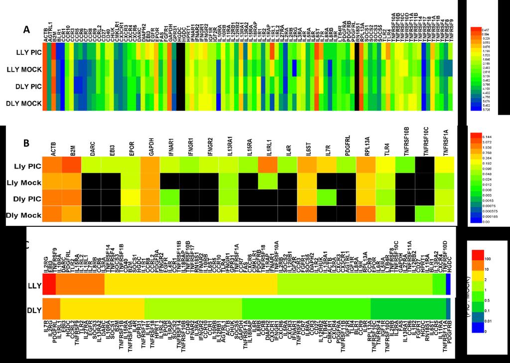
20 4.0 RESULTS 4.1 CYTOKINE RECEPTOR EXPRESSION PROFILES OF HUMAN LECS To understand better the abilities of LECs to sense the local immune environment and cytokine milieu, we screened 2D cultured HMVEC-dLy and HMVEC-LLy cell populations with poly I:C (PIC) or without (MOCK) and examined the total RNAs from these cells for the expression of 84 cytokine receptor related genes using the ExProfile TM Human Cytokine Receptor Related Gene qpcr Array (GeneCopoeia) (Figure 1). In the microarray, ATCB (β-actin), B2M (β-2- Microglobin), Ribosomal Protein L13a (RPL13A), Hypoxanthine Phosphoribosyl transferase 1 (HPRT1) and Glyceraldehyde 3-phosphate dehydrogenase (GAPDH) served as endogenous controls. In addition, two HGDCs served as genomic DNA controls to detect genomic DNA contamination. In Figure 1A, all genes except for RN18S1 have higher 2 - ΔCT values (relative to GAPDH) than HGDCs and could be regarded theoretically as having positive expression. According to that we conclude that at the RNA level LECs have a wide range of the expression of cytokine receptors though most of them are at a relatively low level (Figure 1A), and poly I:C can change the expression of several cytokine receptor related genes (Figure 1C). Through a narrowing of the range by setting the cutoff value of 2 - ΔCT relative to GAPDH at 0.05, we identified the most abundantly expressed cytokine receptor mrnas in Figure 1B. 10
21 11
22 Figure 1. HMVEC-dLy (Dly) or HMVEC-LLy (Lly) expression of cytokine receptors with or without the stimulation by poly I:C GeneCopoeia real-time RT-PCR assay. (A) The heat map shows the expression levels of 84 cytokine receptor related mrnas and 7 control genes in poly I:C treated(25µg/ml, 24h) (PIC) or untreated (MOCK) HMVEC-dLy (Dly) or HMVEC-LLy (Lly) cells. Different colors represent the values of log 10 (2 -ΔCT relative to GAPDH). (B) The heat map shows a collection of abundantly expressed cytokine receptor mrnas in (A). Cutoff value of 2 -ΔCT relative to GAPDH is set to Different colors represent the values of 2 -ΔCT relative to GAPDH, and black blocks represent data lower than (C) Fold-change form of (A). Different colors represent the ratios of the values of 2 -ΔCT relative to GAPDH of poly I:C treated samples to the values of 2 -ΔCT relative to GAPDH of mock treated samples. Data represent one experiment. The heat map was made using Heatmap Illustrator (Huazhong University of Science and Technology, China). 12
23 Among the relatively highly expressed cytokine receptors related genes were the Duffy Antigen Receptor for Chemokines (DARC) or Atypical chemokine receptor 1 (ACKR1), which is a glycosylated membrane protein having affinities to more than 20 inflammatory chemokines. It is mainly expressed in blood cells and endothelial cells, mainly functioning in non-signal-induced binding with chemokines to regulate chemokine gradients. [39] ACKR1 is utilized by malarial parasites Plasmodium vivax and Plasmodium knowlesi as a receptor [40, 41]. A pervious study has shown that ACKR1 functions to inhibit tumor growth and metastasis [42]. DARC expression was mainly observed in the DARC expression was mainly observed in the HMVEC-LLy cells treated with poly I:C. Epstein-Barr virus-induced gene 3 (EBI3) was originally found as a soluble hematopoietin receptor that were upregulated by EBV infection [43]. It is now known as a unit forming the heterodimer of the IL12 cytokine family members IL12, IL27, IL35 [43-45]. The expression of EBI3 was also mainly observed in the HMVEC-LLy cells treated with poly I:C. Erythropoietin receptor (EPOR) which is mainly expressed on erythrocytic progenitors and precursors in bone marrow to mediate red blood cell production [46]. It is highly expressed in all samples and slightly upregulated (1.46 folds change detected by GeneCopoeia, Figure 3) in HMVEC-dLy cells after poly I:C treatment. Three members of the tumor necrosis factor receptor superfamily were relatively highly expressed by LECs, including TNFRSF1A or TNFR1, TNFRSF10B or Death Receptor 5 (DR5), and TNFRSF10C or TRAIL receptor 3 (TRAILR3) (Figure 1B). TNFRSF1A, which is widely expressed in a number of cell types [47] is expressed by all four LECs populations, with a slight increase (2.5 folds change detected by GeneCopoeia, Figure 3) in expression observed in HMVEC- 13
24 LLy cells after poly I:C treatment. TNFRSF10B was abundantly expressed in poly I:C treated HMVEC-LLy cells and TNFRSF10C was expressed more in poly I:C treated HMVEC-dLy cells. IL13RA1 and IL4R(α) which together form the receptor complex of IL13 and IL4 [48] were both expressed (Figure 1B). IL13RA had relatively high expression in all four samples, with expression upregulated (1.47 fold change, Figure 3) in HMVEC-LLy cells and downregulated (0.36 fold change, Figure 3) in HMVEC-dLy cells after poly I:C treatment. High IL4R expression was only observed in HMVEC-LLy cells treated with poly I:C. IL15RA (IL15Rα), together with IL-2Rβ and γ, forms the IL15 receptor [49]. IL15Rα expression was only abundant in the HMVEC-LLy population treated with poly I:C. IL7R, together with IL2RG (IL2Rγ) which is a shared receptor by IL-2, 4, 7, 21[50, 51], forms the IL7 signaling receptor. IL7R expression was upregulated in both HMVEC-LLy and HMVEC-dLy populations after poly I:C stimulation. Two members of Toll-like receptor superfamily were highly expressed by LECs, including TLR4 and Interleukin 1 Receptor-Like 1 (IL1RL1). The high expression of TLR4 was observed in all four samples, which is consist with our pervious results [19]. The expression of TLR4 in HMVEC-dLy cells was downregulated (0.48 fold change, Figure 3) by poly I:C treatment, whereas the expression of TLR4 in HMVEC-LLy cells was upregulated (2.75 fold change, Figure 3) by poly I:C. The expression of IL1RL1 was observed mostly in HMVEC-LLy cells and it was highly upregulated (23.9 fold, Figure 3) by poly I:C treatment. A member of type I interferon receptors, Interferon (Alpha, Beta and Omega) receptor 1 (IFNAR1) which forms type I IFN receptor when combined with IFNAR2 [52], were abundantly expressed by LECs (Figure 1B). And two members of type II interferon receptors, interferon gamma receptor 1 (IFNGR1) and interferon gamma receptor 2 (IFNGR2), were also highly 14
25 expressed by LECs (Figure 1B). IFNAR1 had boarder and higher expression level compared with other two interferon receptors. Interesting, the expression of IFNGR1 was upregulated (2.69 fold change, Figure 3) after poly I:C treatment in HMVEC-LLy cells but downregulated (0.74 fold change, Figure 3) in HMVEC-dLy cells, whereas IFNGR1 and IFNGR2 were relatively highly expressed in HMVEC-LLy cells treated with poly I:C. Platelet-Derived Growth Factor Receptor-Like (PDGFRL) and platelet-derived growth factor receptor beta (PDGFRB) have high similarity with the respect of their coding sequences [53]. Again, the highest expression was observed in HMVEC-LLy cells treated with poly I:C. There overall was high expression of interleukin 6 signal transducer (IL6ST or gp130) in all four LEC samples. IL6ST is a signal transduction receptor shared by interleukin (IL)-6 family cytokines including IL-6, IL-11, leukemia inhibitory factor (LIF), oncostatin M (OSM), ciliary neurotrophic factor (CNTF), cardiotrophin-1 (CT-1), cardiotrophin-like cytokine (CLC), neuropoietin (NPN), IL-27, and IL-31 [54]. Given the high expression of IL6ST, we next included several IL6 family cytokine receptors in a focused real-time RT-PCR follow-up analysis. Using Taqman real-time RT-PCR we followed up on the screening analysis and extended the analyses to include 14 cytokine receptor mrnas (eight non-il6 family receptors, including CCR6, DARC, IL2RG, IL7R, TLR3, IFNAR1, EPOR, PDGFRA, and six IL6 family receptors, including IL6ST, IL6Ra, CNTFR, LIFR, OSMR, IL11Ra) in HMVEC-dLy, HMVEC-LLy, and htert-hdlec cell populations (Figure 2). The results obtained from the GeneCopoeia realtime RT-PCR array generally were similar with the results obtained from Taqman assay (Figure 3). A number of differences were obtained, though. Compared with the Taqman data, the GeneCopoeia array results showed an opposing effect of poly I:C treatment on the expression of IL6ST in HMVEC-dLy cells, lower expression of DARC in HMVEC-dLy cells, and higher 15
26 expression of EPOR and an opposite effect of poly I:C treatment on EPOR expression in both HMVEC-LLy and HMVEC-dLy cell populations. The differences between these findings might result from the reduced specificity of the SYBR-green-based real-time RT-PCR technique used in the GeneCopoeia real-time RT-PCR array, and the fact that the array screening was performed only once. Also, the acquisition of a new aliquot of HMVEC-dLy cells (new low passage cells from the same company) after the performance of the GeneCopoeia array and before the performance of the Taqman assays could have contributed to the differences obtained with the two approaches. Surprisingly, we found using Taqman assays and comparing the different LEC populations, that the HMVEC-dLy, HMVEC-LLy, and htert-hdlec cells overall have similar expression patterns for the 14 cytokine receptor mrnas in both untreated and poly I:C treated environments. (Figure 2). Poly I:C treated HMVEC-LLy and HMVEC-dLy cells expressed CCR6 mrna to low levels and we did not observe CCR6 expression in the three untreated LEC populations (Figure 2A). 16
27 Figure 2. HMVEC-dLy (DLY), HMVEC-LLy (LLY) and htert-hdlec (HTERT) expression of cytokine receptors with or without the stimulation by poly I:C Taqman assay. The graphs show the values of 2-ΔCT relative to β-gus obtained by Taqman assay in poly I:C treated (PIC) (balck) or untreated (MOCK) (white) HMVEC-dLy (DLY), HMVEC-LLy (LLY) and htert-hdlec (HTERT). (A) Eight non-il6 family cytokine receptors expression profile in the three LEC populations. (B) Six IL6 family cytokine receptors expression profile in the three LEC populations. Symbol * means significant difference (P<0.05) is observed between the two groups, and n.s. means nonsignificant (P>0.05). Paired t-tests ware perform to calculate P-values. U.d. means undetected which indicates the CT value of its genome DNA control (No RT control) subtract the CT value of the indicated sample 3. The function of the Taqman primers and probes of the undetected groups were examed on positive control samples (RNA isolates form human cerebral cortex for CNTFR; RNA isolates from human CCR6 transfected murine L1.2 cells for CCR6). Error bars indicate SD. The data were compiled from three independent experiments. 17
28 18
29 Figure 3. Comparison between Taqman and GeneCopoeia data. The graphs show the values of 2 -ΔCT relative to β-gus obtained by Taqman assay (Taqman) or the values of 2 -ΔCT relative to GAPDH obtained from GeneCopoeia assay (GeneCopoeia) in poly I:C treated (PIC) or untreated (MOCK) HMVEC-dLy (DLY), HMVEC-LLy (LLY). Figure (A) shows The Comparison between Taqman and GeneCopoeia data for seven non-il6 family cytokine receptors. Figure (B) shows The Comparison between Taqman and GeneCopoeia data for three IL6 family cytokine receptors. Symbol * means significant difference (P<0.05) is observed between the two groups, and n.s. means nonsignificant (P>0.05). Paired t-tests ware perform to calculate P- values. Error bars indicate SD. The data were compiled from three (Taqman assay) or one (GeneCopoeia assay) independent experiment(s). Platelet-derived growth factor-receptor α (PDGFRA/PDGFR-α) was expressed in the three LEC populations and the expression was upregulated after poly I:C treatment (Figure 2A). PDGFR-α forms heterodimers with platelet-derived growth factor-receptor β (PDGFR-β) to recognize all five PDGFs (PDGF-AA, AB, BB, CC, and DD), it can also form homodimers to recognize PDGF-AB and BB [55, 56]. The expression of PDGFR-α in LECs gives LECs the potential to respond to all PDGFs. In addition to the well-studied vascular endothelial growth factors (VEGFs), the interactions between PDGFs and ECs have been given growing attention in recent years. A recent study has shown that some tumors can express PDGFs [57]. In addition, PDGF-AA, AB, BB, CC are able to function on lymphatic vessels and induce VEGF-C/- D/VEGFR-3 independent lymphangiogenesis in vivo [58]. The observation that PDGFR-α is expressed in LECs and is upregulated by poly I:C treatment at the mrna level (Figure 3) strengthens the evidence of LECs ability to interact with PDGFs and give cue about the viral infection may increase the impact of PDGFs on LEC and possibly increase PDGFs induced lymphangiogenesis. Erythropoietin (EPO) can induce erythropoiesis of hematopoietic precursor cells [59]. EPOR is inducible in multiple cell types and EPO can affect cell types such as cardiac cells [59], neuronal cells [60] and endothelial cells [61, 62]. EPOR expression in human LECs was shown previously [28]and our data confirm this finding. It was also shown that EPO can induce the 19
30 lymphangiogenesis of LECs [28]. Here we have provided additional evidence that LECs express EPOR (Figure 2A). 4.2 TRANSCRITIONAL AND CELL SURFACE EXPRESSION OF IL-6 FAMILY CYTOKINE RECEPTORS The follow-up Taqman analyses of IL6 family receptor mrna levels confirmed the abundant expression of IL6ST in all four LEC populations (Figure 2B). This is consistent with the evidence of IL6ST s wide expression [63]. As for the expression of the specific receptors that are able to form receptor complexes with IL6ST for the specific recognition of IL6 family cytokines, IL6Rα, LIFR, OSMR, IL11Rα but not CNTFRα were observed to be expressed at the mrna level in HMVEC-dLy (DLY), HMVEC-LLy (LLY) and htert-hdlec (HTERT) LEC populations (Figure 2B). Poly I:C increased the expression of IL6ST, LIFR, OSMR and decreased the expression of IL6RA and IL11RA in the three LEC populations. Interestingly, IL6ST, LIFR and OSMR are all signal transducing receptors, whereas IL6Rα and IL11Rα are non-signaling receptors [64]. IL6 and IL11 signal through IL6ST homodimers after initially binding with their own α-receptor (IL6Rα or IL11Rα, respectively), whereas human herpes virus 8 (HHV8) secreted viral IL-6 (vil6) can directly signal through IL6ST homodimers without IL6Rα [65]. LIF, OSM, CT-1 directly bind and signal through the heterodimer of IL6ST/ LIFR. CNTF and CLC signal through the heterodimer of IL6ST/LIFR after binding to CNTF α-receptor (CNTFRα). OSM can also signal by binding with the heterodimer of IL6ST/OSMR [64]. IL-27 has been shown to signal through the gp130/il27r heterodimeric receptor [66]. The Taqman data indicate that poly I:C 20
31 stimulation might increase LEC potential to respond to LIF and OSM but decrease the potential to respond to IL6 and IL11. To extend the mrna analyses, we used flow cytometry to examine cell surface expression of several IL6 family cytokine receptors (Figure 4). The cell surface expression of the indicated proteins were detected using flow cytometry, with gating strategies based on unstained and isotype antibody control staining (Figure 16). Different from the mrna level results (Figure 2), only IL6ST was observed to be highly expressed on the cell surface for all LEC populations. In contrast, IL6R, OSMR, LIFR and IL27R had very low or no surface expression for the three untreated LEC populations. Poly I:C treatment led to slight shifts of positively stained LECs (red) compared to isotype antibody stained cells (blue) for IL6Rα, LIFR, OSMR and IL27R for all the three LECs populations. Different from the mrna results, poly I:C treatment here slightly reduced the percent of IL6ST positive cells in the three populations. As a positive control, all cell populations stained strongly for podoplanin with nearly all cells showing strong staining (Figure 4). 21
32 Figure 4. HMVEC-dLy (DLY), HMVEC-LLy (LLY) and htert-hdlec (HTERT) expression of IL6 family cytokine receptors with or without the stimulation by poly I:C flow cytometry assay. Surface expression of five IL6 family cytokine receptors IL6ST, IL6R(a), OSMR, LIFR, IL27R and one LEC surface marker Podoplanin(PDPN) in poly I:C treated (PIC) or untreated (MOCK) HMVEC-dLy (DLY), HMVEC-LLy (LLY) and htert-hdlec (HTERT) were examined using flow cytometry. Red curves indicate positive staining, while blue curves indicate isotype control. The gates represent the percentage of cells positive for cytokine receptor expression (red) and were set at 1% of the isotype controls (blue). Figure shows representative data out of at least two independent repeats. 22
33 4.3 FUNCTIONAL RESPONSE OF LECS TO IL-6 FAMILY CYTOKINES IL6 is one of the most important and pleiotropic pro- and anti-inflammatory factors [67]. The ubiquitous expression of IL6ST, and the wide expression of IL6 as well as the existence of soluble IL6Rα, enable IL6 to interact and function on many cell types including endothelial cells [68]. Previously, IL6 was shown to induce the expression of VEGF-C in conditionally immortalized murine LECs through Src-mediated ERK1/2 and p38mapk [69]; Leukemia inhibitory factor (LIF) is a member of the IL6 family of cytokines and is commonly used in stem cell culture media to inhibit the spontaneous difference of embryonic stem cells [70]. It has been shown to have contradictory effects on endothelial cells. LIF inhibits the angiogenesis and proliferation in vitro of bovine aortic endothelial cells and bovine microvascular endothelial cells [71-73], whereas when combined with basic FGF, LIF was noted to enhance the formation of capillary-like structures of embryonic endothelial cell [74]. OSM is also a member of the IL6 family of cytokines with multiple functions in hematopoiesis, mesenchymal stem cell differentiation, liver regeneration, heart remodeling, nociception, inflammation and metabolism [75]. Regarding the interaction between OSM and endothelial cells, OSM was shown to induce the expression of cytokines including IL6, CXCL-1, -2, -5, and adhesion molecules including ICAM-1 and VCAM-1 in human umbilical vein endothelial cells (ECs) [76]. OSM also induces cytokine CCL21 expression in human lung microvascular ECs and human dermal microvascular ECs [77], and affects the transcription of CCL20, CCL21, CXCL10, CXCL12, ICAM-1, VCAM-1 in HMVEC-dLy, HMVEC-LLy and htert-hdlec [19]. To examine further the expression and function of IL6 family cytokine receptors by LECs, three IL6 family cytokines (human recombinant IL6, LIF, and OSM) were used to stimulate 23
 and was firstly defined as a nuclear factor that can be activated by IL-6 [78].")
34 HMVEC-dLy (DLY), HMVEC-LLy (LLY) and htert-hdlec (HTERT) cells. STAT3 is a proximal signaling molecule activated by IL6 that was once called acute-phase response factor (APRF) and was firstly defined as a nuclear factor that can be activated by IL-6 [78]. All IL6 family cytokines have the potential to active STAT3 because they all share the IL6ST (gp130) signaling receptor [63]. In addition, OSM [75] and LIF [79] STAT3 activation have been wellstudied. To determine whether LECs are responsive to IL6 family cytokines, immunoblotting was performed for total STAT3 and phosphorylated STAT3 (Tyr705) to examine LEC responsiveness (Figure 5). Figure 5. Multiple IL6 family cytokines (IL6, LIF and OSM) induce the phosphorylation of STAT3 in Dly, Lly and htert-lec. Human recombinant IL6, LIF, or OSM (10 ng/ml) were used to treat HMVEC-dLy (DLY), HMVEC-LLy (LLY) and htert-hdlec (HTERT) cells for 30 or 60min. Immunoblotting was performed to detect the phosphorylation (Tyr705) of STAT3. In parallel, the loading control a-tubulin and total STAT-3 were detected. Figure shows representative data out of at least two independent repeats. Immunoblotting for STAT3-P (Figure 5), revealed that IL6, LIF, OSM were all able to activate STAT3 in HMVEC-dLy, HMVEC-LLy and htert-hdlec cell populations. The activation had different patterns across ligands and LEC populations. The amounts of STAT3-P were universally higher at 30min of treatment compared to 60min of treatment for all ligands. In addition, OSM treatment induced the strongest and longest STAT-P expression in HMVEC-dLy, 24
35 HMVEC-LLy and htert-hdlec compared with other two ligands. Furthermore, IL6 or LIF induced STAT3-P expression usually resolved after 60min of treatment, except for LIF induced STAT3-P expression in HMVEC-LLy, which was sustained after 60min of treatment. The quick drop-off of STAT3-P has been proved before as the result of SOCS3 regulation [80]. Importantly, these data provide strong evidence that LECs can respond to IL6 directly without addition of IL6R, particularly given that there are reports stating the lack of IL6R expression in endothelial cells [81-83]. 4.4 DEVELOPMENT OF ORGANOTYPIC CULTURE MODELS CONTAINING LYMPHATIC ENDOTHELIAL CELLS Network formation by LECs on Matrigel TM It has been reported that LECs can form a network-like structure when cultured on the Matrigel TM extracellular matrix (ECM) substrate [13, 15, 23, 24]. We used this capillary tube formation assay described in [13] to begin to examine the effects that 3D culture might have on LEC phenotype and function and to study the factors affecting LEC network formation. We note that there are no assays that well represent and recapitulate LEC function, although this network formation assay provides some level of functional analysis. We seeded htert-hdlecs on the top of Matrigel TM coated plates (Figure 6A) and observed rapid network formation (Figure 6A-E). Up to 16h after plating, we observed a rapid process of network formation (Figure 6A-D). Around 16h (Figure 6D), the networks were fully formed and additional re-organization stopped. At 18h the outline of the network started to dim, and some network components started to disappear 25

36 suggesting a dissolution of the network (Figure 6E). After 3-4 days, even with media being changed every two days, the network ultimately aggregated and involuted (data not shown). Figure 6. htert-hdlec formed a network-like structure on MatrigelTM coated plates. Matrigel TM was plated on 24-wells plates for 30 minutes. Then htert-hdlecs were seeded on the top of Matrigel TM. Pictures were taken at 0h(A), 2h(B), 4h(C), 16h(D), 18h(E) under microscopy after htert-hdlec was seeded. Pictures show one representative experiment out of at least three independent repeats. We also performed this assay on additional populations of primary LECs, including HMVEC-dLy, HMVEC-LLy, ferret lung LECs (FeLg-LEC) and macaque jejuna LECs (R24J), these last two derived by Dr. Stella Berendam in our laboratory [84]. Although to differing degrees, all of these LECs formed network-like structures on Matrigel TM (Figure 7). The networks formed by HMVEC-dLy (Figure 7A) and FeLg-LECs (Figure 7D) were similar to the networks formed by htert-hdlecs (Figure 6D). At 16h HMVEC-LLy (Figure 7B) and R24J LECs (Figure 7C) only started to form connections and the network structure looked more like htert-hdlec networks at 4h (Figure 6C). This might result from the different growth or migration rates of different cell types. 26
, human lung lymphatic microvascular endothelial cells (HMVEC-LLy, Fig B), ferret lung isolated")
37 Figure 7. Other kinds of LECs also form the network structure on MatrigelTM. After being seeded for 16h, Human dermal microvascular lymphatic endothelial cells (HMVEC-dLy, Fig A), human lung lymphatic microvascular endothelial cells (HMVEC-LLy, Fig B), ferret lung isolated LECs (FeLg-LEC, Fig C) and macaque jejuna isolated LECs (R24J, Fig D) all formed network-like structures to different degrees on Matrigel TM coated plates. Pictures show one representative experiment out of at two independent repeats. To examine whether network formation on Matrigel TM is unique to LECs, we cultured the human bronchial epithelial cell line 16HBE14o- and the human lung fibroblast cell line MRC5 on Matrigel TM for 16h. These two cell types did not form networks. This result indicated that network formation on Matrigel TM observed with LECs is not a property shared by all cell types. 27
 and human lung fibroblasts (MRC5, Fig B) did not form networks on Matrigel TM coated plates.")
38 Figure 8. Human epithelial cells and lung fibroblasts do not form the networks on Matrigel TM. After being seeded for 16h, human bronchial epithelial cells (16HBE14o-, Fig A) and human lung fibroblasts (MRC5, Fig B) did not form networks on Matrigel TM coated plates. Pictures show one representative experiment out of at least two independent repeats. For better tracking of LECs in these and downstream models, we used a lipophilic live cell dye called CM-dil to fluorescently label htert-hdlecs (dil-htert-lecs). We performed the network formation assay on Matrigel TM with dil-htert-lecs and found that the dye efficiently labeled LECs without visibly affecting their network forming activity on Matrigel TM (Figure 9). 28
 and TRIC channel (right).")
39 Figure 9. CM-dil labeled htert-hdlecs (dil-htert-lecs) formed the networks on Matrigel TM. After being seeded on Matrigel TM coated plates for 16h, CM-dil labeled TERT-HDLECs were observed to form network-like structures under both white filed (left) and TRIC channel (right). (A) 40X magnification. (B) 100X magnification. Pictures show one representative experiment out of at least two independent repeats Develop of 3D mucosal tissue models containing LECs To provide a strong tool to study LECs in a tissue-like environment in vitro, we adapted a 3D lung mucosal tissue model with the long life-span LEC htert-hdlec population inside by modifying an established tissue model [21]. We held the hypothesis that the 3D lung mucosal tissue model will give LECs an in vivo-like environment that will support LEC survival and immune potential and that might provide the ability, like Matrigel TM, to support LEC network forming. The tissue models were created in transwells set on 6-wells plates. The basic models developed [21] were made of three layers (from bottom to top): a collagen layer as a base, a 29
40 collagen and fibroblast (MRC5) mixture layer, and an epithelial (human bronchial epithelial cells, 16HBE14o - ) layer. The tissue models were made by adding layer by layer from the bottom to the top with different incubation periods. After all layers were added, a 7-days air exposure was performed to mimic the physiological conditions in lung with the air/liquid interface. (Figure 10 A and B basic ). Two kinds of models containing LEC were attempted: (1) htert-hdlecs were mixed within the collagen & fibroblasts (MRC5) mixture layer, and then the model was cultured as basic model (Figure 10 A and B containing LECs (inside) ); (2) htert-hdlecs were seeded on the top of collagen & fibroblasts (MRC5) mixture layer 7 days after the seeding of the mixture layer, and then the model was cultured for 3-4 days before adding 16HEB cells (Figure 10 A and B containing LECs (inside) ). For obtaining the best culture condition, two kinds of media were tried: DMEM media, which is a basic media containing basic nutrition, and EGM2 media, which indicates a 1:1 mixed media of DMEM and EGM2-MV here (among them EGM-2MV is the media used for LECs culture and contains several growth factors in addition to the basic nutrition). 30

41 Figure 10. Setup of the 3D tissue model. (A) Flowcharts of 3D models setup. From left to right, the flow chart represents the building process of the three kinds of 3D lung mucosal tissue model: incubation time and the main component/operation of each layer are shown. (B) Schematic diagrams of 3D models setup: models are built in transwells, different components are indicated. Immunohistochemistry was done to analyze the structure of the basic 3D tissue models. As an imitation of real in vivo tissue, the basic in vitro lung mucosal tissue model has an epithelial layer, and stromal components on top of the membrane of a transwell insert (Figure 11). As for the models with LECs, Figure 12B shows the culture process of a model containing LECs 31
 of a 10-μm cryosection of the 3D lung tissue model without LECs is shown (magnification X200).")
42 (between) when cultured in DMEM media under microscope. It indicates even under the basic nutrition environment, the model looked very healthy during the culture process (Figure 12). Figure 11. 3D lung tissue model without LECs. Immunohistochemical staining (with hematoxylin counterstaining) of a 10-μm cryosection of the 3D lung tissue model without LECs is shown (magnification X200). (A) IHC using isotype control. (B) IHC for vimentin (MRC5 marker). The distribution of MRC5 (brown) in the stroma is indicated in (B). The epithelial layer (16HBE14o-) and the membrane are also indicated by arrows. Pictures show one representative experiment out of at least two independent repeats. 32
.")
43 Figure 12. Growth process of the 3D lung mucosal tissue model containing LECs (between) cultured in DMEM media. Growth process of a tissue model containing LECs (between) when cultured in DMEM. Phase contrast microscopy images of the middle part of the 3D lung mucosal tissue model with LECs during the culturing process are shown (magnification X40). The first row of images show the growth of MRC5 in the collagen-and-fibroblasts mixture layer after the layer is seeded on the top of the collagen layer at day 1. The second row of images show the growth of htert-lec after addition at day 7. The third row of images show the growth of 16HBE14o- cells after added at day 11. Newly added cells are indicated. In each row of pictures, the healthy growth of newly added cells can be observed (the shape changes and amount increase of newly added can be observed). The fourth row of images show the 7-dayslength air exposure. Pictures show one representative experiment out of three independent repeats. Then Taqman real-time RT-PCR showed that at RNA level the tissue models expressed two kinds of growth factors (VEGF-A, VEGF-C) in all culture conditions (Figure 13). The expression of VEGF-A and VEGF-C in the tissue models gave primary evidences that the models 33
44 have potentials to support the survival of LECs because VEGF-A and VEGF-C were reported to be essential for the survival of LECs [3]. In Figure 13, two patterns can be observed: (1) Comparing between the models having the same structure (e.g. comparing between DMEM+LEC(inside) and EGM+LEC(inside) ), the models cultured in DMEM (e.g. DMEM+LEC(inside) ) always had higher expression levels of VEGF-A and VEGF-C than the models cultured in EGM (e.g. EGM+LEC(inside) ). This may be because that the growth factors in EGM (which contains VEGF-A but not VEGF-C) have performed feed-back inhibitions on the expressions of related growth factors including VEGF-A and VEGF-C in the model. (2) Comparing among the models cultured in the same media (e.g. comparing among DMEM+LEC(inside), DMEM+LEC(outside) and DMEM-LEC ), the models containing LECs (inside) (e.g. DMEM+LEC(inside) ) always had either the highest VEGF-A expression levels (Figure 13A) or the lowest VEGF-C expression levels (Figure 13B), while the VEGF-A and VEGF-C expression levels in models containing LECs (between) (e.g. DMEM+LEC(between) ) and models not containing LECs (e.g. DMEM-LEC ) were similar (Figure 13A and B). Combining with the data showing in 2D culture htert-hdlecs expressed high-level VEGF-A but very low-level VEGF-C (Figure 13C), the data can be interpreted as the model containing LEC (inside) gave LECs the best survival status to (1) contribute the expression of VEGF-A of the whole model, as well as (2) to pull down the expression of VEGF-C of the model because the adding of LEC diluted the percent of VEGF-C RNA in the whole RNA of the model. 34
45 Figure 13. The 3D tissue models are able to express growth factors to support the survival of LECs. After 7 days air exposure. total RNA were extracted from the tissue models. The histograms show the values of 2- ΔCT relative to β-gus obtained by Taqman assay. (A) The expression of VEGFA in different types of tissue models. (B) The expression of VEGFC in different types of tissue models. (C) The expression of VEGFA and VEGFC in 2D cultured (monolayer) htert-hdlecs. DMEM : tissue model was cultured in DMEM media; EGM : tissue model was cultured in a 1:1 mixed media of DMEM and EGM2-MV; + or - : model contains or does not contain htert-hdlec; LEC(between) : htert-hdlecs were seeded between stroma and epithelial layer; LEC(inside) : htert-hdlecs were seeded inside stroma layer. Data represent more than two (model) or one (2D htert-hdlec) independent experiment(s). Symbol * means significant difference (P<0.05) is observed between the two columns. (Paired t-test was done to calculate P-values). Error bars indicate the SD value. To analyze the distribution, morphology and survival status of LECs in the model, htert- HDLECs were pre-stained with the lipophilic live cell dye CM-dil before being added into the tissue models. Then live or fixed models were analyzed using fluorescent microscopy (Figure 14, Figure 15). 35
 (A,C) and tissue models with LECs(inside) (B,D) were analyzed for their structures.")
46 Figure 14. 3D lung tissue models containing LECs: Section views of fixed tissues and Top views of live tissues. After 7 days air exposure, DMEM media cultured tissue models with LECs(between) (A,C) and tissue models with LECs(inside) (B,D) were analyzed for their structures. (A-B) 10-μm cryosections of 4%PFA fixed 3D lung tissue model with CM-Dil per-stained htert-hdlecs (red) were stained with DAPI (blue) and imaged under fluorescent microscopy (magnification X200). (C-D) live tissue models with CM-Dil per-stained htert-hdlecs (red) were directly imaged under fluorescent microscopy (FITC channel, magnification X200). Pictures show one representative experiment out of at least two independent repeats. 36
 or EGM media cultured (B and D) tissue models that contain LECs (inside) (C and D) or LECs (between) (A and B) were analyzed for their")
47 Figure 15. 3D lung tissue models containing LECs: 3D views of live tissues. After 7 days air exposure, DMEM media cultured (A and C) or EGM media cultured (B and D) tissue models that contain LECs (inside) (C and D) or LECs (between) (A and B) were analyzed for their structures under fluorescent confocal microscope (Z-stack, under bright field and TRIC channel). Figures are representative data out of at least two independent repeats. In the images of live tissues (under both phase contract fluorescent microscope and confocal microscope), clear round cell shapes of CM-dil labeled htert-hdlecs were observed in models containing LECs (inside) cultured in both media (Figure 14C, Figure 15C and D); While for models containing LECs (between), red spots that are smaller than the size of normal cells were observed (Figure 14D, Figure 15A and B), these small red spots may be regarded as either dead LEC particles or lipid containing organelles of live LECs. For both the models containing LECs (between) (Figure 15A and B) and the models containing LECs (inside) (Figure 15C and D), EGM culture condition (Figure 15B and D) always enabled the tissue model to have more cells. 37
CD34 + VEGFR-3 + progenitor cells have a potential to differentiate towards lymphatic endothelial cells
 CD34 + VEGFR-3 + progenitor cells have a potential to differentiate towards lymphatic endothelial cells Tan YZ et al. J Cell Mol Med. (2014 Mar;18(3):422-33) Denise Traxler-Weidenauer April 2014 Introduction
Supplementary Materials. for Garmy-Susini, et al, Integrin 4 1 signaling is required for lymphangiogenesis and tumor metastasis
 Supplementary Materials for Garmy-Susini, et al, Integrin 4 1 signaling is required for lymphangiogenesis and tumor metastasis 1 Supplementary Figure Legends Supplementary Figure 1: Integrin expression
Supplementary Figure 1. SA-β-Gal positive senescent cells in various cancer tissues. Representative frozen sections of breast, thyroid, colon and
 Supplementary Figure 1. SA-β-Gal positive senescent cells in various cancer tissues. Representative frozen sections of breast, thyroid, colon and stomach cancer were stained with SA-β-Gal and nuclear fast
SUPPLEMENTARY INFORMATION
 DOI:.38/ncb3399 a b c d FSP DAPI 5mm mm 5mm 5mm e Correspond to melanoma in-situ Figure a DCT FSP- f MITF mm mm MlanaA melanoma in-situ DCT 5mm FSP- mm mm mm mm mm g melanoma in-situ MITF MlanaA mm mm
Lymphoid architecture & Leukocyte recirculation. Thursday Jan 26th, 2017
 Lymphoid architecture & Leukocyte recirculation Thursday Jan 26th, 2017 Topics The life of immune cells Where are they born? Where are they educated? Where do they function? How do they get there? The
Cytokines modulate the functional activities of individual cells and tissues both under normal and pathologic conditions Interleukins,
 Cytokines http://highered.mcgraw-hill.com/sites/0072507470/student_view0/chapter22/animation the_immune_response.html Cytokines modulate the functional activities of individual cells and tissues both under
Immune response to infection
 Immune response to infection Dr. Sandra Nitsche (Sandra.Nitsche@rub.de ) 20.06.2018 1 Course of acute infection Typical acute infection that is cleared by an adaptive immune reaction 1. invasion of pathogen
To compare the relative amount of of selected gene expression between sham and
 Supplementary Materials and Methods Gene Expression Analysis To compare the relative amount of of selected gene expression between sham and mice given renal ischemia-reperfusion injury (IRI), ncounter
Nature Immunology: doi: /ni Supplementary Figure 1. Production of cytokines and chemokines after vaginal HSV-2 infection.
 Supplementary Figure 1 Production of cytokines and chemokines after vaginal HSV-2 infection. C57BL/6 mice were (a) treated intravaginally with 20 µl of PBS or infected with 6.7x10 4 pfu of HSV-2 in the
Supplemental Table 1. Primer sequences for transcript analysis
 Supplemental Table 1. Primer sequences for transcript analysis Primer Sequence (5 3 ) Primer Sequence (5 3 ) Mmp2 Forward CCCGTGTGGCCCTC Mmp15 Forward CGGGGCTGGCT Reverse GCTCTCCCGGTTTC Reverse CCTGGTGTGCCTGCTC
T-cell activation T cells migrate to secondary lymphoid tissues where they interact with antigen, antigen-presenting cells, and other lymphocytes:
 Interactions between innate immunity & adaptive immunity What happens to T cells after they leave the thymus? Naïve T cells exit the thymus and enter the bloodstream. If they remain in the bloodstream,
Supplementary Figure 1:
 Supplementary Figure 1: (A) Whole aortic cross-sections stained with Hematoxylin and Eosin (H&E), 7 days after porcine-pancreatic-elastase (PPE)-induced AAA compared to untreated, healthy control aortas
SUPPLEMENTARY INFORMATION
 DOI: 10.1038/ncb3021 Supplementary figure 1 Characterisation of TIMPless fibroblasts. a) Relative gene expression of TIMPs1-4 by real time quantitative PCR (RT-qPCR) in WT or ΔTimp fibroblasts (mean ±
Suppl Video: Tumor cells (green) and monocytes (white) are seeded on a confluent endothelial
 and monocytes (white) are seeded on a confluent endothelial") Supplementary Information Häuselmann et al. Monocyte induction of E-selectin-mediated endothelial activation releases VE-cadherin junctions to promote tumor cell extravasation in the metastasis cascade
Supplementary Information Häuselmann et al. Monocyte induction of E-selectin-mediated endothelial activation releases VE-cadherin junctions to promote tumor cell extravasation in the metastasis cascade
Cytokines, adhesion molecules and apoptosis markers. A comprehensive product line for human and veterinary ELISAs
 Cytokines, adhesion molecules and apoptosis markers A comprehensive product line for human and veterinary ELISAs IBL International s cytokine product line... is extremely comprehensive. The assays are
1.The metastatic cascade. 2.Pathologic features of metastasis. 3.Therapeutic ramifications
 Metastasis 1.The metastatic cascade 2.Pathologic features of metastasis 3.Therapeutic ramifications Sir James Paget (1814-1899) British Surgeon/ Pathologist Paget s disease of bone Paget s disease of the
Adaptive immune responses: T cell-mediated immunity
 MICR2209 Adaptive immune responses: T cell-mediated immunity Dr Allison Imrie allison.imrie@uwa.edu.au 1 Synopsis: In this lecture we will discuss the T-cell mediated immune response, how it is activated,
PBS Class #2 Introduction to the Immune System part II Suggested reading: Abbas, pgs , 27-30
 PBS 803 - Class #2 Introduction to the Immune System part II Suggested reading: Abbas, pgs. 15-25, 27-30 Learning Objectives Compare and contrast the maturation of B and T lymphocytes Compare and contrast
Supporting Information
 Supporting Information M1 macrophage-derived nanovesicles potentiate the anticancer efficacy of immune checkpoint inhibitors Yeon Woong Choo, 1, Mikyung Kang, 2, Han Young Kim, 1 Jin Han, 1 Seokyung Kang,
Supplementary Table S1. Primers used for quantitative real-time polymerase chain reaction. Marker Sequence (5 3 ) Accession No.
 Accession No.") Supplementary Tables Supplementary Table S1. Primers used for quantitative real-time polymerase chain reaction Marker Sequence (5 3 ) Accession No. Angiopoietin 1, ANGPT1 A CCCTCCGGTGAATATTGGCTGG NM_001146.3
Supplementary Tables Supplementary Table S1. Primers used for quantitative real-time polymerase chain reaction Marker Sequence (5 3 ) Accession No. Angiopoietin 1, ANGPT1 A CCCTCCGGTGAATATTGGCTGG NM_001146.3
TITLE: Notch in Pathological Angiogenesis and Lymphangiogenesis
 Award Number: W81XWH-10-1-0304 TITLE: Notch in Pathological Angiogenesis and Lymphangiogenesis PRINCIPAL INVESTIGATOR: Minji Kim CONTRACTING ORGANIZATION: Columbia University New York, NY 10032 REPORT
Supplementary Figure 1. Deletion of Smad3 prevents B16F10 melanoma invasion and metastasis in a mouse s.c. tumor model.
 A B16F1 s.c. Lung LN Distant lymph nodes Colon B B16F1 s.c. Supplementary Figure 1. Deletion of Smad3 prevents B16F1 melanoma invasion and metastasis in a mouse s.c. tumor model. Highly invasive growth
Thomas HAIDER Journal Club
 Thomas HAIDER Journal Club 20.10.2014 Background Immunology of the CNS - History Ehrlich, 1885 & 1904 dye did not stain brain -> BBB Shirai, Y. (1921) On the transplantation of the rat sarcoma in adult
Endogenous TNFα orchestrates the trafficking of neutrophils into and within lymphatic vessels during acute inflammation
 SUPPLEMENTARY INFORMATION Endogenous TNFα orchestrates the trafficking of neutrophils into and within lymphatic vessels during acute inflammation Samantha Arokiasamy 1,2, Christian Zakian 1, Jessica Dilliway
Lymphoid System: cells of the immune system. Answer Sheet
 Lymphoid System: cells of the immune system Answer Sheet Q1 Which areas of the lymph node have most CD3 staining? A1 Most CD3 staining is present in the paracortex (T cell areas). This is towards the outside
Pathologic Stage. Lymph node Stage
 ASC ASC a c Patient ID BMI Age Gleason score Non-obese PBMC 1 22.1 81 6 (3+3) PBMC 2 21.9 6 6 (3+3) PBMC 3 22 84 8 (4+4) PBMC 4 24.6 68 7 (3+4) PBMC 24. 6 (3+3) PBMC 6 24.7 73 7 (3+4) PBMC 7 23. 67 7 (3+4)
Putting it Together. Stephen Canfield Secondary Lymphoid System. Tonsil Anterior Cervical LN s
 Putting it Together Stephen Canfield smc12@columbia.edu Secondary Lymphoid System Tonsil Anterior Cervical LN s Axillary LN s Mediastinal/Retroperitoneal LN s Thoracic Duct Appendix Spleen Inguinal LN
CYTOKINE RECEPTORS AND SIGNAL TRANSDUCTION
 CYTOKINE RECEPTORS AND SIGNAL TRANSDUCTION What is Cytokine? Secreted popypeptide (protein) involved in cell-to-cell signaling. Acts in paracrine or autocrine fashion through specific cellular receptors.
(A) RT-PCR for components of the Shh/Gli pathway in normal fetus cell (MRC-5) and a
 RT-PCR for components of the Shh/Gli pathway in normal fetus cell (MRC-5) and a") Supplementary figure legends Supplementary Figure 1. Expression of Shh signaling components in a panel of gastric cancer. (A) RT-PCR for components of the Shh/Gli pathway in normal fetus cell (MRC-5) and
Supplementary figure legends Supplementary Figure 1. Expression of Shh signaling components in a panel of gastric cancer. (A) RT-PCR for components of the Shh/Gli pathway in normal fetus cell (MRC-5) and
Analysis on the mechanism of reduced nephron number and the pathological progression of chronic renal failure in Astrin deficient rats
 Analysis on the mechanism of reduced nephron number and the pathological progression of chronic renal failure in Astrin deficient rats Summary of Doctoral Thesis Hidenori Yasuda Graduate School of Veterinary
1. The metastatic cascade. 3. Pathologic features of metastasis. 4. Therapeutic ramifications. Which malignant cells will metastasize?
 1. The metastatic cascade 3. Pathologic features of metastasis 4. Therapeutic ramifications Sir James Paget (1814-1899) British Surgeon/ Pathologist Paget s disease of Paget s disease of the nipple (intraductal
ECM1 controls T H 2 cell egress from lymph nodes through re-expression of S1P 1
 ZH, Li et al, page 1 ECM1 controls T H 2 cell egress from lymph nodes through re-expression of S1P 1 Zhenhu Li 1,4,Yuan Zhang 1,4, Zhiduo Liu 1, Xiaodong Wu 1, Yuhan Zheng 1, Zhiyun Tao 1, Kairui Mao 1,
Chapter 3, Part A (Pages 37-45): Leukocyte Migration into Tissues
: Leukocyte Migration into Tissues") Allergy and Immunology Review Corner: Chapter 3, Part A (pages 37-45) of Cellular and Molecular Immunology (Seventh Edition), by Abul K. Abbas, Andrew H. Lichtman and Shiv Pillai. Chapter 3, Part A (Pages
Allergy and Immunology Review Corner: Chapter 3, Part A (pages 37-45) of Cellular and Molecular Immunology (Seventh Edition), by Abul K. Abbas, Andrew H. Lichtman and Shiv Pillai. Chapter 3, Part A (Pages
Time course of immune response
 Time course of immune response Route of entry Route of entry (cont.) Steps in infection Barriers to infection Mf receptors Facilitate engulfment Glucan, mannose Scavenger CD11b/CD18 Allows immediate response
(a) Significant biological processes (upper panel) and disease biomarkers (lower panel)
 Significant biological processes (upper panel) and disease biomarkers (lower panel)") Supplementary Figure 1. Functional enrichment analyses of secretomic proteins. (a) Significant biological processes (upper panel) and disease biomarkers (lower panel) 2 involved by hrab37-mediated secretory
Supplementary Figure 1. Functional enrichment analyses of secretomic proteins. (a) Significant biological processes (upper panel) and disease biomarkers (lower panel) 2 involved by hrab37-mediated secretory
Tissue repair. (3&4 of 4)
") Tissue repair (3&4 of 4) What will we discuss today: Regeneration in tissue repair Scar formation Cutaneous wound healing Pathologic aspects of repair Regeneration in tissue repair Labile tissues rapid
Tissue repair (3&4 of 4) What will we discuss today: Regeneration in tissue repair Scar formation Cutaneous wound healing Pathologic aspects of repair Regeneration in tissue repair Labile tissues rapid
HOMEWORK RUBRICS MECHANOTRANSDUCTION UNIT: HOMEWORK #1 (20 pts towards your grade)
") HOMEWORK RUBRICS MECHANOTRANSDUCTION UNIT: HOMEWORK #1 (20 pts towards your grade) 1. Mesenchymal stem cells (MSC) cultured on extracellular matrices with different stiffness exhibit diverse lineage commitment
HOMEWORK RUBRICS MECHANOTRANSDUCTION UNIT: HOMEWORK #1 (20 pts towards your grade) 1. Mesenchymal stem cells (MSC) cultured on extracellular matrices with different stiffness exhibit diverse lineage commitment
Effective activity of cytokine-induced killer cells against autologous metastatic melanoma including cells with stemness features
 Effective activity of cytokine-induced killer cells against autologous metastatic melanoma including cells with stemness features Loretta Gammaitoni, Lidia Giraudo, Valeria Leuci, et al. Clin Cancer Res
Irf1 fold changes (D) 24h 48h. p-p65. t-p65. p-irf3. t-irf3. β-actin SKO TKO 100% 80% 60% 40% 20%
 24h 48h. p-p65. t-p65. p-irf3. t-irf3. β-actin SKO TKO 100% 80% 60% 40% 20%") Irf7 Fold changes 3 1 Irf1 fold changes 3 1 8h h 8h 8h h 8h p-p6 p-p6 t-p6 p-irf3 β-actin p-irf3 t-irf3 β-actin TKO TKO STKO (E) (F) TKO TKO % of p6 nuclear translocation % % 1% 1% % % p6 TKO % of IRF3
Irf7 Fold changes 3 1 Irf1 fold changes 3 1 8h h 8h 8h h 8h p-p6 p-p6 t-p6 p-irf3 β-actin p-irf3 t-irf3 β-actin TKO TKO STKO (E) (F) TKO TKO % of p6 nuclear translocation % % 1% 1% % % p6 TKO % of IRF3
Macrophages form functional vascular mimicry channels in vivo. SI Figures and Legend
 Macrophages form functional vascular mimicry channels in vivo Authors: *Faith H. Barnett, *Mauricio Rosenfeld, Malcolm Wood, William Kiosses, Yoshihiko Usui, Valentina Marchetti, Edith Aguilar, and Martin
Scott Abrams, Ph.D. Professor of Oncology, x4375 Kuby Immunology SEVENTH EDITION
 Scott Abrams, Ph.D. Professor of Oncology, x4375 scott.abrams@roswellpark.org Kuby Immunology SEVENTH EDITION CHAPTER 11 T-Cell Activation, Differentiation, and Memory Copyright 2013 by W. H. Freeman and
stem cell products Basement Membrane Matrix Products Rat Mesenchymal Stem Cell Growth and Differentiation Products
 stem cell products Basement Membrane Matrix Products Rat Mesenchymal Stem Cell Growth and Differentiation Products Stem Cell Qualified Extracellular Matrix Proteins Stem cell research requires the finest
Supplementary Figure 1.
 Supplementary Figure 1. Increased expression of cell cycle pathway genes in insulin + Glut2 low cells of STZ-induced diabetic islets. A) random blood glucose measuers of STZ and vehicle treated MIP-GFP
The development of T cells in the thymus
 T cells rearrange their receptors in the thymus whereas B cells do so in the bone marrow. The development of T cells in the thymus The lobular/cellular organization of the thymus Immature cells are called
Generation of post-germinal centre myeloma plasma B cell.
 Generation of post-germinal centre myeloma. DNA DAMAGE CXCR4 Homing to Lytic lesion activation CD38 CD138 CD56 Phenotypic markers Naive Secondary lymphoid organ Multiple myeloma is a malignancy of s caused
Supplementary Information
 Nature Immunology doi:1.138/ni.2477 Supplementary Information Capillary and arteriolar pericytes attract innate leukocytes exiting through venules and instruct them with pattern recognition and motility
Supplemental Table I.
 Supplemental Table I Male / Mean ± SEM n Mean ± SEM n Body weight, g 29.2±0.4 17 29.7±0.5 17 Total cholesterol, mg/dl 534.0±30.8 17 561.6±26.1 17 HDL-cholesterol, mg/dl 9.6±0.8 17 10.1±0.7 17 Triglycerides,
Chapter 2 (pages 22 33): Cells and Tissues of the Immune System. Prepared by Kristen Dazy, MD, Scripps Clinic Medical Group
: Cells and Tissues of the Immune System. Prepared by Kristen Dazy, MD, Scripps Clinic Medical Group") Allergy and Immunology Review Corner: Cellular and Molecular Immunology, 8th Edition By Abul K. Abbas, MBBS; Andrew H. H. Lichtman, MD, PhD; and Shiv Pillai, MBBS, PhD. Chapter 2 (pages 22 33): Cells and
Allergy and Immunology Review Corner: Cellular and Molecular Immunology, 8th Edition By Abul K. Abbas, MBBS; Andrew H. H. Lichtman, MD, PhD; and Shiv Pillai, MBBS, PhD. Chapter 2 (pages 22 33): Cells and
<10. IL-1β IL-6 TNF + _ TGF-β + IL-23
 3 ns 25 ns 2 IL-17 (pg/ml) 15 1 ns ns 5 IL-1β IL-6 TNF
Supplementary Figure 1 IMQ-Induced Mouse Model of Psoriasis. IMQ cream was
 Supplementary Figure 1 IMQ-Induced Mouse Model of Psoriasis. IMQ cream was painted on the shaved back skin of CBL/J and BALB/c mice for consecutive days. (a, b) Phenotypic presentation of mouse back skin
Review Questions: Janeway s Immunobiology 8th Edition by Kenneth Murphy
 Review Questions: Janeway s Immunobiology 8th Edition by Kenneth Murphy Chapter 11 (pages 429-460): Dynamics of Adaptive Immunity prepared by Kelly von Elten, Walter Reed National Military Medical Center,
Supplementary Figure 1: STAT3 suppresses Kras-induced lung tumorigenesis
 Supplementary Figure 1: STAT3 suppresses Kras-induced lung tumorigenesis (a) Immunohistochemical (IHC) analysis of tyrosine 705 phosphorylation status of STAT3 (P- STAT3) in tumors and stroma (all-time
Supplementary Figure 1. Normal T lymphocyte populations in Dapk -/- mice. (a) Normal thymic development in Dapk -/- mice. Thymocytes from WT and Dapk
 Normal thymic development in Dapk -/- mice. Thymocytes from WT and Dapk") Supplementary Figure 1. Normal T lymphocyte populations in Dapk -/- mice. (a) Normal thymic development in Dapk -/- mice. Thymocytes from WT and Dapk -/- mice were stained for expression of CD4 and CD8.
Supplementary Figure 1. Normal T lymphocyte populations in Dapk -/- mice. (a) Normal thymic development in Dapk -/- mice. Thymocytes from WT and Dapk -/- mice were stained for expression of CD4 and CD8.
Immunology - Lecture 2 Adaptive Immune System 1
 Immunology - Lecture 2 Adaptive Immune System 1 Book chapters: Molecules of the Adaptive Immunity 6 Adaptive Cells and Organs 7 Generation of Immune Diversity Lymphocyte Antigen Receptors - 8 CD markers
Supplementary information for: Community detection for networks with. unipartite and bipartite structure. Chang Chang 1, 2, Chao Tang 2
 Supplementary information for: Community detection for networks with unipartite and bipartite structure Chang Chang 1, 2, Chao Tang 2 1 School of Life Sciences, Peking University, Beiing 100871, China
Basis of Immunology and
 Basis of Immunology and Immunophysiopathology of Infectious Diseases Jointly organized by Institut Pasteur in Ho Chi Minh City and Institut Pasteur with kind support from ANRS & Université Pierre et Marie
Type of file: PDF Size of file: 0 KB Title of file for HTML: Supplementary Information Description: Supplementary Figures
 Type of file: PDF Size of file: 0 KB Title of file for HTML: Supplementary Information Description: Supplementary Figures Supplementary Figure 1 mir-128-3p is highly expressed in chemoresistant, metastatic
Chapter 11 CYTOKINES
 Chapter 11 CYTOKINES group of low molecular weight regulatory proteins secreted by leukocytes as well as a variety of other cells in the body (8~30kD) regulate the intensity and duration of the immune
TCR, MHC and coreceptors
 Cooperation In Immune Responses Antigen processing how peptides get into MHC Antigen processing involves the intracellular proteolytic generation of MHC binding proteins Protein antigens may be processed
Supplementary Figure 1: Func8onal Network Analysis of Kinases Significantly Modulated by MERS CoV Infec8on and Conserved Across All Time Points
 A. B. 8 4 Supplementary Figure : Func8onal Network Analysis of Kinases Significantly Modulated by MERS CoV Infec8on and Conserved Across All Time Points Examined. A) Venn diagram analysis of kinases significantly
NTD Vaccine Design Toolkit and Training Workshop Providence, RI January 05, 2011 Cytokines Leslie P. Cousens, PhD EpiVax, Inc.
 NTD Vaccine Design Toolkit and Training Workshop Providence, RI January 05, 2011 Cytokines Leslie P. Cousens, PhD EpiVax, Inc. Cytokines Properties of Cytokines Cytokines are proteins with specific roles
ANGPTL2 increases bone metastasis of breast cancer cells through. Tetsuro Masuda, Motoyoshi Endo, Yutaka Yamamoto, Haruki Odagiri, Tsuyoshi
 Masuda et al. Supplementary information for ANGPTL2 increases bone metastasis of breast cancer cells through enhancing CXCR4 signaling Tetsuro Masuda, Motoyoshi Endo, Yutaka Yamamoto, Haruki Odagiri, Tsuyoshi
Supplemental Material
 Supplemental Material Supplementary Fig. 1. EETs stimulate primary tumor growth. a) Schematic presentation of genetic and pharmacological tools used to manipulate endogenous EET levels. b) Endothelial
ACTIVATION OF T LYMPHOCYTES AND CELL MEDIATED IMMUNITY
 ACTIVATION OF T LYMPHOCYTES AND CELL MEDIATED IMMUNITY The recognition of specific antigen by naïve T cell induces its own activation and effector phases. T helper cells recognize peptide antigens through
Supplementary Figure 1. EC-specific Deletion of Snail1 Does Not Affect EC Apoptosis. (a,b) Cryo-sections of WT (a) and Snail1 LOF (b) embryos at
 Cryo-sections of WT (a) and Snail1 LOF (b) embryos at") Supplementary Figure 1. EC-specific Deletion of Snail1 Does Not Affect EC Apoptosis. (a,b) Cryo-sections of WT (a) and Snail1 LOF (b) embryos at E10.5 were double-stained for TUNEL (red) and PECAM-1 (green).
Supplementary Figure 1. EC-specific Deletion of Snail1 Does Not Affect EC Apoptosis. (a,b) Cryo-sections of WT (a) and Snail1 LOF (b) embryos at E10.5 were double-stained for TUNEL (red) and PECAM-1 (green).
Newly Recognized Components of the Innate Immune System
 Newly Recognized Components of the Innate Immune System NOD Proteins: Intracellular Peptidoglycan Sensors NOD-1 NOD-2 Nod Protein LRR; Ligand Recognition CARD RICK I-κB p50 p65 NF-κB Polymorphisms in Nod-2
Figure 1. Possible role of oncogene activation, receptor, G-protein mutation, or tumor
 Figures Part of introduction Figure 1. Possible role of oncogene activation, receptor, G-protein mutation, or tumor supressor gene deletion in the induction of thyroid carcinoma. ( by James A Fagin, M.D.)
Supplementary Figure 1: TSLP receptor skin expression in dcssc. A: Healthy control (HC) skin with TSLP receptor expression in brown (10x
 skin with TSLP receptor expression in brown (10x") Supplementary Figure 1: TSLP receptor skin expression in dcssc. A: Healthy control (HC) skin with TSLP receptor expression in brown (10x magnification). B: Second HC skin stained for TSLP receptor in brown
Supplementary Figure 1: TSLP receptor skin expression in dcssc. A: Healthy control (HC) skin with TSLP receptor expression in brown (10x magnification). B: Second HC skin stained for TSLP receptor in brown
Immunology 2017: Lecture 12 handout. Secondary lymphoid organs. Dr H Awad
 Immunology 2017: Lecture 12 handout Secondary lymphoid organs Dr H Awad INTRODUCTION So far we discussed the cells of the immune system and how they recognize their antigens and get stimulated. The number
Hepatitis virus immunity. Mar 9, 2005 Rehermann and Nascimbeni review Crispe review
 Hepatitis virus immunity Mar 9, 2005 Rehermann and Nascimbeni review Crispe review HBV & HCV infection outcomes Both viruses cause immune-mediated active and chronic hepatitis HBV Vertical transmission
A. Incorrect! It s not correct. Synergism of cytokines refers to two or more cytokines acting together.
 Immunology - Problem Drill 11: Cytokine and Cytokine Receptors Question No. 1 of 10 1. A single cytokine can act on several different cell types, which is known as. Question #1 (A) Synergism (B) Pleiotropism
Supplementary Figure 1. NAFL enhanced immunity of other vaccines (a) An over-the-counter, hand-held non-ablative fractional laser (NAFL).
 An over-the-counter, hand-held non-ablative fractional laser (NAFL).") Supplementary Figure 1. NAFL enhanced immunity of other vaccines (a) An over-the-counter, hand-held non-ablative fractional laser (NAFL). (b) Depiction of a MTZ array generated by NAFL. (c-e) IgG production
Supplementary Figure 1. NAFL enhanced immunity of other vaccines (a) An over-the-counter, hand-held non-ablative fractional laser (NAFL). (b) Depiction of a MTZ array generated by NAFL. (c-e) IgG production
Lymphoid Organs and Lymphocyte Trafficking. Dr. Issa Abu-Dayyeh
 Lymphoid Organs and Lymphocyte Trafficking Dr. Issa Abu-Dayyeh Invader recognition Where does invader recognition take place?? Secondary lymphoid organs: Lymph nodes Spleen Mucosal-associated lymphoid
PREPARED FOR: U.S. Army Medical Research and Materiel Command Fort Detrick, Maryland
 AD Award Number: W81XWH-08-1-0367 TITLE: Regulatory role of the NF-kB pathway in lymphangiogenesis and breast cancer mestatasis PRINCIPAL INVESTIGATOR: Michael Flister CONTRACTING ORGANIZATION: Southern
Animal Models to Understand Immunity
 Animal Models to Understand Immunity Hussein El Saghire hesaghir@sckcen.be Innate Adaptive immunity Immunity MAPK and NF-kB TLR pathways receptors Fast Slow Non-specific Specific NOD-like receptors T-cell
FGL2 A new biomarker for cancer in a simple blood test
 FGL2 A new biomarker for cancer in a simple blood test WHO IS FGL2 Human gene (chromosome 7) is 7 kb long, 2 exons, monomer protein 70 KD, tetramer in solution. Fibrinogen-like protein 2 (Fgl2), a member
Supplementary Information
 Supplementary Information mediates STAT3 activation at retromer-positive structures to promote colitis and colitis-associated carcinogenesis Zhang et al. a b d e g h Rel. Luc. Act. Rel. mrna Rel. mrna
Chapter 22: The Lymphatic System and Immunity
 Bio40C schedule Lecture Immune system Lab Quiz 2 this week; bring a scantron! Study guide on my website (see lab assignments) Extra credit Critical thinking questions at end of chapters 5 pts/chapter Due
INNATE IMMUNE POTENTIAL OF PRIMARY LYMPHATIC ENDOTHELIAL CELLS. Stella Joan Berendam
 INNATE IMMUNE POTENTIAL OF PRIMARY LYMPHATIC ENDOTHELIAL CELLS by Stella Joan Berendam B.Sc. Microbiology (Honours), University of Malaya, Malaysia, 2004 Submitted to the Graduate Faculty of Graduate School
Supplementary Figures
 Supplementary Figures Supplementary Fig. 1. Galectin-3 is present within tumors. (A) mrna expression levels of Lgals3 (galectin-3) and Lgals8 (galectin-8) in the four classes of cell lines as determined
Tissue renewal and Repair. Nisamanee Charoenchon, PhD Department of Pathobiology, Faculty of Science
 Tissue renewal and Repair Nisamanee Charoenchon, PhD Email: nisamanee.cha@mahidol.ac.th Department of Pathobiology, Faculty of Science Topic Objectives 1. Describe processes of tissue repair, regeneration
Question 1. Kupffer cells, microglial cells and osteoclasts are all examples of what type of immune system cell?
 Abbas Chapter 2: Sarah Spriet February 8, 2015 Question 1. Kupffer cells, microglial cells and osteoclasts are all examples of what type of immune system cell? a. Dendritic cells b. Macrophages c. Monocytes
Ephrin receptor A2 is an epithelial cell receptor for Epstein Barr virus entry
 SUPPLEMENTARY INFORMATION Letters https://doi.org/10.1038/s41564-017-0080-8 In the format provided by the authors and unedited. Ephrin receptor A2 is an epithelial cell receptor for Epstein Barr virus
An Investigation into the Effects of the Addition of Synthetic Receptor on Chemokine Induced Jurkat T-Cell Migration
 An Investigation into the Effects of the Addition of Synthetic Receptor on Chemokine Induced Jurkat T-Cell Migration Jessica Jurado, Jianfang Hu, Avery August, PhD PSU Undergraduate Animal Bioscience July
Afferent lymph-derived T cells and dendritic cells use different CCR7-dependent routes for lymph node entry and intranodal migration
 Braun et al. Supplementary Information 1 Supplementary Information Afferent lymph-derived T cells and dendritic cells use different CCR7-dependent routes for lymph node entry and intranodal migration Asolina
SUPPLEMENTARY FIGURES AND TABLES
 SUPPLEMENTARY FIGURES AND TABLES Supplementary Figure S1: CaSR expression in neuroblastoma models. A. Proteins were isolated from three neuroblastoma cell lines and from the liver metastasis of a MYCN-non
As outlined under External contributions (see appendix 7.1), the group of Prof. Gröne at the
, the group of Prof. Gröne at the") 3 RESULTS As outlined under External contributions (see appendix 7.1), the group of Prof. Gröne at the DKFZ in Heidelberg (Dept. of Cellular and Molecular pathology) contributed to this work by performing
3 RESULTS As outlined under External contributions (see appendix 7.1), the group of Prof. Gröne at the DKFZ in Heidelberg (Dept. of Cellular and Molecular pathology) contributed to this work by performing
Spheroid-based engineering of a human vasculature in mice
 Spheroid-based engineering of a human vasculature in mice Abdullah Alajati, Anna M. Laib, Holger Weber, Anja M. Boos, Arne Bartol, Kristian Ikenberg, Thomas Korff, Hanswalter Zentgraf, Cynthia Obodozie,
Subject Index. Bcl-2, apoptosis regulation Bone marrow, polymorphonuclear neutrophil release 24, 26
 Subject Index A1, apoptosis regulation 217, 218 Adaptive immunity, polymorphonuclear neutrophil role 31 33 Angiogenesis cancer 178 endometrium remodeling 172 HIV Tat induction mechanism 176 inflammatory
Intracellular MHC class II molecules promote TLR-triggered innate. immune responses by maintaining Btk activation
 Intracellular MHC class II molecules promote TLR-triggered innate immune responses by maintaining Btk activation Xingguang Liu, Zhenzhen Zhan, Dong Li, Li Xu, Feng Ma, Peng Zhang, Hangping Yao and Xuetao
FOR OPTIMAL GUT HEALTH KEMIN.COM/GUTHEALTH
 FOR OPTIMAL GUT HEALTH KEMIN.COM/GUTHEALTH ALETA A SOURCE OF 1,3-BETA GLUCANS Aleta is highly bioavailable, offering a concentration greater than 5% of 1,3-beta glucans. Aleta provides a consistent response
activation with anti-cd3/cd28 beads and 3d following transduction. Supplemental Figure 2 shows
 Supplemental Data Supplemental Figure 1 compares CXCR4 expression in untreated CD8 + T cells, following activation with anti-cd3/cd28 beads and 3d following transduction. Supplemental Figure 2 shows the
Mary ET Boyle, Ph. D. Department of Cognitive Science UCSD
 ? Mary ET Boyle, Ph. D. Department of Cognitive Science UCSD Christian S Lobsiger & Don W Cleveland (2007) Nature Neuroscience 10, 1355-1360 Astrocytes: interlinked gatekeepers of glutamate astrocytes
Test Bank for Basic Immunology Functions and Disorders of the Immune System 4th Edition by Abbas
 Test Bank for Basic Immunology Functions and Disorders of the Immune System 4th Edition by Abbas Chapter 04: Antigen Recognition in the Adaptive Immune System Test Bank MULTIPLE CHOICE 1. Most T lymphocytes
Bone Marrow Stroma in Myelodysplastic Syndromes
 Bone Marrow Stroma in Myelodysplastic Syndromes Universidad de Salamanca Prof Mª M Consuelo del Cañizo Hematology Dept. University Hospital, Salamanca SPAIN Bone marrow stroma in MDS Introduction Mesenchymal
Regulation of anti-tumor immunity through migration of immune cell subsets within the tumor microenvironment Thomas F. Gajewski, M.D., Ph.D.
 Regulation of anti-tumor immunity through migration of immune cell subsets within the tumor microenvironment Thomas F. Gajewski, M.D., Ph.D. Professor, Departments of Pathology and Medicine Program Leader,
Immunology MIMM-314 MID-TERM II EXAMINATION. 1 hour between 8:30 a.m. - 10:00 a.m. McIntyre Medical Rm 504 (Martin Amphitheatre)
") GROUP (Version) 1 Annotated version April 8, 2011, RGEP DEPARTMENT OF MICROBIOLOGY AND IMMUNOLOGY Immunology MIMM-314 MID-TERM II EXAMINATION Course Coordinator: Dr. Roger Palfree Date: Thursday, March
GROUP (Version) 1 Annotated version April 8, 2011, RGEP DEPARTMENT OF MICROBIOLOGY AND IMMUNOLOGY Immunology MIMM-314 MID-TERM II EXAMINATION Course Coordinator: Dr. Roger Palfree Date: Thursday, March
The Skinny of the Immune System
 The Skinny of the Immune System Robert Hostoffer, DO, FACOP, FAAP Associate Professor of Pediatrics Case Western Reserve University, Cleveland, Ohio Overview 1. Immune system of the skin 2. Immune Players
VWF other roles than hemostasis. Summary 1: VWF & hemostasis synthesis 11/4/16. Structure/function relationship & functions kDa.
 VWF other roles than hemostasis Len$ng PJ, Casari C et al JTH 2012 Summary 1: VWF & hemostasis synthesis Structure/function relationship & functions (HMWM) 20.000kDa multimerization propeptide FVIII GPIb