ACE s Essentials of Exercise Science for Fitness Professionals. Chapter 1: Human Anatomy
|
|
|
- Melinda Lawson
- 6 years ago
- Views:
Transcription


1 ACE s Essentials of Exercise Science for Fitness Professionals Chapter 1: Human Anatomy
2 Learning Objectives This chapter covers the seven physiological systems of the human body that all fitness professionals must understand: the cardiovascular, respiratory, digestive, skeletal, neuromuscular, muscular, and endocrine systems. Upon completion of this chapter, you will be able to: Understand basic anatomical terminology Describe the functional anatomy of the heart and blood flow through the heart List the components of the respiratory system Describe the function of the skeletal system Explain the structure of, and type of movements allowed by, joints Describe the role of the nervous system in muscular actions List the fundamental movements of the human body Explain muscle names and locations List the principal endocrine glands
3 Introduction A working knowledge of human anatomy requires an understanding of the body s structures. It also requires knowledge of how these structures operate in various systems. Knowledge of the important directional and regional terms associated with the structures of the body help in learning the names of anatomical structures.

4 Anatomical Position Anatomical position is the reference point for describing structures of the body in relation to each other. Anatomical position refers to a person standing erect with the head, eyes, and palms facing forward.
5 Anatomical, Directional, and Regional Terms
6 Anatomical Terminology Knowing the meaning of common root words will help in understanding the structures and terminology.
7 Structural Levels of the Body There are four structural levels of the body: cells, tissues, organs, and systems. Cells are the most basic structure and combine to form tissue. Two or more tissues make up an organ. Organs that function together make up a system. The fitness professional must understand the cardiovascular, respiratory, digestive, skeletal, nervous, muscular, and endocrine systems.

8 Cardiovascular System The cardiovascular system, also called the circulatory system, is composed of the heart, blood vessels, and blood. Blood is the fluid component that transports necessary substances throughout the body. Blood is composed of plasma and formed elements: red blood cells, white blood cells, and platelets. Blood is transported via blood vessels: arteries, veins, and capillaries.
9 The Heart Blood travels continuously through the heart into the arteries, then to the capillaries and into the veins, and then back to the heart. The heart, which is about the size of an adult fist, pumps blood throughout the body. It is divided into four chambers: right atrium, right ventricle, left atrium, and left ventricle. The atria are the receiving chambers and the ventricles are the propulsion chambers. Valves are necessary to prevent backflow between the atria and ventricles, and between the ventricles and the pulmonary arteries and aorta.
 to give off carbon dioxide and pick up fresh oxygen.")
10 Blood Flow Through the Heart The pathway of blood through the heart Oxygen-poor blood coming from the body (via the veins) enters the right atrium. From the right atrium, it is pumped to the right ventricle, which sends it to the lungs (via the pulmonary arteries) to give off carbon dioxide and pick up fresh oxygen. Oxygenated blood returns to the heart (via the pulmonary veins) entering the left atrium. It is then pumped to the left ventricle, which pumps it through the aorta to the rest of the body (except the lungs).
11 The Cardiac Cycle The series of cardiovascular events occurring from the beginning of one heartbeat to the beginning of the next is called the cardiac cycle. The left and right sides of the heart work simultaneously. When the heart beats, both atria contract. Approximately 0.1 second after the atria contract, both ventricles contract. The repeated contraction and relaxation is known as systole and diastole. Systole: contraction phase (ventricles contract) Diastole: relaxation phase (ventricles fill)
12 Respiratory System The functions of the respiratory system include: Replacing oxygen and removing carbon dioxide from the blood Vocalization Regulation of the acid-base balance during exercise Components of the respiratory system include the nose, nasal cavity, pharynx, larynx, trachea, bronchi, and lungs. They form a passage that filters air and transports it to the lungs. Gas exchange occurs in the lungs in the alveoli.
13 Air Flow Through the Respiratory System Air flow Air enters through the mouth and nostrils, where it is warmed and then passed through the pharynx (throat) and the larynx. Air continues through the trachea (windpipe) to the right and left primary bronchi, which divide further into: Secondary bronchi (in each lobe) Tertiary bronchi Tiny bronchioles Terminal bronchioles Smaller respiratory bronchioles Clusters of alveoli (approximately 300 million) The breathing rate through the nose increases from 5 to 6 liters of air per minute at rest to 20 to 30 liters per minute during exercise. During exercise, additional muscles are recruited to aid in both inspiration and expiration.

14 Digestive System The digestive system is responsible for: Moving food along the digestive tract Preparing food for digestion Chemically digesting food Absorbing food Eliminating waste products Digested food molecules may be reassembled into proteins, carbohydrates, and fats. Also may be used in the production of energy to support body activity
15 Skeletal System The human skeleton performs the following functions: Supports soft tissues and provides attachment sites for muscles Movement at joints when muscles are contracted Protects organs (e.g., the skull encases the brain) Stores calcium, phosphorus, fat, sodium, potassium, and other minerals Production of blood cells The skeletal system is divided into two parts: The axial skeleton The appendicular skeleton An illustration of the skeletal system is presented on the following slide.

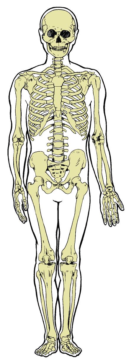
16 Skeletal System Illustration
 and osteoblasts (cells that build bone).")
17 Bones While bones take on different shapes, the majority of bones are long bones. Bone is continuously being remodeled via osteoclasts (cells that break down bone) and osteoblasts (cells that build bone). Wolff s law states that changes in bone structure coincide with changes in bone function. Form follows function
18 Movement of the Skeleton There are three main types of joints: Fibrous joints Cartilaginous joints Synovial joints Synovial joint movement occurs within the three planes of motion: sagittal, frontal, and transverse. Movement occurs along the joint s axis of rotation, where the plane of movement is generally perpendicular to the axis. Uniplanar joints (hinge joints) allow movement in only one plane. Biplanar joints allow movement in two planes that are perpendicular to each other. Multiplanar joints allow movement in all three planes.


19 Movement of Synovial Joints
20 Movement in the Sagittal Plane The sagittal plane runs anterior-posterior, dividing the body into left and right sections. Movements that involve rotation about a mediolateral axis occur in the sagittal plane. Examples include: Flexion Extension Dorsiflexion Plantarflexion


21 Movement in the Frontal Plane The frontal plane runs laterally, dividing the body into anterior and posterior sections. Movements that involve rotation about an anteroposterior axis occur in the frontal plane. Examples include: Abduction Adduction Elevation Depression Inversion Eversion


22 Movement in the Transverse Plane The transverse plane runs horizontally, dividing the body into superior and inferior sections. Movements that involve rotation about a longitudinal axis occur in the transverse plane. Examples include: Rotation Pronation Supination Horizontal flexion Horizontal extension
23 Multiplanar Movement Circumduction and opposition are two specific actions that occur in multiple planes. Circumduction: cone motion; combines flexion, extension, abduction, and adduction in sequence Opposition: thumb movement specific to humans and primates
 and peripheral nervous system (PNS).")
24 Nervous System The nervous system connects the muscles to the brain and spinal cord through a network of nerve circuits. Structurally, it is divided into the central nervous system (CNS) and peripheral nervous system (PNS). The CNS consists of the brain and spinal cord, while the PNS consists of all the nerve structures outside the brain and spinal cord. Nerves are made up of multiple nerve cells called neurons. Sensory nerves carry impulses to the CNS, while motor nerves carry impulses from the CNS to the PNS.
25 Proprioception Proprioception is the sense of knowing where the body is in relation to its various segments and the external environment. Receptors in the skin, in and around the joints and muscles, and in the inner ear transmit the information. The primary receptors involved in muscular control and coordination are the Golgi tendon organs (GTO) and muscle spindles.
.")
26 Musculotendinous Receptors Muscle spindle Located in the muscle belly lying parallel to the fibers Causes a reflexive contraction (stretch reflex) in the muscle when the muscle senses a stretch force. Simultaneously causes the antagonist to relax (reciprocal inhibition). GTO Located between the muscle belly and its tendon Causes muscle inhibition (autogenic inhibition) when it senses tension.
27 Muscular System Three types of muscle: Skeletal Attaches to the skeleton via tendons, contracts to move bones Voluntary Striated appearance Smooth Found on the walls of hollow organs and tubes (e.g., stomach and blood vessels) Involuntary Smooth appearance Cardiac Forms the walls of the heart Involuntary Smooth appearance
28 Skeletal Muscle Fiber Types Skeletal fibers can be divided into two general categories based on how quickly they contract. Slow-twitch muscle fibers contain relatively large amounts of mitochondria and are surrounded by more capillaries than fast-twitch fibers. Slow-twitch fibers contract more slowly than fast-twitch fibers. They have lower force outputs, but are more efficient and fatigue-resistant than fasttwitch fibers. Fast-twitch muscle fibers are further subdivided into fast-glycolytic and fastoxidative glycolytic fibers. Type IIx muscle fibers contain a relatively small amount of mitochondria, have a limited capacity for aerobic metabolism, and fatigue more easily than slow-twitch fibers. Type IIx have considerable anaerobic capacity, and are the largest and fastest, and are capable of producing the most force, of all the skeletal muscle fibers. Type IIa muscle fibers possess speed, fatigue, and force-production capabilities somewhere between type I and type IIx fibers. Type IIa fibers are also called intermediate fibers.
29 Comparison of Muscle Fiber Types The following table compares the three types of muscle fiber using the relative terms low, medium, and high. Type I Type IIa Type IIx Speed of contraction Low Medium High Force capacity Low Medium High Fatigue resistance High Medium Low Mitochondrial content High Medium Low Size Low Medium High Efficiency High Medium Low Aerobic capacity High Medium Low Anaerobic capacity Low Medium High
 composed of a series of repeating segments called sarcomeres.")
30 Muscle-fiber Microanatomy Skeletal muscles are made up of many muscle fibers held in place by connective tissue (fascia). Muscle fibers are made up of myofibrils (protein filaments) composed of a series of repeating segments called sarcomeres. Sarcomeres, made up of thick (myosin) and thin (actin) myofilaments, are the functional contracting unit of skeletal muscle.
31 Muscle Contraction Sliding filament model When acetylcholine is released from the CNS and detected, calcium is released. Calcium exposes binding sites along the actin for the myosin to attach. If sufficient ATP is present, cross-bridges are formed and the myosin pulls the actin toward the center, thereby shortening the sarcomere.
32 Connective Tissue There are two types of connective tissue directly related to joint movement: Collagen Made up of proteins that provide tensile strength and relative inextensibility, thereby limiting motion and resisting stretch Found in tendons and ligaments Elastic fibers Made up of amino acids and allow for extensibility Surround the sarcomere and are found in other organs Tendons are tough, cord-like tissues that transmit force from the muscle to the bone, causing movement. Ligaments contain a greater mixture of collagen and elastic fibers, taking on various shapes that support a joint by attaching bone to bone.

33 Factors That Impact Flexibility Soft tissues contribute to the total resistance to joint movement as follows: Joint capsule: 47% Muscle (fasciae): 41% Tendons: 10% Skin: 2% Other factors that impact flexibility include: Age Muscle strength, endurance, flexibility, and agility naturally decrease with age due to muscle atrophy that coincides with increased collagen. Gender In general, females are more flexible than males due to anatomical and physiological differences. Joint structure and past injury The rebuilding of broken bones and the build-up of scar tissue can limit joint movement.


34 The Shoulder Girdle The muscles of the shoulder girdle act on the scapula, primarily to stabilize it. There are six major muscles that anchor the scapula. Four posterior muscles Two anterior muscles
35 Major Muscles That Act at the Shoulder Girdle

.")

36 The Shoulder The shoulder joint is the most mobile joint in the body. There are a total of nine muscles that cross the shoulder joint (inserting on the humerus). Seven muscles originate from each scapula. Two muscles originate from the axial skeleton (no attachment on the scapula).
.")
37 The Rotator Cuff Four of the muscles that act at the shoulder are commonly called the rotator cuff. The rotator cuff s primary stabilizing function is to hold the humeral head in the glenoid fossa to prevent subluxation (dislocation). The muscles of the rotator cuff can be remembered using the acronym SITS: Supraspinatus Infraspinatus Teres minor Subscapularis

38 Major Muscles That Act at the Shoulder


39 The Elbow Flexion and extension of the elbow are controlled by muscles in the upper arm. Pronation and supination of the forearm are controlled by muscles in the upper arm, as well as several muscles in the forearm.
40 The Wrist The majority of the muscles that act at the wrist cross the elbow and are responsible for flexion and extension of the wrist and pronation and supination of the forearm. Flexion muscles originate primarily from or near the medial epicondyle. Extension muscles originate primarily from or near the lateral epicondyle.

41 Major Muscles That Act at the Elbow and Forearm

42 Major Muscles That Act at the Wrist


43 The Trunk The major muscles of the trunk support, stabilize, and move the spine. The abdominal wall, made up of the rectus abdominis, obliques, and transverse abdominis, has no skeletal support. Its strength comes from the multidirectional layers of muscle.
44 Major Muscles That Act at the Trunk

45 Hip Flexors There are 21 major muscles involved in the actions of the hip joint. Actions of the hip joint include flexion, extension, internal rotation, external rotation, adduction, and abduction. More than half of these muscles are involved in multiple actions.

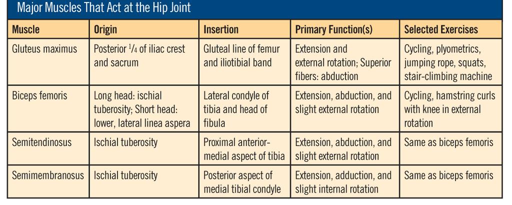
46 Hip Extensors
, and semimembranosus (slight).")
, gluteus medius and minimus (posterior fibers), sartorius, pectineus, and the")
47 Hip Internal and External Rotators The hip internal rotators include the tensor fasciae latae, semitendinosus (slight), and semimembranosus (slight). The hip external rotators include the iliopsoas, gluteus maximus, biceps femoris (slight), gluteus medius and minimus (posterior fibers), sartorius, pectineus, and the six deep external rotators.
48 Hip Adductors

49 Hip Abductors
50 The Knee Joint The muscles of the upper thigh are responsible for movement at the knee.


51 The Anterior Compartment of the Lower Leg The ankle joint allows dorsiflexion and plantarflexion. The subtalar joint allows inversion and eversion of the foot. The muscles of the lower leg control movements of the ankle and foot. The lower leg is divided into three primary compartments: anterior, posterior, and lateral.


52 The Posterior Compartment of the Lower Leg The posterior compartment is made up of muscles that plantarflex the foot and/or flex the toes and is divided further into the superficial posterior and deep posterior compartments: Superficial posterior compartment: gastrocnemius, soleus, and plantaris Deep posterior compartment: flexor hallucis longus, flexor digitorum longus, posterior tibialis, and popliteus
53 The Posterior Compartment of the Lower Leg (cont.)

54 The Lateral Compartment of the Lower Leg The lateral compartment is made up of muscles that plantarflex and evert the foot, including the peroneus longus and peroneus brevis.
55 The Endocrine System The endocrine system, which is made up of various glands throughout the body, is responsible for regulating bodily activities through the production of hormones. The principal glands are as follows: Pituitary Thyroid Parathyroids Adrenals Paradrenals Gonads
56 Major Endocrine Glands and Their Hormones
57 Summary To design safe and effective exercise programs and group fitness classes, fitness professionals must have working knowledge of human anatomy. This session covered: Anatomical terminology Structural levels of the body The cardiovascular, respiratory, digestive, skeletal, neuromuscular, muscular, and endocrine systems Planes of motion Upper- and lower-extremity and trunk muscles Muscle-fiber types
ACE s Essentials of Exercise Science for Fitness Professionals Chapter 1: Human Anatomy
 ACE s Essentials of Exercise Science for Fitness Professionals Chapter 1: Human Anatomy 1 Learning Objectives This session, which is based on Chapter 1 of ACE s Essentials of Exercise Science for Fitness
Chapter 9. The Muscular System
 1 Chapter 9 The Muscular System 2 Introduction Skeletal muscles: movement in environment Smooth muscles: intestines, ureters, veins and arteries Cardiac muscle: pumps blood through heart and blood vessels
Types of Muscle: Skeletal- muscle involved in movement of the skeleton. Striated, has alternating bands of light and dark due to overlapping
 Types of Muscle: Skeletal- muscle involved in movement of the skeleton. Striated, has alternating bands of light and dark due to overlapping filaments within the muscle cell. Skeletal muscle can be consciously
The Human Body. Lesson Goal. Lesson Objectives 9/10/2012. Provide a brief overview of body systems, anatomy, physiology, and topographic anatomy
 The Human Body Lesson Goal Provide a brief overview of body systems, anatomy, physiology, and topographic anatomy Medial Lateral Proximal Distal Superior Inferior Anterior Lesson Objectives Explain the
Unit 7: Skeletal and muscular systems
 Unit 7: Skeletal and muscular systems 1. The locomotor system 2. The skeletal system 2.1. The human skeleton 2.2. Bones 2.3. Joints 2.4. Tendons and ligaments 3. The muscular system 3.1. Muscles of the
Unit 1: Human body: combination I - IV
 Unit 1: Human body: combination I - IV Study online at quizlet.com/_1kzmm2 1. alveoli 6. bronchioles microscopic air sacs in the lung where diffusion of the respiratory gases, oxygen and carbon dioxide
Level 2 Anatomy and Physiology for Exercise and Fitness Instructors (K/616/7823) - Sample Assessment Student: XXXXXX Sample 3
 - Sample Assessment Student: XXXXXX Sample 3") MULTIPLE CHOICE QUESTION PAPER Paper number: SAMPLE 3 Please ensure that this paper number is referenced on your candidate answer sheet Title: Student: XXXXXX Sample 3 Special Instructions: Level 2 Anatomy
MULTIPLE CHOICE QUESTION PAPER Paper number: SAMPLE 3 Please ensure that this paper number is referenced on your candidate answer sheet Title: Student: XXXXXX Sample 3 Special Instructions: Level 2 Anatomy
Anatomy and Physiology for Exercise and Health Level 3 A/600/9051 Mock Paper March 1 st 2015 August 31 st 2015
 Anatomy and Physiology for Exercise and Health Level 3 A/600/9051 Mock Paper March 1 st 2015 August 31 st 2015 There are 40 questions within this paper. To achieve a pass you will need to score 28 out
Anatomy. Anatomy deals with the structure of the human body, and includes a precise language on body positions and relationships between body parts.
 Anatomy deals with the structure of the human body, and includes a precise language on body positions and relationships between body parts. Proper instruction on safe and efficient exercise technique requires
or Everything you ever wanted to know about Muscles, but were afraid to ask!!!
 The Muscular System or Everything you ever wanted to know about Muscles, but were afraid to ask!!! Did you know that? - more than 50% of body weight is muscle! - And muscle is made up of proteins and water
Muscles of the Hip 1. Tensor Fasciae Latae O: iliac crest I: lateral femoral condyle Action: abducts the thigh Nerve: gluteal nerve
 Muscles of the Hip 1. Tensor Fasciae Latae O: iliac crest I: lateral femoral condyle Action: abducts the thigh Nerve: gluteal nerve 2. Gluteus Maximus O: ilium I: femur Action: abduct the thigh Nerve:
Certified Personal Trainer Re-Certification Manual
 Certified Personal Trainer Re-Certification Manual Section II 1 Anatomy & Physiology Terms Anatomy and physiology are closely related fields of study: anatomy is the study of form, and physiology is the
Applied Human Biology for Exercise and Fitness Level 3 J/615/3220 MOCK PAPER
 Applied Human Biology for Exercise and Fitness Level 3 J/615/3220 MOCK PAPER There are 40 questions within this paper. To achieve a pass you will need to score 28 out of 40 marks. All questions are multiple
Temporalis Elevates & retracts mandible. Masseter Elevates mandible. Sternocleidomastoid Neck flexion. Trapezius Elevates & depresses shoulders
 Anterior Posterior Temporalis Elevates & retracts mandible Masseter Elevates mandible Sternocleidomastoid Neck flexion Trapezius Elevates & depresses shoulders Masseter Elevates mandible Temporalis Elevates
The Muscular System PART C. PowerPoint Lecture Slide Presentation by Patty Bostwick-Taylor, Florence-Darlington Technical College
 PowerPoint Lecture Slide Presentation by Patty Bostwick-Taylor, Florence-Darlington Technical College The Muscular System 6 PART C Five Golden Rules of Skeletal Muscle Activity Table 6.2 Muscles and Body
Muscle stations Answers
 Muscle Unit Muscle stations Answers A: What #is: C = 3 F = 5 E = 6 D = 1 B =4 A =2 B 5. superior 6. Inferior 4. anterior C: 1. What # is a,b,c,d 2. What muscle group #1? Quads 3. What muscle is #5? Gastrocnemius
The Muscular System. Myology the study of muscles
 The Muscular System Myology the study of muscles Functions of muscles: 1. Movement 2. Stability /support posture 3. Heat production 85% of our body heat 4. Communication 5. Constriction of organs and vessels
Energy for Muscle Contractions: Direct phosphorylation. Creatine phosphate loses a phosphate to ADP to create ATP
 Energy for Muscle Contractions: Direct phosphorylation Aerobic respiration Anaerobic respiration (lactic acid fermentation) Creatine phosphate loses a phosphate to ADP to create ATP Requires oxygen to
The Muscular System. Chapter 10 Part D. PowerPoint Lecture Slides prepared by Karen Dunbar Kareiva Ivy Tech Community College
 Chapter 10 Part D The Muscular System Annie Leibovitz/Contact Press Images PowerPoint Lecture Slides prepared by Karen Dunbar Kareiva Ivy Tech Community College Table 10.14: Muscles Crossing the Hip and
STUDY GUIDE a comprehensive review of the:
 STUDY GUIDE a comprehensive review of the: NFPT PERSONAL FITNESS TRAINERMANUAL Study & Reference: The Fundamentals for the CERTIFIED PERSONAL TRAINERR (CPT) Third Edition 2015 All Rights Reserved National
Level 2 Anatomy and Physiology Internal Practice Paper
 Level 2 Anatomy and Physiology Internal Practice Paper Time allocated: 60 minutes. 30 questions, multiple choice answers. Select A,B,C or D only select one answer. You are required to achieve 22 correct
Mock Paper Level 2 Anatomy and Physiology for Exercise. Unit Reference Number H/600/9013
 MULTIPLE CHOICE QUESTION PAPER Paper number MPAPEH2.01 Please insert this reference number in the appropriate boxes on your candidate answer sheet Title Time allocation 60 minutes Mock Paper Level 2 Anatomy
Muscles of the Cat. N Deltoid MUSCLES OF THE CHEST. Pectoralis major. (This muscle is superior to Pectoralis minor) MUSCLES OF THE CHEST
 MUSCLES OF THE CHEST") MUSCLES OF THE CHEST Pectoralis major (This muscle is superior to Pectoralis minor) 1. MUSCLES OF THE CHEST Pectoralis minor (This muscle is inferior to Pectoralis major) 2. MUSCLES OF THE ARM Deltoid
MUSCLES OF THE CHEST Pectoralis major (This muscle is superior to Pectoralis minor) 1. MUSCLES OF THE CHEST Pectoralis minor (This muscle is inferior to Pectoralis major) 2. MUSCLES OF THE ARM Deltoid
Chapter 10: Muscular System: Gross Anatomy
 Chapter 10: Muscular System: Gross Anatomy I. General Principles A. General Terminology 1. Tendons attach 2. What is an aponeurosis? 3. The points of muscle attachment are called & 4. How is the "origin"
Level 2 Mock Paper Anatomy and Physiology For Exercise. Unit Accreditation Number H/600/9013
 MULTIPLE CHOICE QUESTION PAPER Paper number MPAPEH Please insert this reference number in the appropriate boxes on your candidate answer sheet Title Time allocation 60 minutes Level 2 Mock Paper Anatomy
Muscle. Dr. Carmen E. Rexach Anatomy 35 Mt San Antonio College
 Muscle Dr. Carmen E. Rexach Anatomy 35 Mt San Antonio College Functions Movements of bones and soft body parts Movements of fluids through a tube (blood, digestive) Functions Maintain posture Support soft
MOCK PAPER Level 3 Anatomy and Physiology For Exercise and Health. Unit Reference Number A/600/9051
 MULTIPLE CHOICE QUESTION PAPER Paper number APEH 3.01 Please insert this reference number in the appropriate boxes on your candidate answer sheet Title Time allocation 60 minutes MOCK PAPER Level 3 Anatomy
Anatomy and Physiology for Exercise Level 2
 Anatomy and Physiology for Exercise Level 2 H/600/9013 Mock Paper There are 30 questions within this paper To achieve a pass you will need to score 21 out of 30 marks All questions are multiple choice
CHAPTER 3 What Is Anatomy?
 CHAPTER 3 What Is Anatomy? Kinesiology Books Publisher 1 TABLE OF CONTENTS The Language of Anatomy Anatomical Position Directional Terms Body Planes Movements Musculoskeletal System Human Skeleton Types
Level 2 Anatomy and Physiology for Exercise and Fitness Instructors (K/616/7823) - Sample Assessment Student: XXXXXX Sample 4
 - Sample Assessment Student: XXXXXX Sample 4") MULTIPLE CHOICE QUESTION PAPER Paper number: SAMPLE 4 Please ensure that this paper number is referenced on your candidate answer sheet Title: Student: XXXXXX Sample 4 Special Instructions: Level 2 Anatomy
MULTIPLE CHOICE QUESTION PAPER Paper number: SAMPLE 4 Please ensure that this paper number is referenced on your candidate answer sheet Title: Student: XXXXXX Sample 4 Special Instructions: Level 2 Anatomy
CHAPTER 3 BASIC ANATOMY AND PHYSIOLOGY
 CHAPTER 3 BASIC ANATOMY AND PHYSIOLOGY SURFACE ANATOMY Surface anatomy is the identification of landmarks on the surface of the skin which allows us to compare our knowledge of our own surface anatomy
Muscular System. IB Sports, exercise and health science 1.2
 Muscular System IB Sports, exercise and health science 1.2 Characteristics Common to Contractility-ability to shorten the muscles length Extensibility-ability to lengthen the muscles length Elasticity-muscle
In which arm muscle are intramuscular injections most often given? (not in text)
") AP1 Lab 9 - Muscles of the Arms and Legs Locate the following muscles on the models and on yourself. Recall anatomical position. Directional terms such as anterior, posterior, lateral, etc. all assume
AP1 Lab 9 - Muscles of the Arms and Legs Locate the following muscles on the models and on yourself. Recall anatomical position. Directional terms such as anterior, posterior, lateral, etc. all assume
Location Terms. Anterior and posterior. Proximal and Distal The term proximal (Latin proximus; nearest) describes where the appendage joins the body.
 describes where the appendage joins the body.") HUMAN ANAT OMY Location Terms Anterior and posterior In human anatomical usage, anterior refers to the front of the individual. Similarly, posterior refers to the back of the subject. In standard anatomical
HUMAN ANAT OMY Location Terms Anterior and posterior In human anatomical usage, anterior refers to the front of the individual. Similarly, posterior refers to the back of the subject. In standard anatomical
Essentials of Human Anatomy & Physiology. The Muscular System
 Essentials of Human Anatomy & Physiology The Muscular System The Muscular System Muscles are responsible for all types of body movement they contract or shorten and are the machine of the body Three basic
Muscles Unit TEST and Final Exam Study Guide May 2017
 Muscles Unit TEST and Final Exam Study Guide May 2017 Part 1 of final exam is pictures, see bottom of the study guide Part 2 of the final exam is only going to cover muscles unit. If you do this study
Year 2004 Paper one: Questions supplied by Megan
 QUESTION 47 A 58yo man is noted to have a right foot drop three days following a right total hip replacement. On examination there is weakness of right ankle dorsiflexion and toe extension (grade 4/5).
Exercise Science Section 3: The Muscular System
 Exercise Science Section 3: The Muscular System An Introduction to Health and Physical Education Ted Temertzoglou Paul Challen ISBN 1-55077-132-9 Major Functions of Muscles Movement Includes: breathing,
5 Specification Content
 5 Specification Content These specifications are set out in the form of teaching modules. Each teaching module is assessed by its associated unit of assessment. 5.1 Module 2562: The Application of Physiological
CHAPTER 8: THE BIOMECHANICS OF THE HUMAN LOWER EXTREMITY
 CHAPTER 8: THE BIOMECHANICS OF THE HUMAN LOWER EXTREMITY _ 1. The hip joint is the articulation between the and the. A. femur, acetabulum B. femur, spine C. femur, tibia _ 2. Which of the following is
CHAPTER 1: 1.1 Muscular skeletal system. Question - text book page 16. Question - text book page 20 QUESTIONS AND ANSWERS. Answers
 QUESTIONS AND ANSWERS CHAPTER 1: 1.1 Muscular skeletal system Question - text book page 16 Using the information on pages 12 to 14 above, complete the table below. joint joint type articulating bones associated
BIOLOGY - CLUTCH CH.49 - MUSCLE SYSTEMS.
 !! www.clutchprep.com BIOLOGY - CLUTCH Muscle system organ system that includes skeletal, cardiac, and smooth muscle Muscle tissue capable of contracting through the interaction of actin and myosin proteins
Applied anatomy and physiology: definitions of key terms
 Applied anatomy and physiology: definitions of key terms See pages 5 46 These are the key terms from Chapter 1. Try cutting them out and then matching the key terms with their definitions, or asking friends
MUSCLES OF THE LOWER LIMBS
 MUSCLES OF THE LOWER LIMBS Naming, location and general function Dr. Nabil khouri ROLES THAT SHOULD NOT BE FORGOTTEN Most anterior compartment muscles of the hip and thigh Flexor of the femur at the hip
Level 2 Anatomy and Physiology for Exercise Assessment Workbook
 Fitness Instructor Level 2 Anatomy and Physiology for Exercise Assessment Workbook IMPORTANT: YOU MUST COMPLETE THE FOLLOWING DETAILS BEFORE RETURNING THIS WORKBOOK TO LIFETIME Name of learner: Email address:
Introduction to Human Body Systems
 The Human Organism: Introduction to Human Body Systems By Deanne Erdmann, MS Levels of Organization in the Body Cells Tissues Epithelial, connective, muscular, nervous Organs Examples include stomach,
REVISION BOOKLET. The Body Systems
 REVISION BOOKLET The Body Systems GCSE PE 2016 Skeletal System Functions of the skeleton Joints for movement Muscle attachment Protection of vital organs Red and white blood cell production platelets Storage
BLUE SKY SCHOOL OF PROFESSIONAL MASSAGE AND THERAPEUTIC BODYWORK. Musculoskeletal Anatomy & Kinesiology MUSCLES, MOVEMENTS & BIOMECHANICS
 BLUE SKY SCHOOL OF PROFESSIONAL MASSAGE AND THERAPEUTIC BODYWORK Musculoskeletal Anatomy & Kinesiology MUSCLES, MOVEMENTS & BIOMECHANICS MSAK101-I Session 7 Learning Objectives: 1. List the three types
MOCK PAPER Level 3 Anatomy and Physiology For Exercise and Health. Unit Accreditation Number A/600/9051
 MULTIPLE CHOICE QUESTION PAPER Paper number APEH 3.01 Please insert this reference number in the appropriate boxes on your candidate answer sheet Title Time allocation 60 minutes MOCK PAPER Level 3 Anatomy
11/15/2018. Temporalis Elevates & retracts mandible. Masseter = Prime mover of jaw closure. Levator scapulae Supraspinatus Clavicle.
 Due in Lab 10 Lab 8 MUSCLES 2 weeks because of Thanksgiving Prelab #10 Both sides! Homework #8 Both sides! Refer to Muscles 22-23 Examples of Origin & Insertion Naming of muscles Origin Site of muscle
Cadaver Muscular System Practice Practical
 Cadaver Muscular System Practice Practical Station 1 Station 1 1. Specific structure 1. Rectus sheath 2. Red line 2. Linea alba Station 2 Station 2 3. Red muscle 1. Rectus abdominis 4. Red muscle actions
A. All movements require muscle which are organs using chemical energy to contract.
 Ch 8 Muscles Introduction: A. All movements require muscle which are organs using chemical energy to contract. B. The three types of muscle in the body are skeletal, smooth, and cardiac muscle. C. This
SUMMER WORK MRS KANSARA
 Name: A LEVEL PE Anatomy and Physiology SUMMER WORK MRS KANSARA Complete the following activities throughout the summer It is important that you understand and remember all the information included in
Test Bank for The Human Body in Health and Illness 4th Edition by Herlihy
 Test Bank for The Human Body in Health and Illness 4th Edition by Herlihy Chapter 9: Muscular System Test Bank MULTIPLE CHOICE 1. Which of the following muscles is described as striated and involuntary?
Chapter 3: Applied Kinesiology. ACE Personal Trainer Manual Third Edition
 Chapter 3: Applied Kinesiology ACE Personal Trainer Manual Third Edition Introduction Kinesiology is the study of the body s infinite number of movements, positions, and postures and is grounded in the
Prime movers provide the major force for producing a specific movement Antagonists oppose or reverse a particular movement Synergists
 Dr. Gary Mumaugh Prime movers provide the major force for producing a specific movement Antagonists oppose or reverse a particular movement Synergists Add force to a movement Reduce undesirable or unnecessary
OBJECTIVES. Unit 7:5 PROPERTIES OR CHARACTERISTICS OF MUSCLES. Introduction. 3 Kinds of Muscles. 3 Kinds of Muscles 4/17/2018 MUSCULAR SYSTEM
 OBJECTIVES Unit 7:5 MUSCULAR SYSTEM Compare the three main kinds of muscles by describing the action of each Differentiate between voluntary and involuntary muscles List at least three functions of muscles
Outline. Bio 105: Muscular System. Muscular System. Types of Muscles. Smooth Muscle. Cardiac Muscle 4/6/2016
 Outline Bio 105: Muscular System Lecture 11 Chapter 6 Characteristics of muscles 3 types of muscles Functions of muscles Structure of skeletal muscles Mechanics of muscle contraction Energy sources for
Muscles of the lower extremities. Dr. Nabil khouri MD, MSc, Ph.D
 Muscles of the lower extremities Dr. Nabil khouri MD, MSc, Ph.D Posterior leg Popliteal fossa Boundaries Biceps femoris (superior-lateral) Semitendinosis and semimembranosis (superior-medial) Gastrocnemius
Muscles of the Gluteal Region
 Muscles of the Gluteal Region 1 Some of the most powerful in the body Extend the thigh during forceful extension Stabilize the iliotibial band and thoracolumbar fascia Related to shoulders and arms because
Human Anatomy and Physiology I Laboratory
 Human Anatomy and Physiology I Laboratory Gross Anatomy of the Muscular System (Two weeks) 1 This lab involves study of the laboratory exercise Gross Anatomy of the Muscular System. Complete the Review
Human Anatomy Unit 2 MUSCULAR SYSTEM
 Human Anatomy Unit 2 MUSCULAR SYSTEM In Anatomy Today Functions Movements of bones and soft body parts Movements of fluids through a tube (blood, digestive) Functions Maintain posture Support soft organs
Human Anatomy Lab #7: Muscles of the Cadaver
 Human Anatomy Lab #7: Muscles of the Cadaver Table of Contents: Expected Learning Outcomes.... 1 Introduction...... 1 Identifying Muscles on Yourself.... 2 Muscles of the Anterior Trunk and Arm.. 2 Muscles
Unit I Problem 5 Anatomy: Types of Movements and Joints
 Unit I Problem 5 Anatomy: Types of Movements and Joints - Anatomical position: The person is standing erect, with the upper limbs by the sides and the face and palms of the hands directed forward. - Imaginary
Assignment 2: Human Anatomy
 Assignment 2: Human Anatomy Chapter 2 Quiz: How Much Do You Know About Anatomy? 1. Which of the following is not a feature of the anatomical position: A) The body stands erect. B) The body is facing forward.
Due in Lab weeks because of Thanksgiving Prelab #10. Homework #8. Both sides! Both sides!
 Lab 8 MUSCLES Due in Lab 10 2 weeks because of Thanksgiving Prelab #10 Both sides! Homework #8 Both sides! Refer to Muscles 22-23 Naming of muscles Origin Site of muscle attachment that doesn t move during
PHYSICAL EDUCATION. 4º E.S.O. 2nd TERM. The skeletal and muscular systems.
 PHYSICAL EDUCATION 4º E.S.O. 2nd TERM. The skeletal and muscular systems. PARTS OF THE BODY Head Torso / Trunk Dorsal: Back Ventral: Thorax y Abdomen Extremities Superior: Arm Forearm Hand Joint: Shoulder
Personal Training Certificate. Anatomy and Physiology Mock Paper
 Candidate Name Date Personal Training Certificate Anatomy and Physiology Mock Paper Instructions: Make sure your name is in the box at the top, followed by the date you did the test Tick or highlight your
2/4/2018. Identify the two reasons why muscle cells may go through muscle fatigue. Ch.7 Review. Sternocleidomastoid.
 Ch.7 Review Identify the two reasons why muscle cells may go through muscle fatigue Temporalis Depressor anguli oris Sternocleidomastoid Tibialis anterior 1 Gluteus medius Deltoid Adducts & rotates scapula
Chiropractic Technician Class
 Chiropractic Technician Class Presentation By: Dr. Kay Miller. The Role of Exercise as it Relates to Our Musculoskeletal System Introduction to the topic and Preliminary Physical exam Musculoskeletal anatomy:
Level 3 Diploma in Personal Training (Practitioner) - Sample Assessment Materials
 - Sample Assessment Materials") MULTIPLE CHOICE QUESTION PAPER Paper number: SAMPLE 5 Please ensure that this paper number is referenced on your candidate answer sheet Title: Student: XXXXXX Sample 5 Special Instructions: Level 3 Diploma
MULTIPLE CHOICE QUESTION PAPER Paper number: SAMPLE 5 Please ensure that this paper number is referenced on your candidate answer sheet Title: Student: XXXXXX Sample 5 Special Instructions: Level 3 Diploma
NZQA Expiring unit standard version 2 Page 1 of 5. Demonstrate knowledge of exercise physiology and human anatomy
 Page 1 of 5 Title Demonstrate knowledge of exercise physiology and human anatomy Level 3 Credits 10 Purpose People credited with this unit standard are able to: explain the nervous system and its functions;
Anatomy Review-INTRODUCTION. The study of the function of the body parts is called. Examples include:
 Anatomy Review-INTRODUCTION The study of the organs and parts of the body is called Examples include: The study of the function of the body parts is called. Examples include: Use the numbers from the diagram
Circulatory System. and. Respiratory System. Ari Min, Yerim Lee and Min Ji Song THE HEART LUNGS. Monday, May 23, 2011
 Human Anatomy Circulatory System and THE HEART Respiratory System LUNGS Ari Min, Yerim Lee and Min Ji Song Purpose of the Circulatory System Function of circulatory system: exchange gases with cardiovascular
Understanding Leg Anatomy and Function THE UPPER LEG
 Understanding Leg Anatomy and Function THE UPPER LEG The long thigh bone is the femur. It connects to the pelvis to form the hip joint and then extends down to meet the tibia (shin bone) at the knee joint.
Action and Support: The Muscles and Skeleton
 Lesson 15 Action and Support: The Muscles and Skeleton Introduction to Life Processes - SCI 102 1 Muscle Contraction Muscles: tissues that produce movement by contracting Vertebrates have three types of
The Muscular System The Muscular System Muscles are responsible for all types of body movement Three basic muscle types are found in the body
 The Muscular System The Muscular System Muscles are responsible for all types of body movement Three basic muscle types are found in the body Skeletal muscle Cardiac muscle Smooth muscle Characteristics
Human Anatomy Biology 351
 Human Anatomy Biology 351 Lower Limb Please place your name on the back of the last page of this exam. You must answer all questions on this exam. Because statistics demonstrate that, on average, between
7/10/18. Introduction. Muscular System. Anatomy. Physiology. Skeletal Muscle Anatomy. Muscle Fiber
 Introduction Muscular System Chapter 20 Shortening or lengthening of a muscle results from changes in relative positions of one small part of a muscle cell to another To understand contraction, we will
10/4/18. Muscular System. 1 Copyright 2016 by Elsevier Inc. All rights reserved. Introduction. Anatomy. Physiology. Skeletal Muscle Anatomy
 Introduction Muscular System Chapter 20 Shortening or lengthening of a muscle results from changes in relative positions of one small part of a muscle cell to another To understand contraction, we will
Introduction to The Human Body
 1 Introduction to The Human Body FOCUS: The human organism is often examined at seven structural levels: chemical, organelle, cell, tissue, organ, organ system, and the organism. Anatomy examines the structure
Unit 4: The Muscular System REVIEW GUIDE
 NPHS Anatomy & Physiology Questions to answer: 1) List the three functions of the muscular system. Unit 4: The Muscular System REVIEW GUIDE 2) What are the four characteristics of muscle tissue? Briefly
The Human Body. Mrs. Green
 The Human Body Mrs. Green Bell Work Which of the following helps the body to cool down? a) Shivering b) Sweating c) Running a fever d) Taking a deep breath Which of the following is a function of the digestive
Introduction to Human Anatomy. Prepared by:- Mohammad Zmaili
 Introduction to Human Anatomy 1 Prepared by:- Mohammad Zmaili Definitions Anatomy: the science of the structure and shape of living organisms and their parts. Physiology: the science that deals with the
Human Body Systems Study Guide
 Human Body Systems Study Guide Nervous System 1. Brain stem part of nervous system and controls the heartbeat and breathing by controlling the cardiac muscle and diaphragm. Also receives information from
Unit 6: The Muscular System
 Unit 6: The Muscular System I. The Muscular System A. Muscles are responsible for all types of body movement B. Three basic muscle types are found in the body 1. Skeletal muscle 2. Cardiac muscle 3. Smooth
VCE Physical Education. Unit 1 AOS 1: Bodies in Motion
 VCE Physical Education Unit 1 AOS 1: Bodies in Motion What are the major body systems that allow movement to happen? Muscular Skeletal } Musculoskeletal System Cardiovascular Respiratory } Cardiorespiratory
Epicranius (frontal belly) Zygomaticus minor. Zygomaticus major Buccinator
 Zygomaticus minor. Zygomaticus major Buccinator") Epicranius (frontal belly) Zygomaticus minor Zygomaticus major Buccinator Masseter Digastric (posterior belly) Stylohyoid Sternocleidomastoid Trapezius Scalenus Omohyoid (inferior belly) Orbicularis oris
Epicranius (frontal belly) Zygomaticus minor Zygomaticus major Buccinator Masseter Digastric (posterior belly) Stylohyoid Sternocleidomastoid Trapezius Scalenus Omohyoid (inferior belly) Orbicularis oris
VCE PHYSICAL EDUCATION WORKBOOK UNIT 1 BODIES IN MOTION NAME:
 VCE PHYSICAL EDUCATION WORKBOOK UNIT 1 BODIES IN MOTION NAME: SKELETAL SYSTEM List the 5 functions of the skeletal system and complete the following table. FUNCTION DESCRIPTION Label the following features
Muscle Lecture Test Questions Set 1
 Muscle Lecture Test Questions Set 1 Fall 2015 1. Muscle cells exhibit the greatest ability to shrink -- this quality of all protoplasm is: a. voluntary b. involuntary c. fusiform d. contractility e. conductivity
The Language of Anatomy. (Anatomical Terminology)
") The Language of Anatomy (Anatomical Terminology) Terms of Position The anatomical position is a fixed position of the body (cadaver) taken as if the body is standing (erect) looking forward with the upper
The Language of Anatomy (Anatomical Terminology) Terms of Position The anatomical position is a fixed position of the body (cadaver) taken as if the body is standing (erect) looking forward with the upper
The Digestive System: to convert food particles into simpler micro molecules that can be absorbed into the bloodstream and used by the body
 Body Systems The Digestive System: to convert food particles into simpler micro molecules that can be absorbed into the bloodstream and used by the body Major Organs and their Functions: Mouth to chew
VCE PHYSICAL EDUCATION WORKBOOK UNIT 1 BODIES IN MOTION NAME:
 VCE PHYSICAL EDUCATION WORKBOOK UNIT 1 BODIES IN MOTION NAME: SKELETAL SYSTEM List the 5 functions of the skeletal system and complete the following table. FUNCTION DESCRIPTION Label the following features
Assignment 4: Muscle Structure and Function
 Assignment 4: Muscle Structure and Function Unit 2: Chapter 5 Part A Multiple Choice Questions 1. Which of the following statements about skeletal muscle is true: A) Skeletal muscles are usually linked
Main Menu. Joint and Pelvic Girdle click here. The Power is in Your Hands
 1 Hip Joint and Pelvic Girdle click here Main Menu K.6 http://www.handsonlineeducation.com/classes//k6entry.htm[3/23/18, 2:01:12 PM] Hip Joint (acetabular femoral) Relatively stable due to : Bony architecture
THE HUMAN BODY. study of the structure of living organisms. Physiologythe study of how the body works. Ex: studying the structure of the heart.
 HUMAN BODY SYSTEMS Anatomythe study of the structure of living organisms. Ex: studying the structure of the heart. Physiologythe study of how the body works. Ex: how the heart works to pump blood, etc.
The Musculoskeletal System. Chapter 46
 The Musculoskeletal System Chapter 46 Types of Skeletal Systems Changes in movement occur because muscles pull against a support structure Zoologists recognize three types: 1. Hydrostatic skeletons a fluid
36.3 The Integumentary System The Skin. KEY CONCEPT The integumentary system has many tissues that protect the body.
 36.3 The Integumentary System The Skin KEY CONCEPT The integumentary system has many tissues that protect the body. 36.3 The Integumentary System The Skin The integument is the body system that surrounds
High School Biology - Problem Drill 20: Skeletal, Muscular, and Integumentary Systems
 High School Biology - Problem Drill 20: Skeletal, Muscular, and Integumentary Systems Question No. 1 of 10 Which of the following about the skeletal system is true? Question #01 A. There are two types
Human Anatomy Biology 351
 Human Anatomy Biology 351 Lower Limb Please place your name on the back of the last page of this exam. You must answer all questions on this exam. Because statistics demonstrate that, on average, between