Etv4 BU EMM Etv5 BC MMM UI-M-BH3- Elk1 BC MMM
|
|
|
- Naomi Davidson
- 5 years ago
- Views:
Transcription
1 Supplementary Tables In situ hybridization probes Genbank Gene Accession Number Clone ID Cat. No Etv1 BC PCR Etv4 BU EMM Etv5 BC MMM Fev AI UI-M-BH3- AVJ-B-02-0-UI EMM Elk1 BC MMM Antibodies Antigen Species Supplier/ Catalog No. Dilution DAT; Slc6a3 Rat, monoclonal Millipore: MAB369 1:500 TH Rabbit Millipore: AB152 1:500 Vmat2; Slc18a2 Rabbit Millipore: AB1598P 1:500 Nurr1; Nr4a2 Rabbit Santa Cruz: SC :500 Supplementary Table 1. In situ hybridization probes and antibodies. Clones containing probes were obtained from Open Biosystems and the corresponding catalog numbers are given. All antibodies gave staining patterns consistent with the mrna expression of the corresponding gene determined by in situ hybridization in our experiments or in available expression databases. DA pathway antigens also produced signal in known targets of DA innervation, such as the striatum. Ectopic staining outside of these known areas was not observed. 1
2 Allen atlas SN/VTA expression Gene E11.5 E13.5 E15.5 Other E15.5 CNS expression Ehf NE Olf Elf NE NE Elf NE NE Elf NE NE Elf NE NE Elf NE Diffuse? *Elk NE Hip Other Adult CNS expression Elk NE CB, Hip, Olf? Elk4 - - ND NE NE *Etv1/ Er Ctx, Hab, IO CB, Ctx, Hab, Hip Erf NE NE Erg/ V-ets NE NE Ets NE NE *Ets NE Ctx Etv2/ Etsrp NE NE *Etv5/ Erm NE CB, Ctx, Hip, Olf, Str, SN, VTA Etv6/ Tel NE NE Fev/ Pet1 ND ND - NE 5HT Fli1 - - ND NE NE Gabpa NE NE Etv3/ Pe NE NE *Etv4/ Pea NE NE *Spedf/ Pse NE NE Sfpi1/ Pu NE NE Spib NE NE Spic NE NE Supplementary Table 2. Ets gene family expression in developing and adult CNS. 5HT, serotonergic neurons; CB, cerebellum; Ctx, cerebral cortex; Hab, habenula; Hip, hippocampal region; IO, inferior olive; Str, striatum; SN, substantial nigra; VTA, ventral tegmental area. ND, no data; NE, no expression. Mouse Ets family members correspond closely to those identified in 2
3 the human (Oikawa and Yamada, 2003; Hollenhorst et al., 2007), with the exception of Etv7, which does not have an identified mouse transcript in NCBI. *See also Supplemental Figures S3-S6. Hollenhorst PC, Shah AA, Hopkins C, Graves BJ (2007) Genome-wide analyses reveal properties of redundant and specific promoter occupancy within the ETS gene family. Genes Dev 21: Oikawa T, Yamada T (2003) Molecular biology of the Ets family of transcription factors. Gene 303:
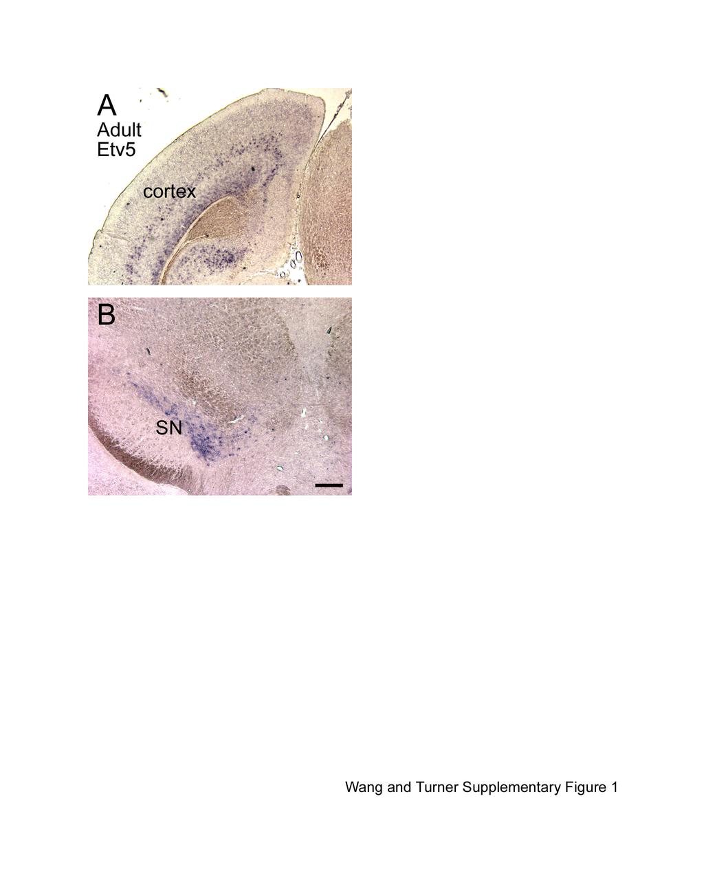
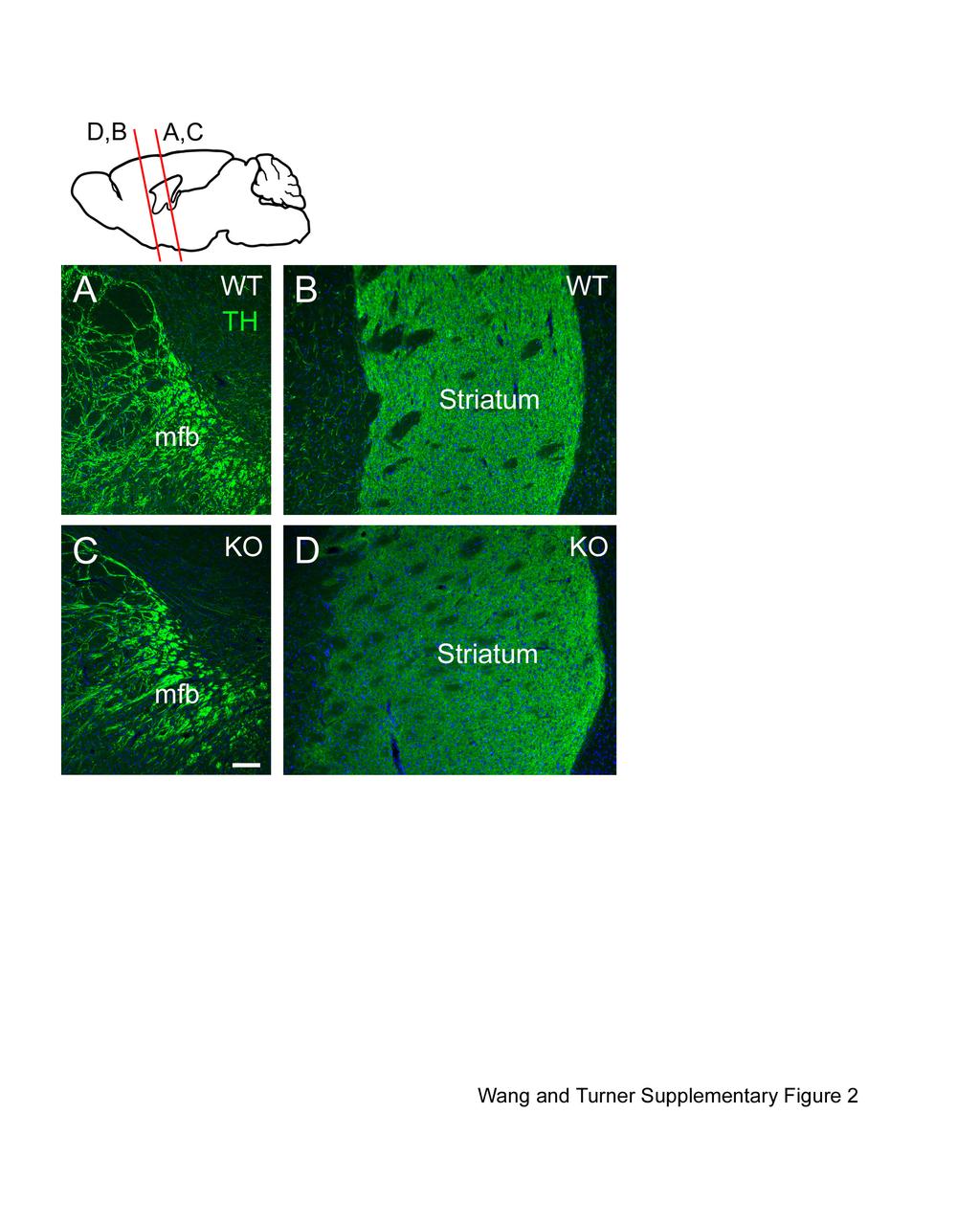
4 Supplementary Figure 1. Etv5 expression in adult mouse brain. The expression of Etv5 in adult mouse brain was visualized by ISH in coronal sections. (A) cerebral cortex, (B) ventral midbrain. SN, substantia nigra. Scale is 250µm. Supplementary Figure 2. Projections of ventral midbrain DA neurons in Etv5 -/- mice. Tyrosine hydroxylase (TH) immunostaining was used to visualize the projections of DA neurons in coronal sections of adult mice (diagram). (A,C) proximal part of medial forebrain bundle in control (WT, A) and Etv5 -/- (KO, C) mice. (B,D) dorsal striatum of control (B) and Etv5 -/- mice (D). Scale is 100µm. Supplementary Figure 3. Expression of Ets family members in GenePaint embryo maps at E14.5. Sagittal sections near the midline are shown. Expression patterns of TH and Nurr1 mrna serve to localize precursors of the DA neurons of the SN/VTA. Expression of Ets family transcription factors, including Etv1, Etv4, Etv5, Spdef, Fev, Ets2 and Elk1 cannot be detected in the ventral midbrain in corresponding sections. Etv5 signal in the cerebral cortex and lung confirms effective hybridization with this probe. Ctx, cerebral cortex; DRG, dorsal root ganglia; Hb, habenula; Hyp, hypothalamus; OB, olfactory bulb; SC, superior colliculus; SG, sympathetic ganglia; SN/VTA, substantia nigra/ventral tegmental area. Supplementary Figure 4. Expression of Ets family transcripts in Allen Developing Mouse Brain Atlas at E11.5. Sagittal sections near the midline are shown. TH and Nurr1 expression identifies precursors of the DA neurons of the SN/VTA. None of these Ets transcription factors 11
5 are detected in the ventral midbrain at this stage. SC, superior colliculus; SG, sympathetic ganglia; SN/VTA, substantia nigra/ventral tegmental area; Tel, telencephalon. Supplementary Figure 5. Expression of Ets family transcripts in Allen Developing Mouse Brain Atlas at E13.5. Sagittal sections near the midline are shown. TH and Nurr1 expression identifies the DA neurons of the SN/VTA. None of these Ets transcription factors are detected in the ventral midbrain at this stage. Hb, habenula; OB, olfactory bulb; SC, superior colliculus; SN/VTA, substantia nigra/ventral tegmental area. Supplementary Figure 6. Expression of Ets family transcripts in Allen Developing Mouse Brain Atlas at E15.5. Sagittal sections near the midline are shown. TH and Nurr1 expression identifies the DA neurons of the SN and VTA. None of these Ets transcription factors are detected in the ventral midbrain at this stage. Hb, habenula; Hyp, hypothalamus; OB, olfactory bulb; SC, superior colliculus; SN/VTA, substantia nigra/ventral tegmental area. 12
6
7
8
9

10
11
Zhu et al, page 1. Supplementary Figures
 Zhu et al, page 1 Supplementary Figures Supplementary Figure 1: Visual behavior and avoidance behavioral response in EPM trials. (a) Measures of visual behavior that performed the light avoidance behavior
9.14 Class 32 Review. Limbic system
 9.14 Class 32 Review Limbic system 1 Lateral view Medial view Brainstem, sagittal section Sensory- Perceptual Motor Behavior Major functional modules of the CNS Motivation Courtesy of MIT Press. Used with
The neurvous system senses, interprets, and responds to changes in the environment. Two types of cells makes this possible:
 NERVOUS SYSTEM The neurvous system senses, interprets, and responds to changes in the environment. Two types of cells makes this possible: the neuron and the supporting cells ("glial cells"). Neuron Neurons
Brainstem. Steven McLoon Department of Neuroscience University of Minnesota
 Brainstem Steven McLoon Department of Neuroscience University of Minnesota 1 Course News Change in Lab Sequence Week of Oct 2 Lab 5 Week of Oct 9 Lab 4 2 Goal Today Know the regions of the brainstem. Know
PROPOSAL: Translational Safety Biomarker Assessment of Neurotoxicity
 PROPOSAL: Translational Safety Biomarker Assessment of Neurotoxicity Andreas Jeromin, PhD Chief Scientific Officer NextGen Sciences DX Boston, MA Emerging Issues Session HESI Annual Meeting 12 June 2012
Brainstem. Amadi O. Ihunwo, PhD School of Anatomical Sciences
 Brainstem Amadi O. Ihunwo, PhD School of Anatomical Sciences Lecture Outline Constituents Basic general internal features of brainstem External and Internal features of Midbrain Pons Medulla Constituents
Chemical Control of Behavior and Brain 1 of 9
 Chemical Control of Behavior and Brain 1 of 9 I) INTRO A) Nervous system discussed so far 1) Specific 2) Fast B) Other systems extended in space and time 1) Nonspecific 2) Slow C) Three components that
BRAIN STEM AND CEREBELLUM..
 Lecture Title: BRAIN STEM AND CEREBELLUM.. (CNS Block, Radiology) Dr. Hamdy Hassan Ass.Prof. Consultant Radiology Department KKHU King Saud University Lecture Objectives.. Students at the end of the lecture
Genesis of cerebellar interneurons and the prevention of neural DNA damage require XRCC1.
 Genesis of cerebellar interneurons and the prevention of neural DNA damage require XRCC1. Youngsoo Lee, Sachin Katyal, Yang Li, Sherif F. El-Khamisy, Helen R. Russell, Keith W. Caldecott and Peter J. McKinnon.
Stanley Pruisinger 1980's
 Neuroanatomy Prion disease cerebellum chapter b/c cerebellar ataxia here as a warning for obvious reasons. Creutzfeldt - Jakob Disease (CJD) "Spongiform" (brain turns to sponge) Jews in Lybia who ate
Neurotransmitter Systems I Identification and Distribution. Reading: BCP Chapter 6
 Neurotransmitter Systems I Identification and Distribution Reading: BCP Chapter 6 Neurotransmitter Systems Normal function of the human brain requires an orderly set of chemical reactions. Some of the
Supplementary Figure 1: Validation of labeling specificity of immature OSNs and presynaptic terminals. (A) (B) (C) (D) (E)
 (B) (C) (D) (E)") Supplementary Figure 1: Validation of labeling specificity of immature OSNs and presynaptic terminals. (A) Confocal images of septal olfactory epithelium of an adult Gγ8-sypGFP-tdTom mouse showing colocalization
Supplementary Figure 1: Validation of labeling specificity of immature OSNs and presynaptic terminals. (A) Confocal images of septal olfactory epithelium of an adult Gγ8-sypGFP-tdTom mouse showing colocalization
Introduction to the Central Nervous System: Internal Structure
 Introduction to the Central Nervous System: Internal Structure Objective To understand, in general terms, the internal organization of the brain and spinal cord. To understand the 3-dimensional organization
Organization of the nervous system 2
 Organization of the nervous system 2 Raghav Rajan Bio 334 Neurobiology I August 22nd 2013 1 Orienting within the brain absolute axes and relative axes SUPERIOR (above) ANTERIOR (in front) Anterior/Posterior,
Bio 3411 Midterm Review:
 Bio 3411 Midterm Review: Structure/Development/Systems/ Plastics/Talents/Diseases/Genes Structure General Overview Wednesday October 26, 2011 1 2 THE BRAIN ATLAS 3 rd ed, p. 8! THE BRAIN ATLAS 3 rd ed,
-80 Figure 1. Identification of dopaminergic neurons in. VTA slices (a) Micrographs demonstrate the location of the VTA with
 Micrographs demonstrate the location of the VTA with") Potential (mv) A B C Potential (mv) D E F Biocytin TH Merge R 12 12 1 1 8 G 6 4 8 6 4 H 2 2-2 -2-4 -4-6 -6 Supplemental -8-8 Figure 1. Identification of dopaminergic neurons in -1 VTA slices. -1 (a) Micrographs
Potential (mv) A B C Potential (mv) D E F Biocytin TH Merge R 12 12 1 1 8 G 6 4 8 6 4 H 2 2-2 -2-4 -4-6 -6 Supplemental -8-8 Figure 1. Identification of dopaminergic neurons in -1 VTA slices. -1 (a) Micrographs
Department of Cognitive Science UCSD
 Department of Cognitive Science UCSD Verse 1: Neocortex, frontal lobe, Brain stem, brain stem, Hippocampus, neural node, Right hemisphere, Pons and cortex visual, Brain stem, brain stem, Sylvian fissure,
I: To describe the pyramidal and extrapyramidal tracts. II: To discuss the functions of the descending tracts.
 Descending Tracts I: To describe the pyramidal and extrapyramidal tracts. II: To discuss the functions of the descending tracts. III: To define the upper and the lower motor neurons. 1. The corticonuclear
Overview of the Nervous System (some basic concepts) Steven McLoon Department of Neuroscience University of Minnesota
 Steven McLoon Department of Neuroscience University of Minnesota") Overview of the Nervous System (some basic concepts) Steven McLoon Department of Neuroscience University of Minnesota 1 Coffee Hour Tuesday (Sept 11) 10:00-11:00am Friday (Sept 14) 8:30-9:30am Surdyk s
Overview of the Nervous System (some basic concepts) Steven McLoon Department of Neuroscience University of Minnesota 1 Coffee Hour Tuesday (Sept 11) 10:00-11:00am Friday (Sept 14) 8:30-9:30am Surdyk s
Bio 3411 Midterm Review:
 Midterm Review: Structure/Development/Systems/ Plastics/Talents/Diseases/Genes Structure General Overview Wednesday 1( 2( THE BRAIN ATLAS 3 rd ed, p. 8! THE BRAIN ATLAS 3 rd ed, p. 9! Mid-line (sagittal)
CNS Developmental. Anke van Eekelen, PhD. Telethon Institute for Child Health Research
 CNS Developmental Anke van Eekelen, PhD Telethon Institute for Child Health Research (Some slides are modified versions of Prof. Alan Harvey s Neuroscience lecture at ANHB and Dr. Joanne Britto s Dev Neuroscience
Medical Neuroscience Tutorial
 Pain Pathways Medical Neuroscience Tutorial Pain Pathways MAP TO NEUROSCIENCE CORE CONCEPTS 1 NCC1. The brain is the body's most complex organ. NCC3. Genetically determined circuits are the foundation
Supplementary Table 1. List of primers used in this study
 Supplementary Table 1. List of primers used in this study Gene Forward primer Reverse primer Rat Met 5 -aggtcgcttcatgcaggt-3 5 -tccggagacacaggatgg-3 Rat Runx1 5 -cctccttgaaccactccact-3 5 -ctggatctgcctggcatc-3
SUPPLEMENTARY INFORMATION. Otx2 controls neuron subtype identity in ventral tegmental area and antagonizes
 Di Salvio et al. 1 SUPPLEMENTARY INFORMATION Otx2 controls neuron subtype identity in ventral tegmental area and antagonizes vulnerability to MPTP Michela Di Salvio, Luca Giovanni Di Giovannantonio, Dario
ErbB4 migrazione I parte. 3- ErbB4- NRG1
 ErbB4 migrazione I parte 3- ErbB4- NRG1 1 In rodent brains postnatal neuronal migration is evident in three main areas: the cerebellum (CB), the hippocampus (Hipp) and the rostral migratory stream (RMS).
Biological Bases of Behavior. 3: Structure of the Nervous System
 Biological Bases of Behavior 3: Structure of the Nervous System Neuroanatomy Terms The neuraxis is an imaginary line drawn through the spinal cord up to the front of the brain Anatomical directions are
SCHOOL OF DISTANCE EDUCATION
 UNIVERSITY OF CALICUT SCHOOL OF DISTANCE EDUCATION QUESTION BANK Complementary Courses in Counselling Psychology (2014 Admission onwards) II Semester Physiological Psychology 1. The central nervous system
Chapter 3. Structure and Function of the Nervous System. Copyright (c) Allyn and Bacon 2004
 Allyn and Bacon 2004") Chapter 3 Structure and Function of the Nervous System 1 Basic Features of the Nervous System Neuraxis: An imaginary line drawn through the center of the length of the central nervous system, from the
Chapter 3 Structure and Function of the Nervous System 1 Basic Features of the Nervous System Neuraxis: An imaginary line drawn through the center of the length of the central nervous system, from the
Internal Organisation of the Brainstem
 Internal Organisation of the Brainstem Major tracts and nuclei of the brainstem (Notes) The brainstem is the major pathway for tracts and houses major nuclei, that contain sensory, motor and autonomics
Broad Integration of Expression Maps and Co-Expression Networks Compassing Novel Gene Functions in the Brain
 Supplementary Information Broad Integration of Expression Maps and Co-Expression Networks Compassing Novel Gene Functions in the Brain Yuko Okamura-Oho a, b, *, Kazuro Shimokawa c, Masaomi Nishimura b,
ErbB4 migrazione II parte
 ErbB4 migrazione II parte Control SVZ cells prefer to migrate on the NRG1 type III substrate the substrate preference of the neuroblasts migrating out of the SVZ explant was evaluated SVZ cells had a strong
Brain anatomy and artificial intelligence. L. Andrew Coward Australian National University, Canberra, ACT 0200, Australia
 Brain anatomy and artificial intelligence L. Andrew Coward Australian National University, Canberra, ACT 0200, Australia The Fourth Conference on Artificial General Intelligence August 2011 Architectures
BMI risk SNPs associate with increased CADM1 and CADM2 expression in the cerebellum of human subjects.
 Supplementary Figure 1 BMI risk SNPs associate with increased CADM1 and CADM2 expression in the cerebellum of human subjects. Boxplots show the 25% and 75% quantiles of normalized mrna expression levels
Dopamine in Ube3a m-/p+ mice. Online Supplemental Material
 Online Supplemental Material S1 Supplemental Figure 1. Schematic of rate-dependent intracranial self-stimulation (ICSS) (A) Mice implanted with monopolar stimulating electrodes to the medial forebrain
Nsci 2100: Human Neuroanatomy 2017 Examination 3
 Name KEY Lab Section Nsci 2100: Human Neuroanatomy 2017 Examination 3 On this page, write your name and lab section. On your bubble answer sheet, enter your name (last name, space, first name), internet
Silencing neurotransmission with membrane-tethered toxins
 nature methods Silencing neurotransmission with membrane-tethered toxins Sebastian Auer, Annika S Stürzebecher, René Jüttner, Julio Santos-Torres, Christina Hanack, Silke Frahm, Beate Liehl & Inés Ibañez-Tallon
Supplemental Information. Otic Mesenchyme Cells Regulate. Spiral Ganglion Axon Fasciculation. through a Pou3f4/EphA4 Signaling Pathway
 Neuron, Volume 73 Supplemental Information Otic Mesenchyme Cells Regulate Spiral Ganglion Axon Fasciculation through a Pou3f4/EphA4 Signaling Pathway Thomas M. Coate, Steven Raft, Xiumei Zhao, Aimee K.
PSY 215 Lecture #5 (01/26/2011) (Anatomy of the Brain) Dr. Achtman PSY 215. Lecture 5 Anatomy of the Brain Chapter 4, pages 86-96
 (Anatomy of the Brain) Dr. Achtman PSY 215. Lecture 5 Anatomy of the Brain Chapter 4, pages 86-96") Corrections: none needed PSY 215 Lecture 5 Anatomy of the Brain Chapter 4, pages 86-96 Announcements: Reminder: The first midterm is in one week! Everyone is encouraged to start studying (recommend 30/night
Corrections: none needed PSY 215 Lecture 5 Anatomy of the Brain Chapter 4, pages 86-96 Announcements: Reminder: The first midterm is in one week! Everyone is encouraged to start studying (recommend 30/night
Role of the ventral striatum in developing anorexia nervosa
 Role of the ventral striatum in developing anorexia nervosa Anne-Katharina Fladung 1 PhD, Ulrike M. E.Schulze 2 MD, Friederike Schöll 1, Kathrin Bauer 1, Georg Grön 1 PhD 1 University of Ulm, Department
Computational Approaches to Transcriptome Signatures in the Human Brain
 Computational Approaches to Transcriptome Signatures in the Human Brain Agilent Technologies eseminar February 18, 2016 Mike Hawrylycz, Ph.D. ALLEN Adult Human Atlas Online tools An Anatomic Transcriptional
Basal Ganglia. Introduction. Basal Ganglia at a Glance. Role of the BG
 Basal Ganglia Shepherd (2004) Chapter 9 Charles J. Wilson Instructor: Yoonsuck Choe; CPSC 644 Cortical Networks Introduction A set of nuclei in the forebrain and midbrain area in mammals, birds, and reptiles.
Brain, Cranial Nerves, and Spinal Cord
 Bio101 Laboratory 13 Neuron/Spinal Cord Histology Brain Anatomy Ear & Eye Anatomy 1 Brain, Cranial Nerves, and Spinal Cord Objectives for today s lab Become familiar with the gross anatomy of the brain
A Cxcl12-Cxcr4 Chemokine Signaling Pathway Defines
 Supplemental Data A Cxcl12-Cxcr4 Chemokine Signaling Pathway Defines the Initial Trajectory of Mammalian Motor Axons Ivo Lieberam, Dritan Agalliu, Takashi Nagasawa, Johan Ericson, and Thomas M. Jessell
DEVELOPMENT OF BRAIN
 Ahmed Fathalla OBJECTIVES At the end of the lecture, students should: List the components of brain stem. Describe the site of brain stem. Describe the relations between components of brain stem & their
Brainstem: Midbrain. 1. Midbrain gross external anatomy 2. Internal structure of the midbrain:
 Brainstem: Midbrain 1. Midbrain gross external anatomy 2. Internal structure of the midbrain: cerebral peduncles tegmentum tectum (guadrigeminal plate) Midbrain Midbrain general features location between
The Nervous System: Sensory and Motor Tracts of the Spinal Cord
 15 The Nervous System: Sensory and Motor Tracts of the Spinal Cord PowerPoint Lecture Presentations prepared by Steven Bassett Southeast Community College Lincoln, Nebraska Introduction Millions of sensory
Student Lab #: Date. Lab: Gross Anatomy of Brain Sheep Brain Dissection Organ System: Nervous Subdivision: CNS (Central Nervous System)
") Lab: Gross Anatomy of Brain Sheep Brain Dissection Organ System: Nervous Subdivision: CNS (Central Nervous System) Student Lab #: Date 1 Objectives: 1. Learn the main components making up a motor neuron.
Lab: Gross Anatomy of Brain Sheep Brain Dissection Organ System: Nervous Subdivision: CNS (Central Nervous System) Student Lab #: Date 1 Objectives: 1. Learn the main components making up a motor neuron.
Systems Neuroscience Dan Kiper. Today: Wolfger von der Behrens
 Systems Neuroscience Dan Kiper Today: Wolfger von der Behrens wolfger@ini.ethz.ch 18.9.2018 Neurons Pyramidal neuron by Santiago Ramón y Cajal (1852-1934, Nobel prize with Camillo Golgi in 1906) Neurons
ARTICLE A Self-Study Tutorial using the Allen Brain Explorer and Brain Atlas to Teach Concepts of Mammalian Neuroanatomy and Brain Function
 ARTICLE A Self-Study Tutorial using the Allen Brain Explorer and Brain Atlas to Teach Concepts of Mammalian Neuroanatomy and Brain Function Bruce G. Jenks Donders Institute for Brain, Cognition and Behaviour,
DISSECTION OF THE SHEEP'S BRAIN
 Sheep Brain Dissection Guide Page 1 DISSECTION OF THE SHEEP'S BRAIN Introduction The purpose of the sheep brain dissection is to familiarize you with the threedimensional structure of the brain and teach
The levels of mrna expression in the mouse brain were measured at 52 dpi after the brains were divided into five regions: cortex, hippocampus,
 Supplemental material Supplemental method RNA extraction, reverse transcription, and real-time PCR The levels of mrna expression in the mouse brain were measured at 52 dpi after the brains were divided
Supplementary Fig. 1 Blocking shh function at the protein level confirms its role as a guidance cue for postcommissural axons.
 Supplementary Fig. 1 Blocking shh function at the protein level confirms its role as a guidance cue for postcommissural axons. As an alternative method to demonstrate the role of shh as a guidance cue
Nature Neuroscience: doi: /nn Supplementary Figure 1
 Supplementary Figure 1 Subcellular segregation of VGluT2-IR and TH-IR within the same VGluT2-TH axon (wild type rats). (a-e) Serial sections of a dual VGluT2-TH labeled axon. This axon (blue outline) has
(a) Significant biological processes (upper panel) and disease biomarkers (lower panel)
 Significant biological processes (upper panel) and disease biomarkers (lower panel)") Supplementary Figure 1. Functional enrichment analyses of secretomic proteins. (a) Significant biological processes (upper panel) and disease biomarkers (lower panel) 2 involved by hrab37-mediated secretory
Supplementary Figure 1. Functional enrichment analyses of secretomic proteins. (a) Significant biological processes (upper panel) and disease biomarkers (lower panel) 2 involved by hrab37-mediated secretory
THE CENTRAL NERVOUS SYSTE M
 THE CENTRAL NERVOUS SYSTE M Structure and Functio n THIRD EDITIO N PER BRODAL A Brief Survey, x i Studying the Structures and Function of the Nervous System, xii i Animal Experiments Crucial for Progress,
Supplementary Fig. 1: TBR2+ cells in different brain regions.
 Hip SVZ OB Cere Hypo Supplementary Fig. 1: TBR2 + cells in different brain regions. Three weeks after the last tamoxifen injection, TBR2 immunostaining images reveal a large reduction of TBR2 + cells in
Page 1 L 58. The University of Connecticut Schools of Medicine and Dental Medicine Humans Systems: Organ Systems /2013 RETICULAR FORMATION
 Page 1 L 58 Douglas L. Oliver, Ph.D. The University of Connecticut Schools of Medicine and Dental Medicine Humans Systems: Organ Systems 1 2012/2013 RETICULAR FORMATION Lecture Lecture: Douglas Oliver
AGS3 and AGS4 in G-protein Signaling. Joe B. Blumer, Ph.D. Cell and Molecular Pharmacology Medical University of South Carolina
 AGS3 and AGS4 in G-protein Signaling Joe B. Blumer, Ph.D. Cell and Molecular Pharmacology Medical University of South Carolina stimulus extracellular Plasma membrane intracellular G-protein G-protein G-protein
Shh signaling guides spatial pathfinding of raphespinal tract axons by multidirectional repulsion
 ORIGINAL ARTICLE Cell Research (2012) 22:697-716. 2012 IBCB, SIBS, CAS All rights reserved 1001-0602/12 $ 32.00 www.nature.com/cr npg Shh signaling guides spatial pathfinding of raphespinal tract axons
processes in the central nervous system (CNS), affecting many of the during the course of ethanol treatment. Ethanol stimulates the release of
, affecting many of the during the course of ethanol treatment. Ethanol stimulates the release of") INTRODUCTION INTRODUCTION Neuroscience research is essential for understanding the biological basis of ethanol-related brain alterations and for identifying the molecular targets for therapeutic compounds
INTRODUCTION INTRODUCTION Neuroscience research is essential for understanding the biological basis of ethanol-related brain alterations and for identifying the molecular targets for therapeutic compounds
Differential Expression of Genes Encoding Subthreshold-Operating Voltage-Gated K Channels in Brain
 The Journal of Neuroscience, July 1, 2001, 21(13):4609 4624 Differential Expression of Genes Encoding Subthreshold-Operating Voltage-Gated K Channels in Brain M. J. Saganich, E. Machado, and B. Rudy Department
Orientation, Development, Gross Anatomy, Blood Supply and Meninges References... 3
 Section I Orientation, Development, Gross Anatomy, Blood Supply and Meninges... 1 1 Orientation... 3 References... 3 2 Development... 7 Early Morphogenesis... 7 FormationoftheBrainRegions... 9 Histogenesis...
SUPPLEMENTARY INFORMATION
 SUPPLEMENTARY INORMATION Activation of the reward system boosts innate and adaptive immunity Tamar L. Ben-Shaanan 1,2, Hilla Azulay-Debby 1,2, Tania Dubovik 1, Elina Starosvetsky 1, Ben Korin 1,2, Maya
The Pitx3-deficient aphakia mouse: a naturally occurring mouse model of dopamine deficiency van den Munckhof, P.
 UvA-DARE (Digital Academic Repository) The Pitx3-deficient aphakia mouse: a naturally occurring mouse model of dopamine deficiency van den Munckhof, P. Link to publication Citation for published version
Supplementary Materials for
 advances.sciencemag.org/cgi/content/full/1/10/e1500775/dc1 Supplementary Materials for Structural-functional connectivity deficits of neocortical circuits in the Fmr1 /y mouse model of autism Matthias
Human Brain and Senses October 13, 2008 Page 1. Examination of the Human Brain
 Human Brain and Senses October 13, 2008 Page 1 Examination of the Human Brain With only a few hours today we can only begin to scratch the surface of a complex subject like neuroanatomy. The purpose of
Nature Neuroscience: doi: /nn.4642
 Supplementary Figure 1 Recording sites and example waveform clustering, as well as electrophysiological recordings of auditory CS and shock processing following overtraining. (a) Recording sites in LC
GPR120 *** * * Liver BAT iwat ewat mwat Ileum Colon. UCP1 mrna ***
 a GPR120 GPR120 mrna/ppia mrna Arbitrary Units 150 100 50 Liver BAT iwat ewat mwat Ileum Colon b UCP1 mrna Fold induction 20 15 10 5 - camp camp SB202190 - - - H89 - - - - - GW7647 Supplementary Figure
Medial View of Cerebellum
 Meds 5371 System Neuroscience D. L. Oliver CEREBELLUM Anterior lobe (spinal) Posterior lobe (cerebral) Flocculonodular lobe (vestibular) Medial View of Cerebellum 1 Ventral View of Cerebellum Flocculus
M555 Medical Neuroscience Lab 1: Gross Anatomy of Brain, Crainal Nerves and Cerebral Blood Vessels
 M555 Medical Neuroscience Lab 1: Gross Anatomy of Brain, Crainal Nerves and Cerebral Blood Vessels Anatomical Directions Terms like dorsal, ventral, and posterior provide a means of locating structures
Detailed protocol Only dissected human brain samples are stored. The microdissection is performed on frozen brains and the samples are kept on -70 C.
 2008 Detailed protocol Only dissected human brain samples are stored. The microdissection is performed on frozen brains and the samples are kept on -70 C. BrainNet Europe II Project Co-ordinator: Prof.
Cranial Nerves. Steven McLoon Department of Neuroscience University of Minnesota
 Cranial Nerves Steven McLoon Department of Neuroscience University of Minnesota 1 Course News Change in Lab Sequence Week of Oct 2 Lab 5 Week of Oct 9 Lab 4 2 Sensory and Motor Systems Sensory Systems:
Nervous System. 1. What N.S. division controls skeletal muscles? 3. What kind of neuroglia myelinates axons in the PNS?
 . What N.S. division controls skeletal muscles? Nervous System SRS Review %. Central nervous system %. Peripheral nervous system %. Afferent division %. Somatic division %. Autonomic division %. Sympathetic
PSY 302: CHAPTER 3 NOTES THE BRAIN (PART II) - 9/5/17. By: Joseline
 - 9/5/17. By: Joseline") PSY 302: CHAPTER 3 NOTES THE BRAIN (PART II) - 9/5/17 By: Joseline Left 3 MAJOR FISSURES : 2HEMISPHERES Right Lateral Ventricle Central Fissure Third Ventricle Sulcus Lateral Fissure Gyros Fissure- Fissures
PSY 302: CHAPTER 3 NOTES THE BRAIN (PART II) - 9/5/17 By: Joseline Left 3 MAJOR FISSURES : 2HEMISPHERES Right Lateral Ventricle Central Fissure Third Ventricle Sulcus Lateral Fissure Gyros Fissure- Fissures
Nervous System: Part IV The Central Nervous System The Brain
 Nervous System: Part IV The Central Nervous System The Brain Can you survive when part of your brain is destroyed? 2 Essential Knowledge 3.D.2 2. Cells communicate with each other through direct contact
Supplementary Materials for. c-abl Activation Plays a Role in α-synucleinopathy Induced Neurodegeneration
 Supplementary Materials for c-abl Activation Plays a Role in α-synucleinopathy Induced Neurodegeneration Saurav Brahmachari, Preston Ge, Su Hyun Lee, Donghoon Kim, Senthilkumar S. Karuppagounder, Manoj
Fig.1: A, Sagittal 110x110 mm subimage close to the midline, passing through the cingulum. Note that the fibers of the corpus callosum run at a
 Fig.1 E Fig.1:, Sagittal 110x110 mm subimage close to the midline, passing through the cingulum. Note that the fibers of the corpus callosum run at a slight angle are through the plane (blue dots with
The Frontal Lobes. Anatomy of the Frontal Lobes. Anatomy of the Frontal Lobes 3/2/2011. Portrait: Losing Frontal-Lobe Functions. Readings: KW Ch.
 The Frontal Lobes Readings: KW Ch. 16 Portrait: Losing Frontal-Lobe Functions E.L. Highly organized college professor Became disorganized, showed little emotion, and began to miss deadlines Scores on intelligence
Visual system invades the endbrain: pathways to striatum and cortex (continued) Why this happened in evolution
 Why this happened in evolution") Visual system invades the endbrain: pathways to striatum and cortex (continued) Why this happened in evolution What were the adaptive advantages? Visual information reaching the striatum directly: Advantages
Visual system invades the endbrain: pathways to striatum and cortex (continued) Why this happened in evolution What were the adaptive advantages? Visual information reaching the striatum directly: Advantages
BIOL212 Biochemistry of Disease. Metabolic Disorders - Obesity
 BIOL212 Biochemistry of Disease Metabolic Disorders - Obesity Obesity Approx. 23% of adults are obese in the U.K. The number of obese children has tripled in 20 years. 10% of six year olds are obese, rising
Neural Recording Methods
 Neural Recording Methods Types of neural recording 1. evoked potentials 2. extracellular, one neuron at a time 3. extracellular, many neurons at a time 4. intracellular (sharp or patch), one neuron at
Primary Mouse Cerebral Cortex Neurons V: 80% TE: 70%
 Primary Mouse Cerebral Cortex Neurons V: 80% TE: 70% Pictures: 9 days after electroporation Red: MAP2 Blue: GFAP Green: GFP The cells were from Embryonic Day 14 Mouse Cerebral Cortex Primary Mouse Hippocampal
Supplemental Information. Th17 Lymphocytes Induce Neuronal. Cell Death in a Human ipsc-based. Model of Parkinson's Disease
 Cell Stem Cell, Volume 23 Supplemental Information Th17 Lymphocytes Induce Neuronal Cell Death in a Human ipsc-based Model of Parkinson's Disease Annika Sommer, Franz Maxreiter, Florian Krach, Tanja Fadler,
Development of Brain Stem, Cerebellum and Cerebrum
 Development of Brain Stem, Cerebellum and Cerebrum The neural tube cranial to the 4th pair of somites develop into the brain. 3 dilatations and 2 flexures form at the cephalic end of the neural tube during
Supplementary Figure 1: si-craf but not si-braf sensitizes tumor cells to radiation.
 Supplementary Figure 1: si-craf but not si-braf sensitizes tumor cells to radiation. (a) Embryonic fibroblasts isolated from wildtype (WT), BRAF -/-, or CRAF -/- mice were irradiated (6 Gy) and DNA damage
SOM Husse et al. Supplementary online material. Synaptotagmin10-Cre, a driver to disrupt clock genes in the SCN
 SOM Husse et al. Supplementary online material Synaptotagmin10-Cre, a driver to disrupt clock genes in the SCN Jana Husse, Xunlei Zhou, Anton Shostak, Henrik Oster and Gregor Eichele SOM Husse et al.,
SUPPLEMENTARY INFORMATION
 DOI: 10.1038/ncb2566 Figure S1 CDKL5 protein expression pattern and localization in mouse brain. (a) Multiple-tissue western blot from a postnatal day (P) 21 mouse probed with an antibody against CDKL5.
The basal forebrain: Questions, chapter 29:
 The basal forebrain: Questions, chapter 29: 7) What is the "basal forebrain", and what is its involvement in Alzheimer' s Disease? The acetylcholine-containing neurons of the nucleus basalis of Meynart
Polarity and Segmentation. Chapter Two
 Polarity and Segmentation Chapter Two Polarization Entire body plan is polarized One end is different than the other Head vs. Tail Anterior vs. Posterior Front vs. Back Ventral vs. Dorsal Majority of neural
Thalamus: VA VM, MD S N. GPi Superior colliculus. compacta reticulata
 . Putamen & Caudate Putamen & Caudate GPe Neocortex Thalamus: VA VM, MD S N GPi Superior colliculus compacta reticulata Substantia Nigra Pedunculopontine nuc. of midbrain ret.form. Satellites of the corpus
Nature Neuroscience: doi: /nn Supplementary Figure 1. Splenic atrophy and leucopenia caused by T3 SCI.
 Supplementary Figure 1 Splenic atrophy and leucopenia caused by T3 SCI. (a) Gross anatomy of representative spleens from control and T3 SCI mice at 28 days post-injury. (b and c) Hematoxylin and eosin
Thalamic nuclei. Each thalamus has several well defined borders: Introduction. Thalamus
 Thalamic nuclei Introduction For the successful completion of any task, some sort of recognition, identification and organisation is needed. Imagine what would happen if employees in a team would just
CLARITY reveals dynamics of ovarian follicular architecture and vasculature in three-dimensions
 CLARITY reveals dynamics of ovarian follicular architecture and vasculature in three-dimensions Yi Feng, Peng Cui, Xiaowei Lu, Brian Hsueh, Fredrik Möller Billig, Livia Zarnescu Yanez, Raju Tomer, Derek
Classes of Neurotransmitters. Neurotransmitters
 1 Drugs Outline 2 Neurotransmitters Agonists and Antagonists Cocaine & other dopamine agonists Alcohol & its effects / Marijuana & its effects Synthetic & Designer Drugs: Ecstasy 1 Classes of Neurotransmitters
Exam 2 PSYC Fall (2 points) Match a brain structure that is located closest to the following portions of the ventricular system
 Match a brain structure that is located closest to the following portions of the ventricular system") Exam 2 PSYC 2022 Fall 1998 (2 points) What 2 nuclei are collectively called the striatum? (2 points) Match a brain structure that is located closest to the following portions of the ventricular system
Exam 2 PSYC 2022 Fall 1998 (2 points) What 2 nuclei are collectively called the striatum? (2 points) Match a brain structure that is located closest to the following portions of the ventricular system
Parts of the Brain. Hindbrain. Controls autonomic functions Breathing, Heartbeat, Blood pressure, Swallowing, Vomiting, etc. Upper part of hindbrain
 Parts of the Brain The human brain is made up of three main parts: 1) Hindbrain (or brainstem) Which is made up of: Myelencephalon Metencephalon 2) Midbrain Which is made up of: Mesencephalon 3) Forebrain
Department of Neurology/Division of Anatomical Sciences
 Spinal Cord I Lecture Outline and Objectives CNS/Head and Neck Sequence TOPIC: FACULTY: THE SPINAL CORD AND SPINAL NERVES, Part I Department of Neurology/Division of Anatomical Sciences LECTURE: Monday,
Emx2 patterns the neocortex by regulating FGF positional signaling
 Emx2 patterns the neocortex by regulating FGF positional signaling Tomomi Fukuchi-Shimogori and Elizabeth A Grove Presented by Sally Kwok Background Cerebral cortex has anatomically and functionally distinct
NS201C Anatomy 1: Sensory and Motor Systems
 NS201C Anatomy 1: Sensory and Motor Systems 25th January 2017 Peter Ohara Department of Anatomy peter.ohara@ucsf.edu The Subdivisions and Components of the Central Nervous System Axes and Anatomical Planes
SUPPLEMENTARY INFORMATION
 SUPPLEMENTARY INFORMATION doi:10.1038/nature19357 Figure 1a Chd8 +/+ Chd8 +/ΔSL Chd8 +/+ Chd8 +/ΔL E10.5_Whole brain E10.5_Whole brain E10.5_Whole brain E14.5_Whole brain E14.5_Whole brain E14.5_Whole