Direct Signaling between Platelets and Cancer Cells Induces an Epithelial-Mesenchymal-Like Transition and Promotes Metastasis
|
|
|
- Della Potter
- 6 years ago
- Views:
Transcription
1 Cancer Cell Article Direct Signaling between Platelets and Cancer Cells Induces an Epithelial-Mesenchymal-Like Transition and Promotes Metastasis Myriam Labelle, 1 Shahinoor Begum, 1 and Richard O. Hynes 1, * 1 Howard Hughes Medical Institute, Koch Institute for Integrative Cancer Research, Massachusetts Institute of Technology, Cambridge, MA 02139, USA *Correspondence: rohynes@mit.edu DOI /j.ccr SUMMARY Interactions of cancer cells with the primary tumor microenvironment are important determinants of cancer progression toward metastasis but it is unknown whether additional prometastatic signals are provided during the intravascular transit to the site of metastasis. Here, we show that platelet-tumor cell interactions are sufficient to prime tumor cells for subsequent metastasis. Platelet-derived TGFb and direct platelettumor cell contacts synergistically activate the TGFb/Smad and NF-kB pathways in cancer cells, resulting in their transition to an invasive mesenchymal-like phenotype and enhanced metastasis in vivo. Inhibition of NF-kB signaling in cancer cells or ablation of TGFb1 expression solely in platelets protects against lung metastasis in vivo. Thus, cancer cells rely on platelet-derived signals outside of the primary tumor for efficient metastasis. INTRODUCTION The dynamic crosstalk between tumors and their microenvironment is increasingly recognized as a key regulator of malignant progression. Tumor cells are known to secrete several cytokines, which activate stromal fibroblasts and induce the recruitment of immune cells to the tumor. In turn, signals provided by the local microenvironment promote the ability of tumor cells to invade and metastasize (Joyce and Pollard, 2009). In primary carcinomas, secreted growth factors and cytokines contributed by stromal cells are key in inducing epithelial-mesenchymal transition (EMT), a transient and reversible process that promotes cell motility, invasion, and dissemination of cancer cells out of the tumor microenvironment (Scheel et al., 2007; Thiery, 2002). Subsequently, tumor cells travel through the bloodstream before arresting and extravasating in a new microenvironment (secondary site). Some current models of tumor progression propose that the metastatic potential of tumor cells is entirely shaped at the primary tumor site, with few or no signaling events occurring during the intravascular transit of tumor cells. Considering that multiple growth factors and cytokines are released in the bloodstream, cancer cells may sense additional signaling cues outside of the primary microenvironment. However, it is unclear whether circulating cancer cells require additional instructive signals for effective metastasis, either while in the circulation or on arrival at the secondary site. Among the multitude of different signaling molecules found in the blood, transforming growth factor-beta (TGFb) is known to promote metastasis by enhancing EMT and invasiveness in primary carcinomas (Oft et al., 1998). Furthermore, inhibition of the ability of tumor cells to respond to TGFb (by overexpression of dominant-negative TGFb receptor II) reduces intravasation and metastatic seeding in the lungs (Biswas et al., 2007; Padua et al., 2008; Siegel et al., 2003) as well as the development of bone metastases (Yin et al., 1999). In particular, upon dissemination to the bones, tumor cells activate osteoclasts to degrade the bone matrix and release the stored TGFb, which in turn leads to enhanced tumor cell malignancy (Kang et al., 2005). However, Significance The host-to-tumor signaling events governing cancer metastasis are poorly understood. We explored the possibility that the metastatic potential of tumor cells is modulated during their transit through the bloodstream. Our findings indicate that direct contact with platelets primes tumor cells for metastasis, and induces an epithelial-mesenchymal-like transition via synergistic activation of both the TGFb and NF-kB pathways. Specific ablation of platelet-derived TGFb or NF-kB signaling in cancer cells prevents metastasis. Globally, our study reveals that the metastatic potential of tumor cells continues to evolve outside the primary tumor site, in response to tumor-host interactions in the bloodstream. Platelet-tumor cell interactions and the signaling pathways that they trigger are therefore crucial determinants of cancer metastasis and potential targets for anti-metastatic therapies. 576 Cancer Cell 20, , November 15, 2011 ª2011 Elsevier Inc.
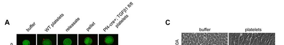
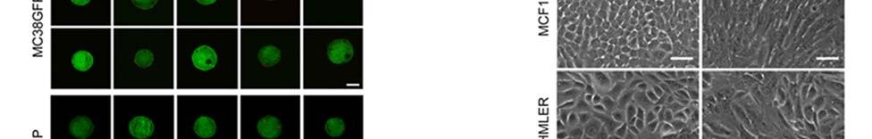
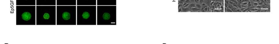
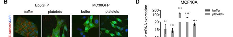
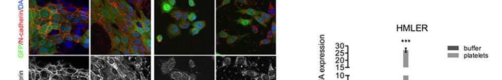
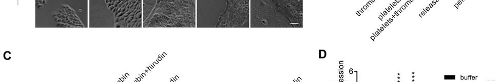
2 Cancer Cell Platelet-to-Tumor Cell Signals Promote Metastasis in the case of metastasis to other tissues such as the lungs, the source of TGFb bioavailable to tumor cells at the site of metastatic seeding remains unknown. Platelets contain a plethora of growth factors and cytokines, including high concentrations of TGFb (Assoian et al., 1983). Thus, platelet-derived factors could potentially be involved in the promotion of a metastatic phenotype. Consistent with a role of platelets in metastasis, defective platelet function or reduced platelet counts have been associated with decreased metastasis formation in various transgenic mouse models (Bakewell et al., 2003; Camerer et al., 2004; Gasic et al., 1968; Kim et al., 1998). The prometastatic effects of platelets have so far been attributed to their ability to promote adhesion or to their capacity to prevent cell death in the circulation by forming a physical shield around tumor cells. This shield protects tumor cells from natural killer cell-mediated lysis (Nieswandt et al., 1999; Palumbo et al., 2005), limits their exposure to shear stress, and promotes their adhesion to the endothelium (Erpenbeck and Schön, 2010; Gay and Felding-Habermann, 2011; Im et al., 2004; Jain et al., 2007; Karpatkin et al., 1988; Sierko and Wojtukiewicz, 2007). In addition, it is possible that platelets provide instructive signals that affect tumor cell behavior and metastatic potential. In this study, we have tested whether platelets can provide a signaling platform for cancer cells outside of the primary tumor in the context of metastasis. RESULTS Platelets Prime Tumor Cells For Metastasis To investigate whether platelets can have a direct impact on tumor cell behavior, we tested whether platelets could prime tumor cells for metastasis. Colon carcinoma cells (MC38GFP; isolated from a grade III carcinoma chemically induced in a C57BL/6 mouse) (Corbett et al., 1975) or breast carcinoma cells (Ep5 [EpRas]; spontaneously immortalized mouse mammary epithelial cell line transformed by the v-ha-ras oncogene) (Oft et al., 1996) were coincubated with purified platelets for 40 hr in vitro. Platelets were then washed away, and tumor cells (substantially devoid of any platelets; see Figure S1A available online) were injected into the tail veins of mice. Pretreating either MC38GFP or Ep5 cells with platelets led to a marked increase in the number of metastatic foci in the lungs 14 days after tail-vein injection (Figure 1A). The increase in metastasis was presumably due to the enhanced capacity of MC38GFP and Ep5 cells to seed the lungs as demonstrated by increased numbers of cells present after 48 hr (Figure 1B). These results indicate that a transient interaction between tumor cells and platelets in vitro is sufficient to increase tumor cell metastatic seeding. Treatment of Tumor Cells with Platelets Induces an Invasive Mesenchymal-Like Phenotype We next sought to define the molecular mechanisms induced by platelet-tumor cell interactions that could mediate the increased metastatic capacity of tumor cells. In tissue culture, both MC38GFP and Ep5 tumor cell lines underwent morphological changes reminiscent of an epithelial-mesenchymal transition (EMT) when treated with platelets for 24 hr (Figure 1C). Analysis of mesenchymal markers and transcription factors involved in EMT revealed that the mrna expression of snail (Snai1), vimentin (Vim), fibronectin (Fn1), and plasminogen activator inhibitor-1 (PAI-1; Serpine1) was consistently upregulated, whereas the epithelial marker claudin 1 (Cldn1) was downregulated in platelet-treated cells (Figure 1D). E-cadherin protein levels were also reduced in platelet-treated Ep5 cells in comparison with controls (Figure 1E; Figure S1B), whereas N-cadherin was relocalized from cell-cell junctions to the cytoplasm (Figure S1B). In addition, zymography revealed increased matrix metalloproteinase-9 (MMP-9) secretion following exposure to platelets (Figure 1F), suggesting a higher capacity to degrade the extracellular matrix (ECM) and to invade the surrounding environment. In agreement with these findings, the human breast epithelial cell lines MCF10A and HMLER also displayed a more mesenchymal morphology upon exposure to platelets (Figure S1C), together with increased expression of EMT-associated genes and MMP-9 secretion (Figures S1D and S1E). Thus, platelets induce EMT-like features also in epithelial cells of human origin. To test directly whether EMT-like morphological and molecular changes promote an invasive behavior, we seeded platelet-treated MC38GFP and Ep5 cells on Matrigel-coated transwells and detected increased invasion in comparison with untreated cells (Figure 1G). Altogether these results show that platelets promote the adoption of a more mesenchymal and invasive phenotype by tumor cells. Platelets Promote Activation of the TGFb/Smad Pathway in Tumor Cells To gain better insight into the signaling pathways involved in platelet-to-tumor cell communication, we defined the plateletinduced gene expression signature by microarray analysis. For this purpose, gene expression profiles of Ep5 cells treated for 24 hr with platelets or buffer alone (untreated) were compared. Any contribution of platelet mrna was excluded from the platelet-induced gene signature by performing a microarray with platelets alone (see Experimental Procedures and Figure 2A). Only 21 mrnas were contributed at significant levels by the platelets and were excluded from subsequent analyses. Among the most highly upregulated genes in tumor cells, we found several genes known to play prominent roles in EMT, ECM remodeling, and the promotion of metastasis (e.g., Mmp9, Serpine1 (PAI-1), Fn1, Jag1, Vegfc, Vim, Edn1, Vegfa, Ctgf) (Tables S1 and S2). Bioinformatic analysis using GeneGo canonical pathway maps (Figure 2B) and gene set enrichment analysis (GSEA) (Table 1) confirmed that platelets strongly activate EMT-related genes and revealed TGFb-dependent pathways as being the most significantly upregulated following platelet treatment (Figure 2B). Interestingly, GSEA analyses further revealed that previously defined gene signatures associated with cancer stem cells, poor prognosis and metastasis are also enriched in platelet-treated cells, suggesting that platelets induce an overall more aggressive phenotype in tumor cells (Table 1). Considering that a prolonged exposure to TGFb promotes EMT in many cancer cell lines, including Ep5 cells (Derynck and Akhurst, 2007; Maschler et al., 2005; Oft et al., 1996), we investigated whether platelet-derived TGFb could activate TGFb/Smad signaling in tumor cells. We found increased levels of active and latent TGFb1 in the medium derived from the coculture of tumor cells and platelets, after the platelets were removed (Figure 2C). Ep5 and MC38GFP cells treated with Cancer Cell 20, , November 15, 2011 ª2011 Elsevier Inc. 577
 Numbers of metastatic foci at the surface of lungs")
3 Cancer Cell Platelet-to-Tumor Cell Signals Promote Metastasis Figure 1. Pretreatment of Tumor Cells with Platelets Promotes Lung Metastasis by Increasing Tumor Cell Seeding and Inducing an EMT-Like Invasive Phenotype (A) Numbers of metastatic foci at the surface of lungs (two largest lobes) 14 days after tail-vein injection of MC38GFP cells or Ep5 cells stably expressing ZsGreen (Ep5-ZsGreen) pretreated with buffer (vehicle) or platelets for 40 hr (n = 5 12). (B) Numbers of tumor cells at the surface of lungs 48 hr after tail-vein injection of MC38GFP or Ep5- ZsGreen cells pretreated with buffer or platelets for 40 hr (n = 6 11). (C) Phase-contrast micrographs of MC38GFP or Ep5 cells treated with buffer or platelets for the times indicated. Scale bar represents 50 mm. (D) Relative fold change in mrna expression in MC38GFP or Ep5 cells treated with buffer or platelets for 40 hr (n = 3). Values are normalized to Gapdh expression. (E) Detection of E-cadherin protein levels by immunoblotting of lysatesof MC38GFPor Ep5cells treated as in (D). Amounts of platelets equal to those used to treatcells were also loaded as control (no cells). b-tubulin was used as loading control. (F) Zymography for MMP-9 in the conditioned medium of MC38GFP or Ep5 cells treated as in (D). Amounts of platelets equal to those used to treat cells were also loaded as control (no cells). (G) MC38GFP and Ep5 cells were added at the top of transwells coated with Matrigel and treated with buffer or platelets. The total number of cells that invaded to the bottom of the transwell was counted after 48 hr (n = 3). For (A), (B), (D), and (G) bars represent the mean ± SEM. *p < 0.05, **p < 0.01, ***p < were determined by Student s t test. See also Figure S1. controls, demonstrating that interaction with platelets induces TGFb/Smad signaling in tumor cells. We next investigated whether platelet-induced tumor cell invasion is dependent on TGFb signaling. Interestingly, adding a TGFbRI inhibitor (SB431542) or a TGFb1 blocking antibody abolished platelet-induced cell invasion (Figure 2F). Treatment with SB also inhibited the upregulation of EMT markers induced by platelets in Ep5 and MC38GFP cells (Figure 2G). Thus, platelet-derived TGFb induces a prometastatic invasive phenotype in tumor cells via activation of the TGFb/ Smad signaling pathway. platelets also showed increased phosphorylation of the TGFb signaling effector Smad2 (Figure 2D) and Smad-binding element (SBE)-dependent transcription (Figure 2E) compared with Platelet-Derived TGFb1 Is Necessary for Metastasis In Vivo To test directly the specific contribution of platelet-derived TGFb1 to metastasis in vivo, Pf4-cre mice (Tiedt et al., 2007) were crossed with TGFb1 fl/fl mice (Li et al., 2007) to generate mice lacking TGFb1 specifically in their megakaryocytes and platelets. Pf4-cre + ; 578 Cancer Cell 20, , November 15, 2011 ª2011 Elsevier Inc.
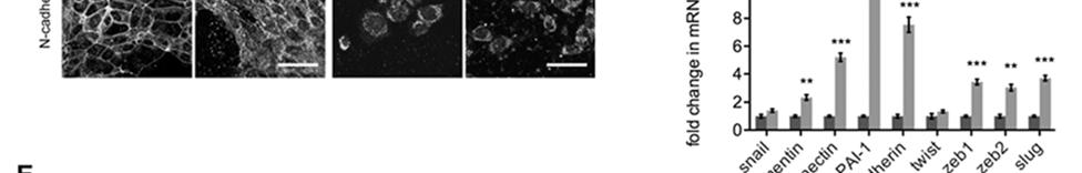
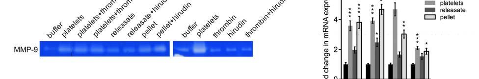
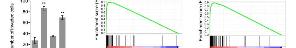
4 Cancer Cell Platelet-to-Tumor Cell Signals Promote Metastasis TGFb1 fl/fl mice had normal bleeding times and platelet counts (Table S3), showing that platelet hemostatic functions are not impaired in these mice. ELISA for TGFb1 showed that platelets from Pf4-cre + ; TGFb1 fl/fl mice contained <1% of the amount of TGFb1 present in platelets from wild-type (WT) mice (Figure 3A), confirming that Pf4-cre + ; TGFb1 fl/fl mice could be used to study the role of platelet-derived TGFb1 in metastasis. Furthermore, the concentration of TGFb1 was significantly lower in plateletrich plasma from Pf4-cre + ; TGFb1 fl/fl mice compared with plasma from WT mice (Figure 3A), suggesting that platelets are a major source of TGFb1 in the circulation. We next tested whether platelets lacking TGFb1 could induce Smad signaling in vitro and found a decrease in SBE-dependent luciferase reporter activity (Figure 3B) and MMP-9 secretion by Ep5 cells, in comparison with treatment with WT platelets (Figure 3C). Thus, although other members of the TGFb family might be secreted by platelets, these results suggest that plateletderived TGFb1 is key for the platelet-induced activation of Smad signaling in cancer cells. To test the role of platelet-derived TGFb1 during metastasis in vivo, MC38GFP cells were injected into Pf4-cre + ; TGFb1 fl/fl or WT mice via the tail vein. Fourteen days after injection, the numbers of metastases present in the lungs of Pf4-cre + ; TGFb1 fl/fl mice were greatly reduced compared with those in littermate controls (WT and Pf4-cre + ; TGFb1 fl/+ mice) (Figures 3D and 3E). Importantly, cells pretreated with platelets from WT mice and injected into Pf4-cre + ; TGFb1 fl/fl mice also formed significantly fewer metastases than platelet-treated cells injected into WT mice (Figure 3F). Conversely, pretreating tumor cells with TGFb1-deficient platelets led to the formation of significantly fewer metastases in WT mice, in comparison to cells pretreated with platelets from WT mice (Figure 3F; see Figure S1A for micrographs of injected cells). Thus, whereas a platelet pretreatment primes tumor cells for metastasis in WT mice in a TGFb1-dependent manner, the presence of plateletderived TGFb1 in the host bloodstream is also required for efficient metastasis. We next investigated the role of platelet-derived TGFb1 in the early steps in the seeding of metastases. Whereas equivalent numbers of cells were found in the lungs 3 hr after injection for both WT and Pf4-cre + ; TGFb1 fl/fl mice (Figure 3G), significantly lower numbers of cells remained in the lungs after 21 hr or 48 hr in Pf4-cre + ; TGFb1 fl/fl mice (Figure 3G). Closer inspection by confocal microscopy and 3D-rendering of lungs revealed that, at 3 hr postinjection, tumor cells were mainly present within the blood vessels (Figures 3H and 3I) in association with platelets (Figure 3J), whereas after 48 hr they were found mainly outside of the blood vessels in both WT and Pf4-cre + ; TGFb1 fl/fl mice (Figures 3H and 3I). Interestingly, at the intermediate time point of 21 hr, tumor cells were found both in the intravascular and extravascular compartments, and a smaller proportion of cells were localized extravascularly in the lungs of Pf4-cre + ; TGFb1 fl/fl mice compared with WT, suggesting that tumor cell extravasation is impaired in Pf4-cre + ; TGFb1 fl/fl mice (Figure 3H). Because another gene (stumpy) was also targeted in the TGFb1 fl/fl mice (Li et al., 2007; Town et al., 2008), we generated Pf4-cre + ; TGFb1 fl/ and TGFb1 fl/ mice, with one null allele of TGFb1, which allows expression of one WT allele of stumpy in these mice. The numbers of metastases observed in lungs of TGFb1 fl/- mice after 14 days were similar to those obtained for WT or Pf4-cre + ; TGFb1 fl/+ mice, whereas Pf4-cre + ; TGFb1 fl/- mice had reduced numbers of metastases (Figure S2). These results show that the reduction in metastasis is not due to the deletion of stumpy, but attributable to the lack of TGFb1. Altogether, our data reveal that platelet-derived TGFb1 plays an important role in promoting metastatic seeding in the lungs possibly by inducing extravasation and invasion into the lung parenchyma. Platelet-Derived TGFb1 and Platelet-Bound Factors Cooperate to Promote Metastasis Considering that TGFb1 is a secreted factor released by platelets upon activation (Assoian and Sporn, 1986), we next asked whether exposure to the releasate from activated platelets would be sufficient to prime tumor cells for metastasis in vivo. Platelets were therefore activated with thrombin and the releasate was separated from the exhausted platelets by centrifugation (neither thrombin nor its inhibitor hirudin affected tumor cell behavior in our system) (Figures S3A S3C). Higher concentrations of active TGFb1 were measured in the conditioned medium of tumor cells treated with the releasate than with the pellet from activated platelets, whereas similar amounts of total TGFb1 were found in the conditioned medium of tumor cells treated with either fraction (Figure 4A). Furthermore, similar levels of Smad2 phosphorylation (Figure 4B) and SBE-based TGFb reporter activity were induced in tumor cells incubated with the releasate or the pellet (Figure 4C), demonstrating that the concentration of TGFb1 present in either fraction is sufficient to induce Smad signaling to comparable levels. However, despite the higher concentrations of TGFb1 present in the conditioned medium of tumor cells treated with the platelet releasate (Figure 4A), increased numbers of lung metastases were observed only when tumor cells were preincubated with the pellet fraction but not with the releasate from activated platelets (Figure 4D). Additionally, tumor cell retention in the lungs was increased 48 hr after tail-vein injection when cells were pretreated with platelets or the pellet fraction but not after treatment with the releasate (Figure 4E). Consistent with these results, cells adopted a more mesenchymal morphology (Figure 4F) and showed higher expression of prometastatic genes (Figure 4G; Figure S3D), increased secretion of MMP-9 (Figure 4H), and increased invasion (Figure S3E) upon incubation with the platelet pellet fraction but not with the platelet releasate. Furthermore, microarray analysis of pellet- or releasatetreated Ep5 cells revealed that treatment with the pellet fraction induced gene expression changes very similar to those observed upon platelet treatment (Figures S3F and S3G). However, treatment with the platelet releasate resulted in only partial gene expression changes in comparison with treatment with either platelets or the pellet fraction (Figures S3F S3I). Gene expression signatures associated with EMT and tumor progression were also robustly enriched in the pellet-treated cells but not in the releasate-treated cells (Table S4). Thus, platelet-induced effects on EMT, invasion, and metastasis are not mediated by secreted TGFb alone, but also require additional platelet-bound factors. These data also suggest that these platelet-bound factors synergize with TGFb to enhance metastasis. Cancer Cell 20, , November 15, 2011 ª2011 Elsevier Inc. 579
5 Cancer Cell Platelet-to-Tumor Cell Signals Promote Metastasis Figure 2. Platelet-Induced Gene Expression Signature Reveals Increased Expression of Prometastatic Genes and Activation of the TGFb Pathway in Tumor Cells (A) Heat map of genes regulated by >4-fold (p < 0.05) in Ep5 cells treated with platelets in comparison with untreated Ep5 cells (line 2). Line 1 and line 2 show Log2 ratios of gene expression compared to untreated Ep5 cells. mrnas present in platelets (line 1) were removed from the list of genes modulated upon platelet treatment of Ep5 cells (line 2) to generate a platelet-induced gene signature (line 3), which is listed in Table S2. (B) Canonical signaling pathways most significantly associated with the list of genes differentially expressed by Ep5 cells upon platelet treatment (plateletinduced gene signature; Table S1; threshold = 2-fold, up and downregulated genes considered, p < 0.05) as determined with GeneGo canonical pathway maps. The 10 pathways with the lowest p values are shown. (C) Concentration of active and total TGFb1 in conditioned medium from MC38GFP or Ep5 cells treated with buffer or platelets for 40 hr. The conditioned medium was collected, centrifuged to remove platelets, and the presence of TGFb1 in the supernatant measured by ELISA. Each bar represents the mean ± SEM of n = 2 6. ***p < as determined by Student s t test. (D) Detection of phospho-smad2 protein levels by immunoblotting of Ep5 or MC38GFP cells treated as in (C). Amounts of platelets equal to those used to treat cells were also loaded as control (no cells). b-tubulin is used as loading control. (E) Relative luciferase activity (RLU) in MC38GFP or Ep5 cells stably expressing a luciferase reporter under the control of an SBE promoter and treated for 40 hr with buffer, platelets or 1ng/ml TGFb1 (positive control) (n = 5 6). Each bar represents the mean ± SEM, and *p < 0.05, **p < 0.01 versus buffer were determined by one-way ANOVA followed by Tukey s posttest. 580 Cancer Cell 20, , November 15, 2011 ª2011 Elsevier Inc.
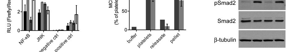
6 Cancer Cell Platelet-to-Tumor Cell Signals Promote Metastasis Table 1. Gene Set Enrichment Analysis for Ep5 Cells Treated with Platelets Gene Sets NES Nominal p Value FDR EMT signatures BLICK_EMT-SIG_UP <0.001 <0.001 TAUBE_EMT_UP TAUBE_EMT_DN <0.001 <0.001 ONDER_CDH1_TARGETS_2_UP < ONDER_CDH1_TARGETS_2_DN < TGFb Signatures GIAMPIERI_TGFB_UP <0.001 <0.001 GIAMPIERI_TGFB_DN <0.001 <0.001 VALCOURT_TGFB_UP < VALCOURT_TGFB_DN <0.001 <0.001 Cancer stem cell signatures CREIGHTON_CSC_UP <0.001 <0.001 CREIGHTON_CSC_DN <0.001 <0.001 Tumor progression and metastasis signatures VANTVEER_BREAST_CANCER_POOR_PROGNOSIS <0.001 <0.001 JAEGER_METASTASIS_UP < Enrichment of gene sets from the literature (see Supplemental Information for references). Positive NES indicates enrichment in platelet-treated Ep5 cells; negative NES indicates enrichment in untreated Ep5 cells. FDR, false discovery rate; NES normalized enrichment score. TGFb1 and Direct Platelet-Tumor Cell Contact Synergize to Promote Prometastatic Gene Expression To test further whether platelets synergize with TGFb signaling, we used the MLEC cell line stably expressing a luciferase reporter based on the promoter region of the TGFb target gene PAI-1 (Abe et al., 1994). These cells provide a specific readout for exogenous TGFb bioavailability and signaling activity, and were therefore used to dissect the specific contribution of TGFb signaling to the effects observed upon platelet treatment. Interestingly, we found that platelets induced higher luciferase activity than achievable with TGFb1 treatment alone (Figure 5A). Furthermore, adding TGFb1 together with platelets from either WT or Pf4-cre + ; TGFb1 fl/fl mice resulted in a synergistic activation of the PAI-1 reporter that was completely blocked by adding either a TGFbRI inhibitor (SB431542) or an anti-tgfb1 blocking antibody (Figure 5B). Moreover, separation of platelets (either resting or activated with thrombin) from the cancer cells by a semipermeable membrane blocked the synergistic activation of the PAI-1 promoter (Figure 5C), inhibited the secretion of MMP-9 (Figure 5D), and abolished the upregulation of prometastatic genes in Ep5 and MC38GFP cells (Figure 5E). Taken together, these results show that platelet-derived TGFb1 is necessary to induce the expression of several prometastatic genes and to promote metastasis in vivo but that, in addition, platelets provide other prometastatic signals that synergize with TGFb signaling upon direct platelet-tumor cell contact. The NF-kB Signaling Pathway Is Activated by Platelets in a Contact-Dependent Manner and Cooperates with TGFb Signaling to Enhance Metastasis We next sought to define the molecular pathway(s) induced upon platelet contact that cooperate with TGFb to induce prometastatic gene expression and behavior. Using a set of luciferase reporter assays for several pathways involved in cancer, we screened for their activation in cancer cells in response to interaction with platelets. Interestingly, coincubation with platelets increased activation of the JNK and NF-kB pathways in Ep5 cells (Figure 6A). Whereas the JNK pathway seemed to be activated by the releasate, activation of the NF-kB pathway was increased only when cells were incubated with either platelets or the pellet fraction from activated platelets (Figure S4A). Ep5 cells stably expressing an NF-kB luciferase reporter also displayed increased luciferase activity upon treatment with platelets or activated platelet pellets but not if treated with the releasate (Figure 6B). Furthermore, secretion of MCP-1, a known target of the NF-kB pathway, was increased in the supernatant of Ep5 and MC38GFP tumor cells treated with platelets or the pellet fraction, further showing that platelet-bound factors activate the NF-kB pathway in tumor cells (Figure S4B). Notably, many of the genes found to be highly upregulated in the platelet-induced gene signature, such as Ccl2 (MCP-1), Mmp9, Vegfc, Tnc, Serpine1, Jag1, and Ncam1 have been previously reported as NF-kB target genes (Figure 2A; Table S1). Furthermore, GSEA analysis revealed that NF-kB-related (F) MC38GFP and Ep5 cells were added at the top of transwells coated with Matrigel and treated with buffer, platelets, platelets + SB (10 mm) or platelets + TGFb1 blocking antibody (6 mg/ml). The total numbers of cells that invaded to the bottom of the transwell were counted after 48 hr (n = 3). Each bar represents the mean ± SEM, and *p < 0.05, **p < 0.01 versus buffer were determined by one-way ANOVA followed by Tukey s posttest. (G) Relative fold change in mrna expression in MC38GFP or Ep5 cells treated with buffer, or platelets +/ SB (10 mm) for 40 hr (n = 3). Values are normalized to Gapdh expression. Each bar represents the mean ± SEM, and *p < 0.05, **p < 0.01, ***p < versus buffer were determined by one-way ANOVA followed by Tukey s posttest. Cancer Cell 20, , November 15, 2011 ª2011 Elsevier Inc. 581
 Concentration of TGFb1 in washed platelets and platelet-rich plasma (PRP) from WT, Pf4-cre + ; TGFb1 fl/fl or Pf4-cre + ; TGFb1 fl/+ mice measured")
7 Cancer Cell Platelet-to-Tumor Cell Signals Promote Metastasis Figure 3. Platelet-Derived TGFb1 Promotes Lung Metastasis (A) Concentration of TGFb1 in washed platelets and platelet-rich plasma (PRP) from WT, Pf4-cre + ; TGFb1 fl/fl or Pf4-cre + ; TGFb1 fl/+ mice measured by ELISA (n = 2). Each bar represents the mean ± SEM, and **p < 0.01 versus WT were determined by one-way ANOVA followed by Tukey s posttest. (B) Relative luciferase activity (RLU) in Ep5 cells stably expressing a luciferase reporter under the control of the SBE promoter and treated with buffer, platelets from WT, Pf4-cre + ; TGFb1 fl/fl,orpf4-cre + ; TGFb1 fl/+ mice, or with WT platelets + SB (10 mm) for 20 hr (n = 3). Each bar represents the mean ± SEM, and *p < 0.05, ***p < versus buffer were determined by one-way ANOVA followed by Tukey s posttest. (C) Zymography for MMP-9 in the conditioned medium from Ep5 cells treated with buffer, platelets from WT, Pf4-cre + ; TGFb1 fl/fl or Pf4-cre + ; TGFb1 fl/+ mice for 40 hr. 582 Cancer Cell 20, , November 15, 2011 ª2011 Elsevier Inc.
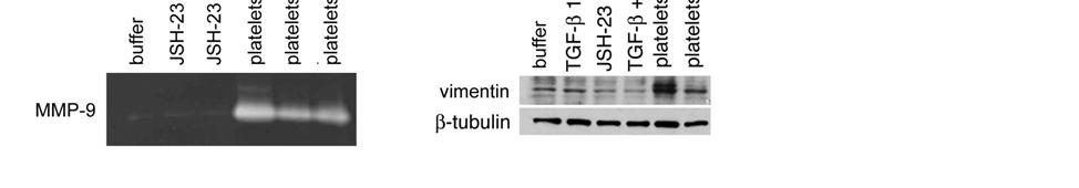
8 Cancer Cell Platelet-to-Tumor Cell Signals Promote Metastasis gene signatures are enriched in platelet- or platelet pellet-treated Ep5 cells, but not in platelet releasate-treated cells (Table S4). Importantly, treatment of tumor cells with either TGFb1-deficient platelets or with WT platelets together with the TGFbRI inhibitor SB still led to activation of the NF-kB pathway reporter (Figure 6C), ruling out the possibility that activation of the NF-kB pathway is TGFb1-dependent in this context. To study whether NF-kB signaling contributes to the prometastatic phenotype observed upon platelet contact, we established Ep5 cells stably expressing an IkBa mutant (IkBaS32A/ S36A) that cannot be phosphorylated or degraded and therefore irreversibly sequesters NF-kB in the cytoplasm, and inhibits its function (IkBa super-repressor; Ep5-IkBSR) (Brown et al., 1995). In contrast to Ep5 expressing a control vector (Ep5- vector), Ep5-IkBSR cells failed to activate an NF-kB-dependent luciferase reporter and did not express increased levels of MCP- 1 following platelet treatment, demonstrating that the NF-kB signaling is blocked in these cells (Figures 6D and 6E). Importantly, pretreatment of Ep5-vector cells with platelets prior to tail-vein injection led to increased numbers of metastases, whereas it failed to enhance metastasis of Ep5-IkBSR cells (Figure 6F). Similarly, retention of Ep5-IkBSR cells in the lungs after 48 hr was not enhanced by a pretreatment with platelets, showing that the NF-kB pathway is necessary for the prometastatic effects of platelets on tumor cells (Figure 6G). Furthermore, Ep5-IkBSR cells did not acquire a mesenchymal morphology (Figure 6H) and did not display increased invasion (Figure 6I) in response to treatment with platelets. These results correlated with lower levels of MMP-9 secretion (Figure 6J) and lower vimentin and fibronectin gene expression (Figure 6K) by Ep5- IkBSR cells following platelet treatment. The inhibition of platelet prometastatic effects was not due to an impairment of TGFb/ Smad signaling pathway by the IkBSR construct because Smad2 phosphorylation levels were intact in Ep5-IkBSR cells (Figure S4C). In addition, inhibition of MMP-9 secretion and vimentin protein expression induced upon platelet treatment were also observed when Ep5 cells were treated with JSH-23, a pharmacological inhibitor of the NF-kB pathway (Figures S4D and S4E). JSH-23 treatment also abolished the synergy observed on PAI-1 reporter activity between TGFb1 and platelets (Figure S4F), but had no effect on SBE-reporter activity (Figure S4G). This finding further demonstrates that platelets activate NF-kB independently of TGFb/Smad signaling, and cooperate with TGFb1 to induce an EMT-like transformation in cancer cells. Thus, the ability of platelets to prime tumor cells for metastasis depends on the synergistic interaction between the NF-kB and TGFb signaling pathways, which is triggered by direct platelet-tumor cell contact (Figure 7). DISCUSSION Signals provided by the primary tumor microenvironment are important modulators of the capacity of tumor cells to invade, access the vasculature, and metastasize (Joyce and Pollard, 2009; Nguyen et al., 2009). However, the metastatic potential of tumor cells may be further defined in response to signals provided during their intravascular transit. Here, we have tested this hypothesis and show that platelets present in the bloodstream actively signal to tumor cells to promote their metastatic potential outside of the primary microenvironment. This effect is independent of any direct contribution of platelets to immunosurveillance, adhesion, or physical shielding functions, as tumor cells can be primed for metastasis by a transient exposure to purified platelets in vitro. Mechanistically, a transient contact between platelets and tumor cells is sufficient to induce a prometastatic gene expression signature, induce an EMT-like transformation and invasive behavior in vitro, and promote metastatic seeding in the lungs in vivo (Figure 7). Considering that tumor cells would normally interact with platelets once in the bloodstream, these results suggest that tumor cells could gain a more mesenchymal phenotype and increased metastatic capacities after leaving the primary tumor microenvironment. This implies that cells that have intravasated without losing their epithelial properties either via leaky blood vessels (Carmeliet and Jain, 2000; Mazzone et al., 2009) or via collective invasion mechanisms (Friedl and Gilmour, 2009) could acquire a mesenchymal phenotype during their transit in the vasculature. In support of this idea, circulating tumor cells have been found to express epithelial markers (EpCAM, cytokeratins), suggesting that EMT is not absolutely required to access the blood flow (Nagrath et al., 2007). Thus, interactions with platelets may be particularly important in mediating extravasation of circulating epithelial tumor cells, and to maintain or further enhance the extravasation potential of circulating mesenchymal tumor cells. In this respect, it would be interesting to define the impact of platelets on gene expression and metastatic potential of circulating tumor cells from cancer patients. Several signaling molecules, including TGFb, PDGF, VEGF and angiopoietin are abundant in platelets (Erpenbeck and Schön, 2010; Sierko and Wojtukiewicz, 2007) and may therefore (D and E) Numbers of metastatic foci at the surface of lungs (two largest lobes) 14 days after tail-vein injection of MC38GFP cells in WT, Pf4-cre + ; TGFb1 fl/fl or Pf4-cre + ; TGFb1 fl/+ mice (n = 7 9), and representative pictures of lungs (E). (D) Each bar represents the mean ± SEM, and **p < 0.01 versus WT was determined by one-way ANOVA followed by Tukey s posttest. (F) Numbers of metastatic foci at the surface of lungs (two largest lobes) 14 days after tail-vein injection of MC38GFP cells pretreated with buffer ( ), platelets from WT mice (WT) or platelets from Pf4-cre + ; TGFb1 fl/fl (fl/fl) and injected into WT or Pf4-cre + ; TGFb1 fl/fl mice (n = 9 14). Each bar represents the mean ± SEM, and *p < 0.05, **p < 0.01 were determined by one-way ANOVA followed by Tukey s posttest. (G) Numbers of tumor cells at the surface of lungs 3 hr, 21 hr, and 48 hr after tail-vein injection of MC38GFP cells in WT or Pf4-cre + ; TGFb1 fl/fl mice. Each point represents the mean ± SEM number of cells/view field (33) (n = 3 14). *p < 0.05, **p < 0.01 were determined by Student s t test. (H) Percentage of intravascular and extravascular MC38GFP cells in lungs of WT or Pf4-cre + ; TGFb1 fl/fl mice 3 hr, 21 hr, and 48 hr after tail-vein injection of tumor cells (n = cells). **p < 0.01 as determined by Fisher s exact test. (I and J) Confocal microscopy of lungs of WT or Pf4-cre + ; TGFb1 fl/fl mice 3 hr and 48 hr after tail-vein injection of tumor cells for MC38GFP cells (green) and either blood vessels (I; PECAM-1 staining; red) or platelets (J; GP1bb staining; red. Note platelet aggregates surrounding tumor cells.). Scale bar represents 50 mm. See also Figure S2 and Table S3. Cancer Cell 20, , November 15, 2011 ª2011 Elsevier Inc. 583
 Concentration of TGFb1 measured by ELISA in the conditioned medium of MC38GFP or Ep5 cells incubated with buffer,")
9 Cancer Cell Platelet-to-Tumor Cell Signals Promote Metastasis Figure 4. Platelet-Derived TGFb1 and Platelet-Bound Factors Cooperate to Promote Metastasis (A) Concentration of TGFb1 measured by ELISA in the conditioned medium of MC38GFP or Ep5 cells incubated with buffer, platelets, releasate from activated platelets (releasate), or the pellet fraction from activated platelets (pellet) for 40 hr (n = 4 6). Each bar represents the mean ± SEM, and ns (p > 0.05), *p < 0.05, ***p < were determined by one-way ANOVA followed by Tukey s posttest. (B) Detection of phospho-smad2 and total Smad2 protein levels by immunoblotting in Ep5 cells treated as in (A). b-tubulin was used as loading control. (C) Relative luciferase activity (RLU) in Ep5 cells stably expressing a luciferase reporter under the control of the SBE promoter and treated as in (A) for 20 hr (n = 2). Each bar represents the mean ± SEM, and ns (p > 0.05) was determined by one-way ANOVA followed by Tukey s posttest. 584 Cancer Cell 20, , November 15, 2011 ª2011 Elsevier Inc.
10 Cancer Cell Platelet-to-Tumor Cell Signals Promote Metastasis impact tumor cell behavior and induce EMT. Our results show that the prometastatic effects of platelets are in large part mediated via activation of the TGFb signaling pathway, and that abrogating either TGFb signaling in tumor cells or TGFb expression by platelets is sufficient to inhibit metastasis and EMT. Although TGFb has been implicated in the induction of a prometastatic phenotype in many contexts (Padua et al., 2008; Siegel et al., 2003), the relevant cellular source of TGFb bioavailable to circulating tumor cells, particularly at the site of metastatic extravasation, was previously unclear. Our results strongly indicate that platelets are an important source of bioavailable TGFb for tumor cells in the circulation and at the site of extravasation. This finding is supported by the observation that platelets contain concentrations of TGFb1 many-fold higher than most cell types (Assoian et al., 1983). Furthermore, the amount of TGFb1 produced by other cells and taken up by platelets seems minimal, as purified platelets from Pf4-cre + ; TGFb1 fl/fl mice contained <1% of the amount of TGFb1 present in platelets from WT mice. Most importantly, we show that abrogation of TGFb1 expression solely in platelets and megakaryocytes is sufficient to inhibit metastasis and prevent the seeding of tumor cells in the lungs. Furthermore, the presence of platelet-derived TGFb1 in situ in the host bloodstream is crucial for metastasis, because pretreating tumor cells with platelets from WT mice fails to enhance metastasis formation in mice lacking TGFb1 in their platelets. Because platelet-tumor cell interactions are transient and occur only within the first 24 hr (Läubli et al., 2006 and data not shown), we propose that platelets could provide a pulse of TGFb1 to circulating tumor cells, which would allow them to gain a more invasive, mesenchymal-like phenotype and extravasate. Along these lines, previous studies have shown that tumor cells transiently exposed to TGFb1 have an enhanced capacity to seed the lungs, whereas cells that are continuously exposed to TGFb1 have decreased metastatic capacity due to the cytostatic effect of TGFb1(Giampieri et al., 2009; Padua et al., 2008). In this respect, specific therapeutic inhibition of platelet-derived TGFb1 might result in the impairment of tumor cell extravasation at the metastatic site. Importantly, Pf4-cre + ; TGFb1 fl/fl mice maintain normal platelet counts and hemostatic functions, suggesting that pharmacological inhibition of platelet-derived TGFb could inhibit metastasis without adverse effects on physiological hemostasis. We also find that, although required for metastasis, activation of TGFb signaling alone is unable to generate effects of the magnitude of those observed with platelets. Indeed, although tumor cells treated for 24 hr with platelets or with the pellet fraction from activated platelets undergo EMT, cells treated with the releasate of activated platelets (that contained a similar concentration of TGFb1) do not. In line with this result, a prolonged treatment with TGFb1 typically 1 week or longer is needed to induce EMT in several epithelial cancer cell lines including Ep5 cells (Labelle et al., 2008; Mani et al., 2008; Maschler et al., 2005). Our data further support the existence of additional platelet-bound factors synergizing with TGFb1. First, platelets induce the TGFb-responsive PAI-1 reporter to levels higher than achievable with TGFb1 alone. Second, combining exogenous TGFb1 with platelets from either WT or Pf4-cre + ; TGFb1 fl/fl mice results in a synergistic activation of the PAI-1 reporter. Last, the synergistic effects on PAI-1 reporter activity as well as the induction of prometastatic genes are blocked if platelets are separated from tumor cells by a semipermeable membrane. Thus, our results clearly demonstrate that additional platelet-bound factors synergize with TGFb1 to promote metastasis, and to induce a prometastatic EMT program in tumor cells. In particular, we show that this synergy is dependent on the activation of the NF-kB pathway, which is specifically triggered upon direct contact between tumor cells and platelets independently from TGFb activity. NF-kB regulates the expression of proinflammatory genes and has been associated with increased metastasis and EMT induction (Huber et al., 2004; Lin and Karin, 2007). For example, NF-kB promotes osteolytic bone metastasis by inducing the proinflammatory cytokine GM-CSF (Park et al., 2007). Notably, the activation of NF-kB has also been proposed to be part of the mechanism allowing TGFb signaling to switch from a cytostatic to a prometastatic signal (Neil and Schiemann, 2008). In support of this idea, we found that NF-kB activation potentiates TGFb-induced prometastatic gene expression, and that NF-kB signaling is necessary for the induction of EMT and efficient metastatic seeding upon platelet-cancer cell interactions. Thus, platelet-tumor cell contacts during metastasis potentiate tumor cell transcriptional responses to TGFb via NF-kB activation. In conclusion, we establish platelets as a crucial source of TGFb bioavailable to tumor cells in the vasculature and necessary for tumor cell extravasation and metastasis formation. Importantly, our study reveals that platelets are more than physical shields and actively signal to tumor cells via the TGFb and NF-kB pathways to potently induce a prometastatic phenotype. We thus propose a model whereby the metastatic potential of tumor cells continues to evolve outside of the primary tumor site, in response to tumor-host interactions in the bloodstream and at the site of metastasis. In particular, we identify platelettumor cell interactions and the signaling pathways that they trigger as fundamental determinants of cancer metastasis that may provide the basis for developing effective anti-metastatic therapies. (D) Numbers of metastatic foci at the surface of lungs (two largest lobes) 14 days after tail-vein injection of MC38GFP or Ep5-ZsGreen cells pretreated with buffer, platelets, releasate from activated platelets (releasate), or the pellet fraction from activated platelets (pellet) for 40 hr (n = 5 17). Each bar represents the mean ± SEM, and *p < 0.05, **p < 0.01 versus buffer were determined by one-way ANOVA followed by Tukey s posttest. (E) Numbers of tumor cells at the surface of lungs 48h after tail-vein injection of MC38GFP or Ep5-ZsGreen cells pretreated as in (D). Each bar represents the mean ± SEM number of cells/view field (33) (n = 5 13). *p < 0.05, **p < 0.01 versus buffer were determined by Student s t test. (F) Phase-contrast micrographs of MC38GFP and Ep5 cells treated as in (A) for 24 hr. Scale bar represents 50 mm. (G) Relative fold change in mrna expression in MC38GFP or Ep5 cells treated as in (A) for 40 hr. Values are normalized to Gapdh expression (n = 3). Each bar represents the mean ± SEM, and *p < 0.05, **p < 0.01, ***p < versus buffer were determined by one-way ANOVA followed by Tukey s posttest. (H) Zymography for MMP-9 in the conditioned medium from Ep5 or MC38GFP cells treated as in (A) for 40 hr. See also Figure S3 and Table S4. Cancer Cell 20, , November 15, 2011 ª2011 Elsevier Inc. 585
11 Cancer Cell Platelet-to-Tumor Cell Signals Promote Metastasis Figure 5. TGFb1 and Direct Platelet-Tumor Cell Contact Synergize to Promote Prometastatic Gene Expression (A) Relative luciferase activity (RLU) in MLEC cells stably expressing a luciferase reporter under the control of a TGFb responsive PAI-1 promoter construct and treated with different concentrations of TGFb1 (left) or platelets (right). Note that the y axis scale is the same for both panels and that platelets give higher stimulation than achievable with TGFb1 alone. (B) Relative luciferase activity (RLU) in MLEC cells stably expressing a luciferase reporter under the control of a PAI-1 promoter construct treated with buffer, platelets from WT or Pf4-cre + ; TGFb1 fl/fl mice, TGFb1 (1 ng/ml) or with combinations of platelets + TGFb1 (1 ng/ml), +/ SB (SB; 10 mm) or +/ TGFb1 blocking antibody (Ab; 6 mg/ml)(n = 3 16). Each bar represents the mean ± SEM, and ***p < were determined by one-way ANOVA followed by Tukey s posttest. (C) Relative luciferase activity (RLU) in MLEC cells stably expressing a luciferase reporter under the control of a PAI-1 promoter construct treated with buffer, platelets, or thrombin-activated platelets +/ TGFb1 (1ng/ml) seeded either at the bottom (direct contact with tumor cells) or in the upper chamber of a transwell (0.4 mm pore size) to prevent direct contact between platelets and tumor cells (n = 2 3). Each bar represents the mean ± SEM, and ***p < versus buffer were determined by one-way ANOVA followed by Tukey s posttest. (D) Zymography for MMP-9 in the conditioned medium from Ep5 or MC38GFP cells treated with buffer, platelets, or thrombin-activated platelets seeded either at the bottom or in the upper chamber of a transwell (0.4 mm pore size). (E) Relative fold change in mrna expression in Ep5 or MC38GFP cells treated as in (D) (n = 3). Values are normalized to Gapdh expression. Each bar represents the mean ± SEM, and *p < 0.05, **p < 0.01, ***p < versus buffer were determined by one-way ANOVA followed by Tukey s posttest. 586 Cancer Cell 20, , November 15, 2011 ª2011 Elsevier Inc.
12 Cancer Cell Platelet-to-Tumor Cell Signals Promote Metastasis Figure 6. The NF-kB Signaling Pathway Is Activated by Platelets in a Contact-Dependent Manner and Cooperates with TGFb Signaling to Induce an EMT-Like Transition and Promote Metastasis (A) Ep5 cells were transfected with pathway-specific firefly luciferase reporters and constitutively expressed control Renilla luciferase reporters. Twenty-four hours after transfection, cells were treated with buffer or platelets for 20 hr, and the relative luciferase activity (RLU) was measured. Firefly luciferase activity was normalized to Renilla luciferase activity. Each bar represents the mean ± SEM of n = 3. *p < 0.05 was determined by Student s t test. (B) Relative luciferase activity (RLU) in Ep5 cells stably expressing an NF-kB luciferase reporter and treated with buffer, platelets, releasate from activated platelets (releasate), or the pellet fraction from activated platelets (pellet) for 20 hr (n = 2). Each bar represents the mean ± SEM, and ***p < versus buffer were determined by one-way ANOVA followed by Tukey s posttest. (C) Relative luciferase activity (RLU) in Ep5 cells stably expressing a NF-kB luciferase reporter and treated with buffer, platelets from WT, Pf4-cre + ; TGFb1 fl/fl,or Pf4-cre + ; TGFb1 fl/+ mice, or with WT platelets + SB (10 mm) for 20 hr (n = 5). Each bar represents the mean ± SEM, and *p < 0.05, **p < 0.01 versus buffer were determined by one-way ANOVA followed by Tukey s posttest. Cancer Cell 20, , November 15, 2011 ª2011 Elsevier Inc. 587
 were transduced into Ep5 cells as described previously (Stern et al., 2008) and in Supplemental Experimental Procedures.")
13 Cancer Cell Platelet-to-Tumor Cell Signals Promote Metastasis Generation of Cell Lines Stably Expressing ZsGreen, IkBSR, and Luciferase-Based Reporters Retroviral vectors coding for ZsGreen or IkBa super-repressor plus GFP (IkBSR) were transduced into Ep5 cells as described previously (Stern et al., 2008) and in Supplemental Experimental Procedures. Ep5 and MC38GFP cell lines stably expressing SBE (AGCCAGACA tandem repeats)-luciferase and NF-kB (GGGACTTTCC tandem repeats)-luciferase reporter constructs were generated by infecting cells with Cignal Lenti Reporter vectors (SABiosciences) according to manufacturers instructions. Preparation of Platelets and Platelet Fractions Mouse blood was collected by cardiac puncture and washed platelets were prepared as described previously (Frenette et al., 1995; Hartwell et al., 1998). To prepare platelet fractions, platelets were activated with thrombin 0.5 U/ml for 15 min at 37 C. The pellet fraction was separated from the releasate (supernatant) by centrifugation at g for 7 min. Figure 7. Platelet-Tumor Cell Contact and Platelet-Derived TGFb1 Synergize to Promote an EMT-Like Transition and Metastasis Platelets secrete TGFb1, which activates the TGFb/Smad pathway in tumor cells. Upon direct platelet-tumor cell contact, the NF-kB pathway is also activated in tumor cells and synergizes with TGFb/Smad signaling to induce a rapid EMT, enhance invasiveness and promote metastasis. Activation of neither the TGFb/Smad nor the NF-kB pathway alone is sufficient to promote metastasis. Thus, platelet-tumor cell contact triggers a synergistic interaction between TGFb/Smad and NF-kB pathways that is necessary for efficient metastasis. The metastatic potential of tumor cells therefore continues to evolve outside of the primary tumor site in response to platelet-to-tumor cell signaling. EXPERIMENTAL PROCEDURES Mice Mice homozygous for the TGFb1 floxed allele (TGFb1 fl/fl ; obtained from R. Flavell) (Li et al., 2007) on a C57BL/6 genetic background were crossed with Pf4-cre mice on a C57BL/6 background (obtained from S. Shattil) (Tiedt et al., 2007). To obtain mice with TGFb1-deficient platelets, Pf4-cre + ; TGFb1 fl/+ mice were bred with TGFb1 fl/fl or TGFb1 fl/+ mice. TGFb1 +/ mice (the null allele was obtained by egfp knockin, which disrupted TGFb1 without affecting stumpy) (Li et al., 2007) were crossed with Pf4-cre + ; TGFb1 fl/fl mice to obtain Pf4-cre + ; TGFb1 fl/- mice. For genotyping primers, see Supplemental Experimental Procedures. All mice were housed and handled in accordance with approved Massachusetts Institute of Technology Division of Comparative Medicine protocols. Treatment of Tumor Cells with Platelets Cells were seeded in DMEM 10% FCS and incubated overnight. Immediately prior to treatment the medium was changed for fresh DMEM. A total of 150,000 platelets/ml and equivalent volumes of releasate and pellet fractions were added. Where indicated, cells were treated with 1 or 10 ng/ml of recombinant TGFb1 (R&D Systems), 10 mm SB (Sigma), or 6 mg/ml anti-tgfb1 blocking antibody (R&D systems). In Vivo Metastasis Assays For lung metastasis assays, cells treated with platelets for 40 hr were washed in PBS, and either trypsinized (Ep5) or lifted with 2mM EDTA in PBS (MC38GFP). Cells were then washed and centrifuged twice to remove platelets, and resuspended in HBSS at a constant number of cells for all mice in a given experiment (250,000 to 1,000,000 cells/injection). One hundred microliters of cell suspension were then injected via the tail vein of syngeneic mice. The numbers of single cells and metastatic foci were determined as described in Supplemental Experimental Procedures. Microarray Analysis Total RNA was isolated from platelet-treated or untreated Ep5 cells (n = 5). In order to detect mrnas contributed by platelets, RNA was also isolated from platelet lysates mixed with untreated Ep5 cells lysates immediately prior to RNA isolation (n = 3) (the concentration of RNA isolated from platelets alone was below detection limits and could therefore not be used for microarray analysis). crna was then synthesized and hybridized onto GeneChip Mouse Exon 1.0 ST Arrays (Affymetrix). Luciferase Assay A total of 15,000 Ep5 cells/50 ml Opti-MEM were plated in a 96-well Cignal Finder Multi Pathway reporter Array plate as recommended by the manufacturer (SABiosciences) and incubated for 24 hr. The medium was (D) Relative luciferase activity (RLU) in Ep5 cells stably expressing a NF-kB luciferase reporter and either a IkB super-repressor (Ep5-IkBSR) or a control vector (Ep5-vector) and treated with buffer or platelets for 20 hr (n = 4). Each bar represents the mean ± SEM, and ***p < versus buffer was determined by two-way ANOVA followed by Bonferroni s posttest. (E) MCP-1 concentration in the conditioned medium from Ep5 cells stably expressing an IkB super-repressor (Ep5-IkBSR) or a control vector (Ep5-vector) and treated with buffer or platelets for 20 hr (n = 2). Each bar represents the mean ± SEM, and **p < 0.01 versus buffer was determined by two-way ANOVA followed by Bonferroni s posttest. (F) Numbers of metastatic foci at the surface of lungs (two largest lobes) 14 days after tail-vein injection of Ep5-IkBSR and Ep5-vector cells pretreated with buffer or platelets for 40h. Each bar represents the mean ± SEM of n = 5 7. *p < 0.05 was determined by Student s t test. (G) Numbers of tumor cells at the surface of lungs 48 hr after tail-vein injection of Ep5-IkBSR and Ep5-vector cells pretreated with buffer or platelets for 40 hr. Each bar represents the mean ± SEM of n = 4 7. **p < 0.01 was determined by Student s t test. (H) Phase-contrast micrographs of Ep5-IkBSR and Ep5-vector cells treated with buffer or platelets for 24 hr. Scale bar represents 50 mm. (I) Ep5-IkBSR and Ep5-vector cells were added at the top of transwells coated with Matrigel and treated with buffer or platelets. The total numbers of cells that invaded to the bottom of the transwell were counted after 48 hr (n = 2). Each bar represents the mean ± SEM, and *p < 0.05 versus buffer was determined by two-way ANOVA followed by Bonferroni s posttest. (J) Zymography for MMP-9 in the conditioned medium from Ep5-IkBSR and Ep5-vector cells treated with buffer or platelets for 40 hr. (K) Relative fold change in mrna expression in Ep5-IkBSR and Ep5-vector cells treated as in (J) (n = 3). Values are normalized to Gapdh expression. Each bar represents the mean ± SEM, and ***p < were determined by one-way ANOVA followed by Tukey s posttest. See also Figure S Cancer Cell 20, , November 15, 2011 ª2011 Elsevier Inc.
14 Cancer Cell Platelet-to-Tumor Cell Signals Promote Metastasis then changed and cells were treated with platelets or platelet fractions. The ratios of Firefly to Renilla Luciferase activities (relative light units [RLU]) were measured in cell lysates with the Dual Luciferase Reporter Assay System (Promega) 24 hr after treatment with platelets. TGFb1 ELISA TGFb1 levels were detected in tissue-culture-conditioned medium (40 hr), washed platelets or platelet-rich plasma either by direct assay (active TGFb1), or following acid treatment to activate latent TGFb1 (total TGFb1) with the Quantikine TGFb1 immunoassay kit (R&D Systems). MCP-1 Detection Concentration of MCP-1 in conditioned medium of Ep5 or MC38GFP cells was analyzed with CBA soluble Flex Set cytometric beads, following manufacturer s instructions (BD Biosciences). Statistics Statistical analyses were performed with the GraphPad Prism Software following guidelines found in Bremer and Doerge (2010) and in GraphPad Prism. Briefly, the Student s t test was used to compare means of two independent groups to each other, whereas one-way ANOVA followed by Tukey s posttest was used to compare the means of more than two independent groups. Two-way ANOVA followed by Bonferroni s posttest was used to compare the means of groups influenced by two independent factors. ACCESSION NUMBERS Microarray data are deposited in Gene Expression Omnibus under accession number GSE SUPPLEMENTAL INFORMATION Supplemental Information includes four figures, four tables, and Supplemental Experimental Procedures and can be found with this article online at doi: /j.ccr ACKNOWLEDGMENTS We are grateful to Patrick Stern for viral vectors driving ZsGreen and IkBSR expression, and to Richard Flavell and Sanford Shattil for mouse strains. We thank Charlie Whittaker (Swanson Biotechnology Center, Koch Institute) for assistance with microarray analysis, members of the Hynes lab for advice, and John Lamar and Patrick Stern for critically reading the manuscript. This work was supported by the Ludwig Center for Molecular Oncology at MIT, by the Koch Institute at MIT, and by the Howard Hughes Medical Institute of which R.O.H. is an Investigator. M.L. was supported by a postdoctoral fellowship from the Anna Fuller Fund. Received: February 21, 2011 Revised: June 28, 2011 Accepted: September 20, 2011 Published: November 14, 2011 REFERENCES Abe, M., Harpel, J.G., Metz, C.N., Nunes, I., Loskutoff, D.J., and Rifkin, D.B. (1994). An assay for transforming growth factor-beta using cells transfected with a plasminogen activator inhibitor-1 promoter-luciferase construct. Anal. Biochem. 216, Assoian, R.K., and Sporn, M.B. (1986). Type beta transforming growth factor in human platelets: release during platelet degranulation and action on vascular smooth muscle cells. J. Cell Biol. 102, Assoian, R.K., Komoriya, A., Meyers, C.A., Miller, D.M., and Sporn, M.B. (1983). Transforming growth factor-beta in human platelets. Identification of a major storage site, purification, and characterization. J. Biol. Chem. 258, Bakewell, S.J., Nestor, P., Prasad, S., Tomasson, M.H., Dowland, N., Mehrotra, M., Scarborough, R., Kanter, J., Abe, K., Phillips, D., and Weilbaecher, K.N. (2003). Platelet and osteoclast beta3 integrins are critical for bone metastasis. Proc. Natl. Acad. Sci. USA 100, Biswas, S., Guix, M., Rinehart, C., Dugger, T.C., Chytil, A., Moses, H.L., Freeman, M.L., and Arteaga, C.L. (2007). Inhibition of TGF-beta with neutralizing antibodies prevents radiation-induced acceleration of metastatic cancer progression. J. Clin. Invest. 117, Bremer, M., and Doerge, R.W. (2010). Statistics at the Bench: A Step-by Step Handbook for Biologists (Cold Spring Harbor: Cold Spring Harbor Laboratory Press). Brown, K., Gerstberger, S., Carlson, L., Franzoso, G., and Siebenlist, U. (1995). Control of I kappa B-alpha proteolysis by site-specific, signal-induced phosphorylation. Science 267, Camerer, E., Qazi, A.A., Duong, D.N., Cornelissen, I., Advincula, R., and Coughlin, S.R. (2004). Platelets, protease-activated receptors, and fibrinogen in hematogenous metastasis. Blood 104, Carmeliet, P., and Jain, R.K. (2000). Angiogenesis in cancer and other diseases. Nature 407, Corbett, T.H., Griswold, D.P., Jr., Roberts, B.J., Peckham, J.C., and Schabel, F.M., Jr. (1975). Tumor induction relationships in development of transplantable cancers of the colon in mice for chemotherapy assays, with a note on carcinogen structure. Cancer Res. 35, Derynck, R., and Akhurst, R.J. (2007). Differentiation plasticity regulated by TGF-beta family proteins in development and disease. Nat. Cell Biol. 9, Erpenbeck, L., and Schön, M.P. (2010). Deadly allies: the fatal interplay between platelets and metastasizing cancer cells. Blood 115, Frenette, P.S., Johnson, R.C., Hynes, R.O., and Wagner, D.D. (1995). Platelets roll on stimulated endothelium in vivo: an interaction mediated by endothelial P-selectin. Proc. Natl. Acad. Sci. USA 92, Friedl, P., and Gilmour, D. (2009). Collective cell migration in morphogenesis, regeneration and cancer. Nat. Rev. Mol. Cell Biol. 10, Gasic, G.J., Gasic, T.B., and Stewart, C.C. (1968). Antimetastatic effects associated with platelet reduction. Proc. Natl. Acad. Sci. USA 61, Gay, L.J., and Felding-Habermann, B. (2011). Contribution of platelets to tumour metastasis. Nat. Rev. Cancer 11, Giampieri, S., Manning, C., Hooper, S., Jones, L., Hill, C.S., and Sahai, E. (2009). Localized and reversible TGFbeta signalling switches breast cancer cells from cohesive to single cell motility. Nat. Cell Biol. 11, Hartwell, D.W., Mayadas, T.N., Berger, G., Frenette, P.S., Rayburn, H., Hynes, R.O., and Wagner, D.D. (1998). Role of P-selectin cytoplasmic domain in granular targeting in vivo and in early inflammatory responses. J. Cell Biol. 143, Huber, M.A., Azoitei, N., Baumann, B., Grünert, S., Sommer, A., Pehamberger, H., Kraut, N., Beug, H., and Wirth, T. (2004). NF-kappaB is essential for epithelial-mesenchymal transition and metastasis in a model of breast cancer progression. J. Clin. Invest. 114, Im, J.H., Fu, W., Wang, H., Bhatia, S.K., Hammer, D.A., Kowalska, M.A., and Muschel, R.J. (2004). Coagulation facilitates tumor cell spreading in the pulmonary vasculature during early metastatic colony formation. Cancer Res. 64, Jain, S., Zuka, M., Liu, J., Russell, S., Dent, J., Guerrero, J.A., Forsyth, J., Maruszak, B., Gartner, T.K., Felding-Habermann, B., and Ware, J. (2007). Platelet glycoprotein Ib alpha supports experimental lung metastasis. Proc. Natl. Acad. Sci. USA 104, Joyce, J.A., and Pollard, J.W. (2009). Microenvironmental regulation of metastasis. Nat. Rev. Cancer 9, Kang, Y., He, W., Tulley, S., Gupta, G.P., Serganova, I., Chen, C.R., Manova- Todorova, K., Blasberg, R., Gerald, W.L., and Massagué, J. (2005). Breast cancer bone metastasis mediated by the Smad tumor suppressor pathway. Proc. Natl. Acad. Sci. USA 102, Cancer Cell 20, , November 15, 2011 ª2011 Elsevier Inc. 589
15 Cancer Cell Platelet-to-Tumor Cell Signals Promote Metastasis Karpatkin, S., Pearlstein, E., Ambrogio, C., and Coller, B.S. (1988). Role of adhesive proteins in platelet tumor interaction in vitro and metastasis formation in vivo. J. Clin. Invest. 81, Kim, Y.J., Borsig, L., Varki, N.M., and Varki, A. (1998). P-selectin deficiency attenuates tumor growth and metastasis. Proc. Natl. Acad. Sci. USA 95, Labelle, M., Schnittler, H.J., Aust, D.E., Friedrich, K., Baretton, G., Vestweber, D., and Breier, G. (2008). Vascular endothelial cadherin promotes breast cancer progression via transforming growth factor beta signaling. Cancer Res. 68, Läubli, H., Stevenson, J.L., Varki, A., Varki, N.M., and Borsig, L. (2006). L-selectin facilitation of metastasis involves temporal induction of Fut7- dependent ligands at sites of tumor cell arrest. Cancer Res. 66, Li, M.O., Wan, Y.Y., and Flavell, R.A. (2007). T cell-produced transforming growth factor-beta1 controls T cell tolerance and regulates Th1- and Th17- cell differentiation. Immunity 26, Lin, W.W., and Karin, M. (2007). A cytokine-mediated link between innate immunity, inflammation, and cancer. J. Clin. Invest. 117, Mani, S.A., Guo, W., Liao, M.J., Eaton, E.N., Ayyanan, A., Zhou, A.Y., Brooks, M., Reinhard, F., Zhang, C.C., Shipitsin, M., et al. (2008). The epithelial-mesenchymal transition generates cells with properties of stem cells. Cell 133, Maschler, S., Wirl, G., Spring, H., Bredow, D.V., Sordat, I., Beug, H., and Reichmann, E. (2005). Tumor cell invasiveness correlates with changes in integrin expression and localization. Oncogene 24, Mazzone, M., Dettori, D., Leite de Oliveira, R., Loges, S., Schmidt, T., Jonckx, B., Tian, Y.M., Lanahan, A.A., Pollard, P., Ruiz de Almodovar, C., et al. (2009). Heterozygous deficiency of PHD2 restores tumor oxygenation and inhibits metastasis via endothelial normalization. Cell 136, Nagrath, S., Sequist, L.V., Maheswaran, S., Bell, D.W., Irimia, D., Ulkus, L., Smith, M.R., Kwak, E.L., Digumarthy, S., Muzikansky, A., et al. (2007). Isolation of rare circulating tumour cells in cancer patients by microchip technology. Nature 450, Neil, J.R., and Schiemann, W.P. (2008). Altered TAB1:I kappab kinase interaction promotes transforming growth factor beta-mediated nuclear factorkappab activation during breast cancer progression. Cancer Res. 68, Nguyen, D.X., Bos, P.D., and Massagué, J. (2009). Metastasis: from dissemination to organ-specific colonization. Nat. Rev. Cancer 9, Nieswandt, B., Hafner, M., Echtenacher, B., and Männel, D.N. (1999). Lysis of tumor cells by natural killer cells in mice is impeded by platelets. Cancer Res. 59, Oft, M., Heider, K.H., and Beug, H. (1998). TGFbeta signaling is necessary for carcinoma cell invasiveness and metastasis. Curr. Biol. 8, Oft, M., Peli, J., Rudaz, C., Schwarz, H., Beug, H., and Reichmann, E. (1996). TGF-beta1 and Ha-Ras collaborate in modulating the phenotypic plasticity and invasiveness of epithelial tumor cells. Genes Dev. 10, Padua, D., Zhang, X.H., Wang, Q., Nadal, C., Gerald, W.L., Gomis, R.R., and Massagué, J. (2008). TGFbeta primes breast tumors for lung metastasis seeding through angiopoietin-like 4. Cell 133, Palumbo, J.S., Talmage, K.E., Massari, J.V., La Jeunesse, C.M., Flick, M.J., Kombrinck, K.W., Jirousková, M., and Degen, J.L. (2005). Platelets and fibrin(ogen) increase metastatic potential by impeding natural killer cellmediated elimination of tumor cells. Blood 105, Park, B.K., Zhang, H., Zeng, Q., Dai, J., Keller, E.T., Giordano, T., Gu, K., Shah, V., Pei, L., Zarbo, R.J., et al. (2007). NF-kappaB in breast cancer cells promotes osteolytic bone metastasis by inducing osteoclastogenesis via GM-CSF. Nat. Med. 13, Scheel, C., Onder, T., Karnoub, A., and Weinberg, R.A. (2007). Adaptation versus selection: the origins of metastatic behavior. Cancer Res. 67, , discussion Siegel, P.M., Shu, W., Cardiff, R.D., Muller, W.J., and Massagué, J. (2003). Transforming growth factor beta signaling impairs Neu-induced mammary tumorigenesis while promoting pulmonary metastasis. Proc. Natl. Acad. Sci. USA 100, Sierko, E., and Wojtukiewicz, M.Z. (2007). Inhibition of platelet function: does it offer a chance of better cancer progression control? Semin. Thromb. Hemost. 33, Stern, P., Astrof, S., Erkeland, S.J., Schustak, J., Sharp, P.A., and Hynes, R.O. (2008). A system for Cre-regulated RNA interference in vivo. Proc. Natl. Acad. Sci. USA 105, Thiery, J.P. (2002). Epithelial-mesenchymal transitions in tumour progression. Nat. Rev. Cancer 2, Tiedt, R., Schomber, T., Hao-Shen, H., and Skoda, R.C. (2007). Pf4-Cre transgenic mice allow the generation of lineage-restricted gene knockouts for studying megakaryocyte and platelet function in vivo. Blood 109, Town, T., Breunig, J.J., Sarkisian, M.R., Spilianakis, C., Ayoub, A.E., Liu, X., Ferrandino, A.F., Gallagher, A.R., Li, M.O., Rakic, P., and Flavell, R.A. (2008). The stumpy gene is required for mammalian ciliogenesis. Proc. Natl. Acad. Sci. USA 105, Yin, J.J., Selander, K., Chirgwin, J.M., Dallas, M., Grubbs, B.G., Wieser, R., Massagué, J., Mundy, G.R., and Guise, T.A. (1999). TGF-beta signaling blockade inhibits PTHrP secretion by breast cancer cells and bone metastases development. J. Clin. Invest. 103, Cancer Cell 20, , November 15, 2011 ª2011 Elsevier Inc.
16 Cancer Cell, 20 Supplemental Information Direct Signaling between Platelets and Cancer Cells Induces an Epithelial-Mesenchymal-Like Transition and Promotes Metastasis Myriam Labelle, Shahinoor Begum, and Richard O. Hynes Inventory of Supplemental Information Figure S1, related to Figure 1 Table S1, related to Figure 2. Provided as an Excel File. Table S2, related to Figure 2 Figure S2, related to Figure 3 Table S3, related to Figure 3 Figure S3, related to Figure 4 Table S4, related to Figure 4 Figure S4, related to Figure 6 Supplemental Experimental Procedures 1
17 SUPPLEMENTAL INFORMATION 2
18 Figure S1, related to Figure 1. Pretreatment of Tumor Cells with Platelets Induces an EMT-Like Phenotype in Mouse and Human Cell Lines. (A) Immunofluorescence staining for platelets (CD41;red) in cell suspension (prepared as for tail-vein injection) of MC38GFP or Ep5 cells stably expressing GFP. Two representative cells for each condition are shown. Note that very few platelets remain attached to tumor cells treated with platelets or the platelet pellet fraction from WT mice, or with platelets from Pf4-cre + ; TGF 1 fl/fl mice. Scale bar=10µm. (B) Immunofluorescence stainings for E-cadherin and N-cadherin (red) in MC38GFP cells or Ep5 cells stably expressing GFP treated with buffer or platelets for 40h. Scale bar=50µm. (C) Phase-contrast micrographs of MCF10A or HMLER cells treated with buffer or platelets for 24h. Scale bar=50µm. (D) Relative fold change in mrna expression in human breast epithelial MCF10A or HMLER human cells treated with buffer or platelets for 40h (n=3). Values are normalized to GAPDH expression. Bars represent the mean SEM. **p<0.01, ***p<0.001 were determined by Student s t-test. (E) Zymography for MMP-9 in the conditioned medium of MCF10A or HMLER human cells treated as in (D). 3
19 Table S1, related to Figure 2. List of Genes Modulated by More than 2 Fold in Ep5 Cells upon Exposure to Platelets (p<0.05) (Provided as an Excel File) Table S2, related to Figure 2. List of Genes Modulated by More than 4 fold in Ep5 Cells upon Exposure to Platelets (p<0.05) Gene Symbol Log2 fold change P value C13Rik E J05Rik E M20Rik Aldh3a E-05 Ankrd E-06 Arg E-05 Atp13a E-08 Atp8b Bhlhe E-08 Bhlhe E-05 C1qtnf E-10 Ccl Chst E-08 Cldn E-08 Clu E-09 Ctsw E-11 Cxcl Edn E-05 Eya E-07 Fermt E-10 Fn E-07 Gm E-07 Gm E-09 Gpr E-05 Gstt E-06 Id Irs E-07 Jag E-06 Klhl E-06 Klk E-06 Lrrc E-08 Mcpt E-05 Mgat Mmp E-06 Mmp E-11 Mt E-05 Mxd E-09 4
20 Ncam E-09 Npnt E-06 Nt5e E-05 Padi E-07 Pdia E-05 Ppl E-05 Rhpn E-07 Rom E-06 Serpinb Serpine E-09 Sgk E-05 Slc25a Slc26a E-06 Slc44a E-05 Styk E-06 Tc2n Tmem Tns E-09 Vegfc E-07 Vim Wisp E-11 5
 Numbers of metastatic foci at the surface of lungs (2 largest lobes) 14 days after tail-vein injection of MC38GFP cells in wild-type (WT), Pf4-cre + ;")
21 Figure S2, related to Figure 3. Stumpy Deletion Does Not Affect Metastasis (A) Numbers of metastatic foci at the surface of lungs (2 largest lobes) 14 days after tail-vein injection of MC38GFP cells in wild-type (WT), Pf4-cre + ; TGF 1 fl/fl, Pf4-cre + ; TGF 1 fl/+ mice, Pf4-cre + ; TGF 1 fl/- mice or TGF 1 fl/- mice. Each bar represents the mean SEM of n=4-9. *p<0.05, **p<0.01 vs WT were determined by one-way ANOVA followed by Tuckey s post test. (B) Micrographs of lungs 14 days after tail-vein injection of MC38GFP cells in wild-type (WT), Pf4-cre + ; TGF 1 fl/fl, Pf4-cre + ; TGF 1 fl/+ mice, Pf4-cre + ; TGF 1 fl/- mice, or TGF 1 fl/- mice. 6
22 Table S3, related to Figure 3. Bleeding Times and Platelet Counts Genotype Bleeding time (s) Platelet count (10 6 /µl) WT (n=18) (n=5) Pf4-cre + ; TGF 1 fl/fl (n=6) (n=4) Pf4-cre + ; TGF 1 fl/ (n=5) (n=5) Pf4-cre + ; TGF 1 fl/ (n=5) (n=2) TGF 1 fl/ (n=7) (n=4) Bleeding times and platelet counts SEM 7

23 8
24 Figure S3, related to Figure 4. Platelet-Derived TGF 1 and Platelet-Bound Factors Cooperate to Promote Metastasis (A) Phase-contrast micrographs of Ep5 cells treated with buffer, platelets, releasate from activated platelets (releasate), or the pellet fraction from activated platelets (pellet) +/- thrombin and hirudin for 24h. The releasate and pellet fractions were generated by treating platelets with thrombin (0.5U/ml) and separated by centrifugation. For some conditions, thrombin was blocked with hirudin (5U/ml) prior dilution in culture medium and co-incubation with the tumor cells. Scale bar=50µm. (B) Relative fold change in PAI-1 mrna expression in Ep5 cells treated as in (A) for 40h (n=3). Values are normalized to Gapdh expression. ns (p>0.05) was determined by one-way ANOVA followed by Tuckey s post test. (C) Zymography for MMP-9 in the conditioned medium of Ep5 cells treated as in (B). (D) Relative fold change in mrna expression in Ep5 cells treated with buffer, platelets, releasate from activated platelets (releasate), or the pellet fraction from activated platelets (pellet) (n=3). Values are normalized to Gapdh expression. Bars represent the mean SEM, and *p<0.05, **p<0.01, ***p<0.001 vs buffer were determined by one-way ANOVA followed by Tuckey s post test. (E) Ep5 cells were added at the top of transwells coated with Matrigel and treated with buffer, platelets, releasate from activated platelets (releasate), or the pellet fraction from activated platelets (pellet). The total numbers of cells that invaded to the bottom of the transwell were counted after 48h. Each bar represents the mean SEM of n=2. **p<0.01 vs buffer were determined by one-way ANOVA followed by Tuckey s post test. (F-I) Enrichment plots for the platelet-induced gene signature (genes upregulated by more than 2 fold; Table S1) in an independent set of microarray data generated with Ep5 cells treated with 9
25 buffer, platelets, releasate from activated platelets (releasate), or the pellet fraction from activated platelets (pellet) (n=3). Enrichment in platelet-, platelet pellet- or releasate-treated cells versus untreated cells (buffer) are shown in F, G and H. Enrichment in the platelet-treated cells in comparison to the releasate-treated cells is presented in I. Each vertical black line represents a platelet-induced gene. The left-to-right position of each line indicates the relative position of the gene within the rank ordering of the 13,243 genes represented in the dataset from the gene most upregulated upon platelet treatment (position 1 on the left) to the most down-regulated (position 13,243 on the right). The genes near the middle are unaffected by the platelet treatment. The platelet-induced gene signature is clearly enriched in the platelet-treated Ep5 cells (E; p<0.001, FDR<0.001), as evidenced by the cluster of vertical black lines at the very left of the distribution and the positive enrichment score marked by the green line, validating the platelet-induced gene signature in this data set. Similarly, the gene signature is also highly enriched in the pellet-treated cells (F; p<0.001, FDR<0.001). Interestingly, while the platelet-induced gene signature is overall also enriched in releasate-treated cells (G; p<0.001, FDR<0.001), there is a subset of genes which are less affected by this treatment and are redistributed towards the right of the plot, suggesting that treatment with the releasate only induces partial gene expression changes in comparison to treatment with platelets in Ep5 cells. The overall lower magnitude of gene expression changes observed in the releasate-treated cells in comparison with platelet-treated cells is further illustrated by the enrichment of the platelet-induced gene signature in platelettreated cells directly compared to releasate-treated cells (H; p<0.001, FDR<0.001). The NES (normalized enrichment score), p-value and FDR (false discovery rate) are indicated at the top of each plot. 10
26 Table S4, related to Figure 4. Gene Set Enrichment Analysis (GSEA) for Ep5 Cells Treated with Platelets, Platelet Pellet or Platelet Releasate Platelets vs Buffer Pellet vs Buffer Releasate vs Buffer Gene sets NES Nominal FDR NES Nominal FDR NES Nominal FDR p-value p-value p-value EMT Signatures BLICK_EMT-SIG_UP TAUBE_EMT_UP TAUBE_EMT_DN < <0.001 < <0.001 <0.001 ONDER_CDH1_TARGETS_2_UP ONDER_CDH1_TARGETS_2_DN < <0.001 < < TGF Signatures GIAMPIERI_TGFB_UP <0.001 < <0.001 < GIAMPIERI_TGFB_DN <0.001 < <0.001 < <0.001 <0.001 VALCOURT_TGFB_UP < VALCOURT_TGFB_DN <0.001 < <0.001 < Cancer Stem Cell Signatures CREIGHTON_CSC_UP CREIGHTON_CSC_DN < Tumor progression and Metastasis Signatures VANTVEER_BREAST_CANCER_POOR_PROGNOSIS JAEGER_METASTASIS_UP NF- B Signatures HINATA_NFKB_TARGETS_KERATINOCYTE_UP SANA_TNF_SIGNALING_UP <0.001 <
27 Enrichment of gene sets from the literature. Positive normalized enrichment score (NES) indicates enrichment in either platelet-, pelletor releasate-treated Ep5 cells; negative NES indicates enrichment in untreated Ep5 cells (buffer). FDR (false discovery rate). NF- B signatures are enriched in platelet- or pellet-treated Ep5 cells, but not in releasate-treated Ep5 cells (note the negative NES for releasate-treated cells), suggesting a dependence on platelet-bound factors for activation of this pathway. Similarly, while genes upregulated during EMT, upon TGF treatment, in cancer stem cells or during tumor progression and metastasis are significantly enriched in platelet- or pellet-treated cells (p<0.05 and/or FDR<0.25), this enrichment is not observed in releasate-treated cells (p>0.05 and/or FDR>0.25). However, genes downregulated upon TGF treatment and during EMT are significantly depleted upon all three treatments, suggesting that partial TGF and EMT responses are maintained in releasate-treated cells. 12

28 13
Supplementary Figure 1. Characterization of NMuMG-ErbB2 and NIC breast cancer cells expressing shrnas targeting LPP. NMuMG-ErbB2 cells (a) and NIC
 and NIC") Supplementary Figure 1. Characterization of NMuMG-ErbB2 and NIC breast cancer cells expressing shrnas targeting LPP. NMuMG-ErbB2 cells (a) and NIC cells (b) were engineered to stably express either a LucA-shRNA
Supplementary Figure 1. Characterization of NMuMG-ErbB2 and NIC breast cancer cells expressing shrnas targeting LPP. NMuMG-ErbB2 cells (a) and NIC cells (b) were engineered to stably express either a LucA-shRNA
In vitro scratch assay: method for analysis of cell migration in vitro labeled fluorodeoxyglucose (FDG)
") In vitro scratch assay: method for analysis of cell migration in vitro labeled fluorodeoxyglucose (FDG) 1 Dr Saeb Aliwaini 13/11/2015 Migration in vivo Primary tumors are responsible for only about 10%
In vitro scratch assay: method for analysis of cell migration in vitro labeled fluorodeoxyglucose (FDG) 1 Dr Saeb Aliwaini 13/11/2015 Migration in vivo Primary tumors are responsible for only about 10%
SUPPLEMENTARY INFORMATION
 DOI: 10.1038/ncb2607 Figure S1 Elf5 loss promotes EMT in mammary epithelium while Elf5 overexpression inhibits TGFβ induced EMT. (a, c) Different confocal slices through the Z stack image. (b, d) 3D rendering
An epithelial-to-mesenchymal transition-inducing potential of. granulocyte macrophage colony-stimulating factor in colon. cancer
 An epithelial-to-mesenchymal transition-inducing potential of granulocyte macrophage colony-stimulating factor in colon cancer Yaqiong Chen, Zhi Zhao, Yu Chen, Zhonglin Lv, Xin Ding, Renxi Wang, He Xiao,
Type of file: PDF Size of file: 0 KB Title of file for HTML: Supplementary Information Description: Supplementary Figures
 Type of file: PDF Size of file: 0 KB Title of file for HTML: Supplementary Information Description: Supplementary Figures Supplementary Figure 1 mir-128-3p is highly expressed in chemoresistant, metastatic
(A) Cells grown in monolayer were fixed and stained for surfactant protein-c (SPC,
 Cells grown in monolayer were fixed and stained for surfactant protein-c (SPC,") Supplemental Figure Legends Figure S1. Cell line characterization (A) Cells grown in monolayer were fixed and stained for surfactant protein-c (SPC, green) and co-stained with DAPI to visualize the nuclei.
Supplemental Figure Legends Figure S1. Cell line characterization (A) Cells grown in monolayer were fixed and stained for surfactant protein-c (SPC, green) and co-stained with DAPI to visualize the nuclei.
Supplementary Figure 1. HOPX is hypermethylated in NPC. (a) Methylation levels of HOPX in Normal (n = 24) and NPC (n = 24) tissues from the
 Methylation levels of HOPX in Normal (n = 24) and NPC (n = 24) tissues from the") Supplementary Figure 1. HOPX is hypermethylated in NPC. (a) Methylation levels of HOPX in Normal (n = 24) and NPC (n = 24) tissues from the genome-wide methylation microarray data. Mean ± s.d.; Student
Supplementary Figure 1. HOPX is hypermethylated in NPC. (a) Methylation levels of HOPX in Normal (n = 24) and NPC (n = 24) tissues from the genome-wide methylation microarray data. Mean ± s.d.; Student
SSM signature genes are highly expressed in residual scar tissues after preoperative radiotherapy of rectal cancer.
 Supplementary Figure 1 SSM signature genes are highly expressed in residual scar tissues after preoperative radiotherapy of rectal cancer. Scatter plots comparing expression profiles of matched pretreatment
Supplementary Figure (OH) 22 nanoparticles did not affect cell viability and apoposis. MDA-MB-231, MCF-7, MCF-10A and BT549 cells were
 22 nanoparticles did not affect cell viability and apoposis. MDA-MB-231, MCF-7, MCF-10A and BT549 cells were") Supplementary Figure 1. Gd@C 82 (OH) 22 nanoparticles did not affect cell viability and apoposis. MDA-MB-231, MCF-7, MCF-10A and BT549 cells were treated with PBS, Gd@C 82 (OH) 22, C 60 (OH) 22 or GdCl
Supplementary Figure 1. Gd@C 82 (OH) 22 nanoparticles did not affect cell viability and apoposis. MDA-MB-231, MCF-7, MCF-10A and BT549 cells were treated with PBS, Gd@C 82 (OH) 22, C 60 (OH) 22 or GdCl
Supplementary Figure 1. The mir-182 binding site of SMAD7 3 UTR and the. mutated sequence.
 Supplementary Figure 1. The mir-182 binding site of SMAD7 3 UTR and the mutated sequence. 1 Supplementary Figure 2. Expression of mir-182 and SMAD7 in various cell lines. (A) Basal levels of mir-182 expression
Supplementary Figure S1 Expression of mir-181b in EOC (A) Kaplan-Meier
 Kaplan-Meier") Supplementary Figure S1 Expression of mir-181b in EOC (A) Kaplan-Meier curves for progression-free survival (PFS) and overall survival (OS) in a cohort of patients (N=52) with stage III primary ovarian
Supplementary Figure S1 Expression of mir-181b in EOC (A) Kaplan-Meier curves for progression-free survival (PFS) and overall survival (OS) in a cohort of patients (N=52) with stage III primary ovarian
Suppl Video: Tumor cells (green) and monocytes (white) are seeded on a confluent endothelial
 and monocytes (white) are seeded on a confluent endothelial") Supplementary Information Häuselmann et al. Monocyte induction of E-selectin-mediated endothelial activation releases VE-cadherin junctions to promote tumor cell extravasation in the metastasis cascade
Supplementary Information Häuselmann et al. Monocyte induction of E-selectin-mediated endothelial activation releases VE-cadherin junctions to promote tumor cell extravasation in the metastasis cascade
Cancer Biology Course. Invasion and Metastasis
 Cancer Biology Course Invasion and Metastasis 2016 Lu-Hai Wang NHRI Cancer metastasis Major problem: main reason for killing cancer patients, without it cancer can be cured or controlled. Challenging questions:
Supplementary Figure 1. The CagA-dependent wound healing or transwell migration of gastric cancer cell. AGS cells transfected with vector control or
 Supplementary Figure 1. The CagA-dependent wound healing or transwell migration of gastric cancer cell. AGS cells transfected with vector control or 3xflag-CagA expression vector were wounded using a pipette
Supplementary Figure 1: STAT3 suppresses Kras-induced lung tumorigenesis
 Supplementary Figure 1: STAT3 suppresses Kras-induced lung tumorigenesis (a) Immunohistochemical (IHC) analysis of tyrosine 705 phosphorylation status of STAT3 (P- STAT3) in tumors and stroma (all-time
Supplementary. presence of the. (c) mrna expression. Error. in naive or
 mrna expression. Error. in naive or") Figure 1. (a) Naive CD4 + T cells were activated in the presence of the indicated cytokines for 3 days. Enpp2 mrna expression was measured by qrt-pcrhr, infected with (b, c) Naive CD4 + T cells were activated
Figure 1. (a) Naive CD4 + T cells were activated in the presence of the indicated cytokines for 3 days. Enpp2 mrna expression was measured by qrt-pcrhr, infected with (b, c) Naive CD4 + T cells were activated
T H E J O U R N A L O F C E L L B I O L O G Y
 Supplemental material Chairoungdua et al., http://www.jcb.org/cgi/content/full/jcb.201002049/dc1 T H E J O U R N A L O F C E L L B I O L O G Y Figure S1. Expression of CD9 and CD82 inhibits Wnt/ -catenin
Paracrine and Autocrine Signals Induce and Maintain Mesenchymal and Stem Cell States in the Breast
 Paracrine and Autocrine Signals Induce and Maintain Mesenchymal and Stem Cell States in the Breast The MIT Faculty has made this article openly available. Please share how this access benefits you. Your
(A) Dose response curves of HMLE_shGFP (blue circle), HMLE_shEcad (red square),
 Dose response curves of HMLE_shGFP (blue circle), HMLE_shEcad (red square),") Supplementary Figures and Tables Figure S1. Validation of EMT-selective small molecules (A) Dose response curves of HMLE_shGFP (blue circle), HMLE_shEcad (red square), and HMLE_Twist (black diamond) cells
Supplementary Figures and Tables Figure S1. Validation of EMT-selective small molecules (A) Dose response curves of HMLE_shGFP (blue circle), HMLE_shEcad (red square), and HMLE_Twist (black diamond) cells
A Long Noncoding RNA Activated by TGF-b Promotes the Invasion-Metastasis Cascade in Hepatocellular Carcinoma
 Article A Long Noncoding RNA Activated by TGF-b Promotes the Invasion-Metastasis Cascade in Hepatocellular Carcinoma Ji-hang Yuan, 1 Fu Yang, 1 Fang Wang, 1 Jin-zhao Ma, 1 Ying-jun Guo, 1 Qi-fei Tao, 2
(a) Significant biological processes (upper panel) and disease biomarkers (lower panel)
 Significant biological processes (upper panel) and disease biomarkers (lower panel)") Supplementary Figure 1. Functional enrichment analyses of secretomic proteins. (a) Significant biological processes (upper panel) and disease biomarkers (lower panel) 2 involved by hrab37-mediated secretory
Supplementary Figure 1. Functional enrichment analyses of secretomic proteins. (a) Significant biological processes (upper panel) and disease biomarkers (lower panel) 2 involved by hrab37-mediated secretory
Protocol for A-549 VIM RFP (ATCC CCL-185EMT) TGFβ1 EMT Induction and Drug Screening
 TGFβ1 EMT Induction and Drug Screening") Protocol for A-549 VIM RFP (ATCC CCL-185EMT) TGFβ1 EMT Induction and Drug Screening Introduction: Vimentin (VIM) intermediate filament (IF) proteins are associated with EMT in lung cancer and its metastatic
Protocol for A-549 VIM RFP (ATCC CCL-185EMT) TGFβ1 EMT Induction and Drug Screening Introduction: Vimentin (VIM) intermediate filament (IF) proteins are associated with EMT in lung cancer and its metastatic
Cell isolation. Spleen and lymph nodes (axillary, inguinal) were removed from mice
 were removed from mice") Supplementary Methods: Cell isolation. Spleen and lymph nodes (axillary, inguinal) were removed from mice and gently meshed in DMEM containing 10% FBS to prepare for single cell suspensions. CD4 + CD25
Supplementary Methods: Cell isolation. Spleen and lymph nodes (axillary, inguinal) were removed from mice and gently meshed in DMEM containing 10% FBS to prepare for single cell suspensions. CD4 + CD25
Supplementary Figure 1. EC-specific Deletion of Snail1 Does Not Affect EC Apoptosis. (a,b) Cryo-sections of WT (a) and Snail1 LOF (b) embryos at
 Cryo-sections of WT (a) and Snail1 LOF (b) embryos at") Supplementary Figure 1. EC-specific Deletion of Snail1 Does Not Affect EC Apoptosis. (a,b) Cryo-sections of WT (a) and Snail1 LOF (b) embryos at E10.5 were double-stained for TUNEL (red) and PECAM-1 (green).
Supplementary Figure 1. EC-specific Deletion of Snail1 Does Not Affect EC Apoptosis. (a,b) Cryo-sections of WT (a) and Snail1 LOF (b) embryos at E10.5 were double-stained for TUNEL (red) and PECAM-1 (green).
Supplementary Figure 1:
 Supplementary Figure 1: (A) Whole aortic cross-sections stained with Hematoxylin and Eosin (H&E), 7 days after porcine-pancreatic-elastase (PPE)-induced AAA compared to untreated, healthy control aortas
Supplementary Figures
 Supplementary Figures Supplementary Figure 1 Characterization of stable expression of GlucB and sshbira in the CT26 cell line (a) Live cell imaging of stable CT26 cells expressing green fluorescent protein
Inflammatory Cells and Metastasis
 Inflammatory Cells and Metastasis Experimentelle Krebsforschung SS 07 Gerhard Christofori Institute of Biochemistry and Genetics Department of Clinical-Biological Sciences Center of Biomedicine University
Supplemental Figure 1. Western blot analysis indicated that MIF was detected in the fractions of
 Supplemental Figure Legends Supplemental Figure 1. Western blot analysis indicated that was detected in the fractions of plasma membrane and cytosol but not in nuclear fraction isolated from Pkd1 null
Interactions between cancer stem cells and their niche govern metastatic colonization
 Correction Interactions between cancer stem cells and their niche govern metastatic colonization Ilaria Malanchi, Albert Santamaria-Martínez, Evelyn Susanto, Hong Peng, Hans-Anton Lehr, Jean-Francois Delaloye
Abnormally differentiating keratinocytes in the epidermis of systemic sclerosis patients show enhanced secretion of CCN2 and S100A9
 Abnormally differentiating keratinocytes in the epidermis of systemic sclerosis patients show enhanced secretion of CCN2 and S1A9 Joanna Nikitorowicz-Buniak UCL Division of Medicine Systemic sclerosis
CHAPTER VII CONCLUDING REMARKS AND FUTURE DIRECTION. Androgen deprivation therapy is the most used treatment of de novo or recurrent
 CHAPTER VII CONCLUDING REMARKS AND FUTURE DIRECTION Stathmin in Prostate Cancer Development and Progression Androgen deprivation therapy is the most used treatment of de novo or recurrent metastatic PCa.
A Novel CTC-Detecting Technique Using TelomeScan and Its Clinical Applications
 A Novel CTC-Detecting Technique Using TelomeScan and Its Clinical Applications Yasuo Urata CEO and President Oncolys BioPharma Inc. February 16, 2013 Telomere Length is a Limiting Factor for Cell Replication
Supplementary Figures
 Supplementary Figures Supplementary Figure 1. Confirmation of Dnmt1 conditional knockout out mice. a, Representative images of sorted stem (Lin - CD49f high CD24 + ), luminal (Lin - CD49f low CD24 + )
RAW264.7 cells stably expressing control shrna (Con) or GSK3b-specific shrna (sh-
 or GSK3b-specific shrna (sh-") 1 a b Supplementary Figure 1. Effects of GSK3b knockdown on poly I:C-induced cytokine production. RAW264.7 cells stably expressing control shrna (Con) or GSK3b-specific shrna (sh- GSK3b) were stimulated
1 a b Supplementary Figure 1. Effects of GSK3b knockdown on poly I:C-induced cytokine production. RAW264.7 cells stably expressing control shrna (Con) or GSK3b-specific shrna (sh- GSK3b) were stimulated
Figure S1. Generation of inducible PTEN deficient mice and the BMMCs (A) B6.129 Pten loxp/loxp mice were mated with B6.
 B6.129 Pten loxp/loxp mice were mated with B6.") Figure S1. Generation of inducible PTEN deficient mice and the BMMCs (A) B6.129 Pten loxp/loxp mice were mated with B6.129-Gt(ROSA)26Sor tm1(cre/ert2)tyj /J mice. To induce deletion of the Pten locus,
Figure S1. Generation of inducible PTEN deficient mice and the BMMCs (A) B6.129 Pten loxp/loxp mice were mated with B6.129-Gt(ROSA)26Sor tm1(cre/ert2)tyj /J mice. To induce deletion of the Pten locus,
mir-509-5p and mir-1243 increase the sensitivity to gemcitabine by inhibiting
 mir-509-5p and mir-1243 increase the sensitivity to gemcitabine by inhibiting epithelial-mesenchymal transition in pancreatic cancer Hidekazu Hiramoto, M.D. 1,3, Tomoki Muramatsu, Ph.D. 1, Daisuke Ichikawa,
Supplemental Figure S1. RANK expression on human lung cancer cells.
 Supplemental Figure S1. RANK expression on human lung cancer cells. (A) Incidence and H-Scores of RANK expression determined from IHC in the indicated primary lung cancer subgroups. The overall expression
In vivo bromodeoxyuridine (BrdU) incorporation was performed to analyze cell
 incorporation was performed to analyze cell") Supplementary Methods BrdU incorporation in vivo In vivo bromodeoxyuridine (BrdU) incorporation was performed to analyze cell proliferation in the heart. Mice were subjected to LI-TAC, and 5 days later
Supplementary Methods BrdU incorporation in vivo In vivo bromodeoxyuridine (BrdU) incorporation was performed to analyze cell proliferation in the heart. Mice were subjected to LI-TAC, and 5 days later
Supplementary Information and Figure legends
 Supplementary Information and Figure legends Table S1. Primers for quantitative RT-PCR Target Sequence (5 -> 3 ) Target Sequence (5 -> 3 ) DAB2IP F:TGGACGATGTGCTCTATGCC R:GGATGGTGATGGTTTGGTAG Snail F:CCTCCCTGTCAGATGAGGAC
Neoplasia 18 lecture 8. Dr Heyam Awad MD, FRCPath
 Neoplasia 18 lecture 8 Dr Heyam Awad MD, FRCPath ILOS 1. understand the angiogenic switch in tumors and factors that stimulate and inhibit angiogenesis. 2. list the steps important for tumor metastasis
1.The metastatic cascade. 2.Pathologic features of metastasis. 3.Therapeutic ramifications
 Metastasis 1.The metastatic cascade 2.Pathologic features of metastasis 3.Therapeutic ramifications Sir James Paget (1814-1899) British Surgeon/ Pathologist Paget s disease of bone Paget s disease of the
well for 2 h at rt. Each dot represents an individual mouse and bar is the mean ±
 Supplementary data: Control DC Blimp-1 ko DC 8 6 4 2-2 IL-1β p=.5 medium 8 6 4 2 IL-2 Medium p=.16 8 6 4 2 IL-6 medium p=.3 5 4 3 2 1-1 medium IL-1 n.s. 25 2 15 1 5 IL-12(p7) p=.15 5 IFNγ p=.65 4 3 2 1
Supplementary Figure 1 CD4 + T cells from PKC-θ null mice are defective in NF-κB activation during T cell receptor signaling. CD4 + T cells were
 Supplementary Figure 1 CD4 + T cells from PKC-θ null mice are defective in NF-κB activation during T cell receptor signaling. CD4 + T cells were isolated from wild type (PKC-θ- WT) or PKC-θ null (PKC-θ-KO)
SUPPLEMENTARY INFORMATION
 SUPPLEMENTARY INFORMATION doi:1.138/nature9814 a A SHARPIN FL B SHARPIN ΔNZF C SHARPIN T38L, F39V b His-SHARPIN FL -1xUb -2xUb -4xUb α-his c Linear 4xUb -SHARPIN FL -SHARPIN TF_LV -SHARPINΔNZF -SHARPIN
Nature Immunology: doi: /ni Supplementary Figure 1. Huwe1 has high expression in HSCs and is necessary for quiescence.
 Supplementary Figure 1 Huwe1 has high expression in HSCs and is necessary for quiescence. (a) Heat map visualizing expression of genes with a known function in ubiquitin-mediated proteolysis (KEGG: Ubiquitin
Supplementary Figure 1. Deletion of Smad3 prevents B16F10 melanoma invasion and metastasis in a mouse s.c. tumor model.
 A B16F1 s.c. Lung LN Distant lymph nodes Colon B B16F1 s.c. Supplementary Figure 1. Deletion of Smad3 prevents B16F1 melanoma invasion and metastasis in a mouse s.c. tumor model. Highly invasive growth
Supplemental Data. TGF-β-mediated mir-181a expression promotes breast cancer metastasis by targeting Bim.
 Supplemental Data TGF-β-mediated mir-181a expression promotes breast cancer metastasis by targeting Bim. Molly A. Taylor 1, Khalid Sossey-Alaoui 2, Cheryl L. Thompson 3, David Danielpour 4, and William
(A) RT-PCR for components of the Shh/Gli pathway in normal fetus cell (MRC-5) and a
 RT-PCR for components of the Shh/Gli pathway in normal fetus cell (MRC-5) and a") Supplementary figure legends Supplementary Figure 1. Expression of Shh signaling components in a panel of gastric cancer. (A) RT-PCR for components of the Shh/Gli pathway in normal fetus cell (MRC-5) and
Supplementary figure legends Supplementary Figure 1. Expression of Shh signaling components in a panel of gastric cancer. (A) RT-PCR for components of the Shh/Gli pathway in normal fetus cell (MRC-5) and
Problem Set 8 Key 1 of 8
 7.06 2003 Problem Set 8 Key 1 of 8 7.06 2003 Problem Set 8 Key 1. As a bright MD/PhD, you are interested in questions about the control of cell number in the body. Recently, you've seen three patients
Supplementary Figure 1. Normal T lymphocyte populations in Dapk -/- mice. (a) Normal thymic development in Dapk -/- mice. Thymocytes from WT and Dapk
 Normal thymic development in Dapk -/- mice. Thymocytes from WT and Dapk") Supplementary Figure 1. Normal T lymphocyte populations in Dapk -/- mice. (a) Normal thymic development in Dapk -/- mice. Thymocytes from WT and Dapk -/- mice were stained for expression of CD4 and CD8.
Supplementary Figure 1. Normal T lymphocyte populations in Dapk -/- mice. (a) Normal thymic development in Dapk -/- mice. Thymocytes from WT and Dapk -/- mice were stained for expression of CD4 and CD8.
1. The metastatic cascade. 3. Pathologic features of metastasis. 4. Therapeutic ramifications. Which malignant cells will metastasize?
 1. The metastatic cascade 3. Pathologic features of metastasis 4. Therapeutic ramifications Sir James Paget (1814-1899) British Surgeon/ Pathologist Paget s disease of Paget s disease of the nipple (intraductal
Postn MCM Smad2 fl/fl Postn MCM Smad3 fl/fl Postn MCM Smad2/3 fl/fl. Postn MCM. Tgfbr1/2 fl/fl TAC
 A Smad2 fl/fl Smad3 fl/fl Smad2/3 fl/fl Tgfbr1/2 fl/fl 1. mm B Tcf21 MCM Tcf21 MCM Smad3 fl/fl Tcf21 MCM Smad2/3 fl/fl Tcf21 MCM Tgfbr1/2 fl/fl αmhc MCM C 1. mm 1. mm D Smad2 fl/fl Smad3 fl/fl Smad2/3
SUPPLEMENTARY INFORMATION
 doi:.38/nature8975 SUPPLEMENTAL TEXT Unique association of HOTAIR with patient outcome To determine whether the expression of other HOX lincrnas in addition to HOTAIR can predict patient outcome, we measured
Supplemental Table S1
 Supplemental Table S. Tumorigenicity and metastatic potential of 44SQ cell subpopulations a Tumorigenicity b Average tumor volume (mm ) c Lung metastasis d CD high /4 8. 8/ CD low /4 6./ a Mice were injected
Supplemental Experimental Procedures
 Cell Stem Cell, Volume 2 Supplemental Data A Temporal Switch from Notch to Wnt Signaling in Muscle Stem Cells Is Necessary for Normal Adult Myogenesis Andrew S. Brack, Irina M. Conboy, Michael J. Conboy,
(A) PCR primers (arrows) designed to distinguish wild type (P1+P2), targeted (P1+P2) and excised (P1+P3)14-
 PCR primers (arrows) designed to distinguish wild type (P1+P2), targeted (P1+P2) and excised (P1+P3)14-") 1 Supplemental Figure Legends Figure S1. Mammary tumors of ErbB2 KI mice with 14-3-3σ ablation have elevated ErbB2 transcript levels and cell proliferation (A) PCR primers (arrows) designed to distinguish
1 Supplemental Figure Legends Figure S1. Mammary tumors of ErbB2 KI mice with 14-3-3σ ablation have elevated ErbB2 transcript levels and cell proliferation (A) PCR primers (arrows) designed to distinguish
SUPPLEMENTARY INFORMATION
 DOI: 1.138/ncb3355 a S1A8 + cells/ total.1.8.6.4.2 b S1A8/?-Actin c % T-cell proliferation 3 25 2 15 1 5 T cells Supplementary Figure 1 Inter-tumoral heterogeneity of MDSC accumulation in mammary tumor
Supplementary Figure 1
 Supplementary Figure 1 YAP negatively regulates IFN- signaling. (a) Immunoblot analysis of Yap knockdown efficiency with sh-yap (#1 to #4 independent constructs) in Raw264.7 cells. (b) IFN- -Luc and PRDs
Supplementary Figure 1 IMQ-Induced Mouse Model of Psoriasis. IMQ cream was
 Supplementary Figure 1 IMQ-Induced Mouse Model of Psoriasis. IMQ cream was painted on the shaved back skin of CBL/J and BALB/c mice for consecutive days. (a, b) Phenotypic presentation of mouse back skin
Therapeutic targeting neuraminidase-1 in multi-stage of tumorigenesis
 Therapeutic targeting neuraminidase-1 in multi-stage of tumorigenesis Myron R. Szewczuk Dept. Biomedical and Molecular Sciences, Queen's University, Kingston, K7L 3N6 Ontario, Canada HIGHLIGHTS. An innovative
Figure S1. Sorting nexin 9 (SNX9) specifically binds psmad3 and not psmad 1/5/8. Lysates from AKR-2B cells untreated (-) or stimulated (+) for 45 min
 specifically binds psmad3 and not psmad 1/5/8. Lysates from AKR-2B cells untreated (-) or stimulated (+) for 45 min") Figure S1. Sorting nexin 9 (SNX9) specifically binds psmad3 and not psmad 1/5/8. Lysates from AKR2B cells untreated () or stimulated () for 45 min with 5 ng/ml TGFβ or 10 ng/ml BMP4 were incubated with
Figure S1. Sorting nexin 9 (SNX9) specifically binds psmad3 and not psmad 1/5/8. Lysates from AKR2B cells untreated () or stimulated () for 45 min with 5 ng/ml TGFβ or 10 ng/ml BMP4 were incubated with
Supplementary Figure 1. SA-β-Gal positive senescent cells in various cancer tissues. Representative frozen sections of breast, thyroid, colon and
 Supplementary Figure 1. SA-β-Gal positive senescent cells in various cancer tissues. Representative frozen sections of breast, thyroid, colon and stomach cancer were stained with SA-β-Gal and nuclear fast
ECM1 controls T H 2 cell egress from lymph nodes through re-expression of S1P 1
 ZH, Li et al, page 1 ECM1 controls T H 2 cell egress from lymph nodes through re-expression of S1P 1 Zhenhu Li 1,4,Yuan Zhang 1,4, Zhiduo Liu 1, Xiaodong Wu 1, Yuhan Zheng 1, Zhiyun Tao 1, Kairui Mao 1,
number Done by Corrected by Doctor Maha Shomaf
 number 21 Done by Ahmad Rawajbeh Corrected by Omar Sami Doctor Maha Shomaf Ability to Invade and Metastasize The metastatic cascade can be subdivided into two phases: 1-invasion of ECM and vascular dissemination:
Supplemental Figure 1
 Supplemental Figure 1 1a 1c PD-1 MFI fold change 6 5 4 3 2 1 IL-1α IL-2 IL-4 IL-6 IL-1 IL-12 IL-13 IL-15 IL-17 IL-18 IL-21 IL-23 IFN-α Mut Human PD-1 promoter SBE-D 5 -GTCTG- -1.2kb SBE-P -CAGAC- -1.kb
EPIGENETIC RE-EXPRESSION OF HIF-2α SUPPRESSES SOFT TISSUE SARCOMA GROWTH
 EPIGENETIC RE-EXPRESSION OF HIF-2α SUPPRESSES SOFT TISSUE SARCOMA GROWTH Supplementary Figure 1. Supplementary Figure 1. Characterization of KP and KPH2 autochthonous UPS tumors. a) Genotyping of KPH2
Fundamental research on breast cancer in Belgium. Rosita Winkler
 Fundamental research on breast cancer in Belgium Rosita Winkler Medline search for «breast cancer» and Belgium limits: english, posted in the last 5 years. Result: 484 papers - fundamental / clinical -
Impact of hyper-o-glcnacylation on apoptosis and NF-κB activity SUPPLEMENTARY METHODS
 SUPPLEMENTARY METHODS 3D culture and cell proliferation- MiaPaCa-2 cell culture in 3D was performed as described previously (1). Briefly, 8-well glass chamber slides were evenly coated with 50 µl/well
Transcription factor ATF3 links host adaptive response to breast cancer metastasis
 Transcription factor ATF3 links host adaptive response to breast cancer metastasis Chris C. Wolford,, Robert L. Sutherland, Tsonwin Hai J Clin Invest. 2013;123(7):2893-2906. https://doi.org/10.1172/jci64410.
Intracellular MHC class II molecules promote TLR-triggered innate. immune responses by maintaining Btk activation
 Intracellular MHC class II molecules promote TLR-triggered innate immune responses by maintaining Btk activation Xingguang Liu, Zhenzhen Zhan, Dong Li, Li Xu, Feng Ma, Peng Zhang, Hangping Yao and Xuetao
Tumor microenvironment Interactions and Lung Cancer Invasiveness. Pulmonary Grand Rounds Philippe Montgrain, M.D.
 Tumor microenvironment Interactions and Lung Cancer Invasiveness Pulmonary Grand Rounds Philippe Montgrain, M.D. February 26, 2009 Objectives Review epithelial mesenchymal transition (EMT), and its implications
SUPPLEMENTARY INFORMATION
 DOI:.38/ncb3399 a b c d FSP DAPI 5mm mm 5mm 5mm e Correspond to melanoma in-situ Figure a DCT FSP- f MITF mm mm MlanaA melanoma in-situ DCT 5mm FSP- mm mm mm mm mm g melanoma in-situ MITF MlanaA mm mm
Reduction of metastatic and angiogenic potency of malignant cancer by Eupatorium. fortunei via suppression of MMP-9 activity and VEGF production
 Supplementary Information Reduction of metastatic and angiogenic potency of malignant cancer by Eupatorium fortunei via suppression of MMP-9 activity and VEGF production Aeyung Kim, Minju Im, Nam-Hui Yim
SUPPLEMENTARY INFORMATION
 doi: 1.138/nature775 4 O.D. (595-655) 3 1 -ζ no antibody isotype ctrl Plated Soluble 1F6 397 7H11 Supplementary Figure 1 Soluble and plated anti- Abs induce -! signalling. B3Z cells stably expressing!
The Angiopoietin Axis in Cancer
 Ang2 Ang1 The Angiopoietin Axis in Cancer Tie2 An Overview: The Angiopoietin Axis Plays an Essential Role in the Regulation of Tumor Angiogenesis Growth of a tumor beyond a limiting size is dependent upon
Biobehavioral Pathways in Epithelial Ovarian Cancer
 Biobehavioral Pathways in Epithelial Ovarian Cancer Susan K. Lutgendorf, Ph.D. Departments of Psychology, Obstetrics and Gynecology, and Urology and Holden Comprehensive Cancer Center University of Iowa
EMT: Epithelial Mesenchimal Transition
 EMT: Epithelial Mesenchimal Transition A phenotypic change that is characteristic of some developing tissues and certain forms of cancer. This is a multistep, key process in embryonic development and metastasis
Cancer and Oncogenes Bioscience in the 21 st Century. Linda Lowe-Krentz
 Cancer and Oncogenes Bioscience in the 21 st Century Linda Lowe-Krentz December 1, 2010 Just a Few Numbers Becoming Cancer Genetic Defects Drugs Our friends and family 25 More mutations as 20 you get older
Src-INACTIVE / Src-INACTIVE
 Biology 169 -- Exam 1 February 2003 Answer each question, noting carefully the instructions for each. Repeat- Read the instructions for each question before answering!!! Be as specific as possible in each
Supplementary Information Titles Journal: Nature Medicine
 Supplementary Information Titles Journal: Nature Medicine Article Title: Corresponding Author: Supplementary Item & Number Supplementary Fig.1 Fig.2 Fig.3 Fig.4 Fig.5 Fig.6 Fig.7 Fig.8 Fig.9 Fig. Fig.11
The Avatar System TM Yields Biologically Relevant Results
 Application Note The Avatar System TM Yields Biologically Relevant Results Liquid biopsies stand to revolutionize the cancer field, enabling early detection and noninvasive monitoring of tumors. In the
Metabolic ER stress and inflammation in white adipose tissue (WAT) of mice with dietary obesity.
 of mice with dietary obesity.") Supplementary Figure 1 Metabolic ER stress and inflammation in white adipose tissue (WAT) of mice with dietary obesity. Male C57BL/6J mice were fed a normal chow (NC, 10% fat) or a high-fat diet (HFD,
Supplementary Figure 1 Metabolic ER stress and inflammation in white adipose tissue (WAT) of mice with dietary obesity. Male C57BL/6J mice were fed a normal chow (NC, 10% fat) or a high-fat diet (HFD,
Supplemental Table 1. Primer sequences for transcript analysis
 Supplemental Table 1. Primer sequences for transcript analysis Primer Sequence (5 3 ) Primer Sequence (5 3 ) Mmp2 Forward CCCGTGTGGCCCTC Mmp15 Forward CGGGGCTGGCT Reverse GCTCTCCCGGTTTC Reverse CCTGGTGTGCCTGCTC
Introduction: 年 Fas signal-mediated apoptosis. PI3K/Akt
 Fas-ligand (CD95-L; Fas-L) Fas (CD95) Fas (apoptosis) 年 了 不 度 Fas Fas-L 力 不 Fas/Fas-L T IL-10Fas/Fas-L 不 年 Fas signal-mediated apoptosis 度降 不 不 力 U-118, HeLa, A549, Huh-7 MCF-7, HepG2. PI3K/Akt FasPI3K/Akt
Supplementary Figure 1
 CD31 FN Supplementary Figure 1 a Multivariate Cox regression analysis of predicting factors for disease-free and overall survival in 435 HNSCC patients b FN staining in whole sections of HNSCC c FN expression
HOMEWORK RUBRICS MECHANOTRANSDUCTION UNIT: HOMEWORK #1 (20 pts towards your grade)
") HOMEWORK RUBRICS MECHANOTRANSDUCTION UNIT: HOMEWORK #1 (20 pts towards your grade) 1. Mesenchymal stem cells (MSC) cultured on extracellular matrices with different stiffness exhibit diverse lineage commitment
HOMEWORK RUBRICS MECHANOTRANSDUCTION UNIT: HOMEWORK #1 (20 pts towards your grade) 1. Mesenchymal stem cells (MSC) cultured on extracellular matrices with different stiffness exhibit diverse lineage commitment
CytoSelect Tumor- Endothelium Adhesion Assay
 Product Manual CytoSelect Tumor- Endothelium Adhesion Assay Catalog Number CBA- 215 100 assays FOR RESEARCH USE ONLY Not for use in diagnostic procedures Introduction Cancer metastasis comprises several
% of live splenocytes. STAT5 deletion. (open shapes) % ROSA + % floxed
 % ROSA + % floxed") Supp. Figure 1. a 14 1 1 8 6 spleen cells (x1 6 ) 16 % of live splenocytes 5 4 3 1 % of live splenocytes 8 6 4 b 1 1 c % of CD11c + splenocytes (closed shapes) 8 6 4 8 6 4 % ROSA + (open shapes) % floxed
Supp. Figure 1. a 14 1 1 8 6 spleen cells (x1 6 ) 16 % of live splenocytes 5 4 3 1 % of live splenocytes 8 6 4 b 1 1 c % of CD11c + splenocytes (closed shapes) 8 6 4 8 6 4 % ROSA + (open shapes) % floxed
Supplementary Figures
 Supplementary Figures Supplementary Figure 1 Correlation between LKB1 and YAP expression in human lung cancer samples. (a) Representative photos showing LKB1 and YAP immunohistochemical staining in human
Supplementary Information
 Supplementary Information mediates STAT3 activation at retromer-positive structures to promote colitis and colitis-associated carcinogenesis Zhang et al. a b d e g h Rel. Luc. Act. Rel. mrna Rel. mrna
Figure S1. ERBB3 mrna levels are elevated in Luminal A breast cancers harboring ERBB3
 Supplemental Figure Legends. Figure S1. ERBB3 mrna levels are elevated in Luminal A breast cancers harboring ERBB3 ErbB3 gene copy number gain. Supplemental Figure S1. ERBB3 mrna levels are elevated in
Supplementary information. MARCH8 inhibits HIV-1 infection by reducing virion incorporation of envelope glycoproteins
 Supplementary information inhibits HIV-1 infection by reducing virion incorporation of envelope glycoproteins Takuya Tada, Yanzhao Zhang, Takayoshi Koyama, Minoru Tobiume, Yasuko Tsunetsugu-Yokota, Shoji
Can we prevent metastasis?
 Can we prevent metastasis? A research example to translate from the bench to the bedside Diane Palmieri, Ph.D. Women s Cancers Section Laboratory of Molecular Pharmacology CCR, NCI Some Basic Truths Most
Supplementary data Supplementary Figure 1 Supplementary Figure 2
 Supplementary data Supplementary Figure 1 SPHK1 sirna increases RANKL-induced osteoclastogenesis in RAW264.7 cell culture. (A) RAW264.7 cells were transfected with oligocassettes containing SPHK1 sirna
Does EMT Contribute to Radiation Resistance in Human Breast Cancer?
 AD Award Number: W81XWH-10-1-0592 TITLE: Does EMT Contribute to Radiation Resistance in Human Breast Cancer? PRINCIPAL INVESTIGATOR: Anupama Munshi, Ph.D CONTRACTING ORGANIZATION: University of Oklahoma
Supplementary Figure 1. A. Bar graph representing the expression levels of the 19 indicated genes in the microarrays analyses comparing human lung
 Supplementary Figure 1. A. Bar graph representing the expression levels of the 19 indicated genes in the microarrays analyses comparing human lung immortalized broncho-epithelial cells (AALE cells) expressing
Foxm1 Transcription Factor is Required for Lung Fibrosis and Epithelial to Mesenchymal Transition.
 Manuscript EMBO-2012-82682 Foxm1 Transcription Factor is Required for Lung Fibrosis and Epithelial to Mesenchymal Transition. David Balli, Vladimir Ustiyan, Yufang Zhang, I-Ching Wang, Alex J. Masino,
W/T Itgam -/- F4/80 CD115. F4/80 hi CD115 + F4/80 + CD115 +
 F4/8 % in the peritoneal lavage 6 4 2 p=.15 n.s p=.76 CD115 F4/8 hi CD115 + F4/8 + CD115 + F4/8 hi CD115 + F4/8 + CD115 + MHCII MHCII Supplementary Figure S1. CD11b deficiency affects the cellular responses
Supplementary Figure 1 IL-27 IL
 Tim-3 Supplementary Figure 1 Tc0 49.5 0.6 Tc1 63.5 0.84 Un 49.8 0.16 35.5 0.16 10 4 61.2 5.53 10 3 64.5 5.66 10 2 10 1 10 0 31 2.22 10 0 10 1 10 2 10 3 10 4 IL-10 28.2 1.69 IL-27 Supplementary Figure 1.
Introduction. Cancer Biology. Tumor-suppressor genes. Proto-oncogenes. DNA stability genes. Mechanisms of carcinogenesis.
 Cancer Biology Chapter 18 Eric J. Hall., Amato Giaccia, Radiobiology for the Radiologist Introduction Tissue homeostasis depends on the regulated cell division and self-elimination (programmed cell death)