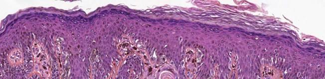
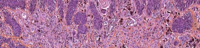
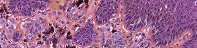
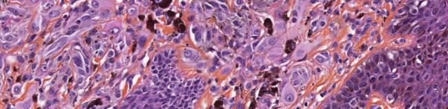
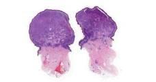
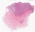
21/07/2017. The «gray zone» of diagnosis is visible. Nevus Atypical nevus Melanoma. Melanoma ex-blue nevus
|
|
|
- Godfrey Randall Wilkerson
- 5 years ago
- Views:
Transcription

1 Update on the Clinico- Pathological and Molecular Diagnosis of Melanocytic Lesions None to declare Conflicts of interest Belfast pathology Arnaud de la Fouchardière MD, PhD Lyon, France What is new? Today s Menu Realm of «Molecular Pathology» What is new? Realm of «Molecular Pathology» New concepts New techniques applied to FFPE New entities What is new? Realm of «Molecular Pathology» What is new? Realm of «Molecular Pathology» New concepts New concepts Step by step histo-molecular progression from nevus to melanoma 1


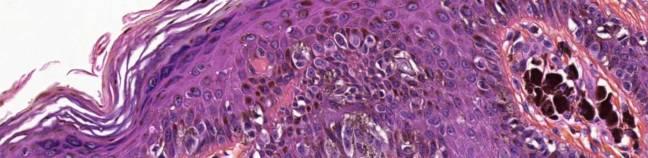
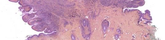
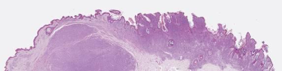
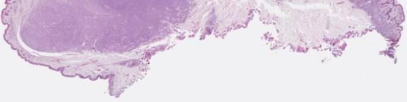
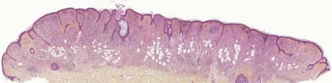
2 In many lesions there is a progressive gain of molecular anomalies linked with incremental microscopic atypia The «gray zone» of diagnosis is visible Benign Atypical Malignant Nevus Atypical nevus Melanoma Blue nevus Atypical blue nevus Melanoma ex-blue nevus Spitz Nevus Atypical Spitz tumor Spitzoid Melanoma DPN Atypical DPN Plexiform Melanoma Wiesner et al. pathology2015 2


3 What is new? Realm of «Molecular Pathology» New concepts Step by step histo-molecular progression from nevus to melanoma «Integrative analysis» Melanoledge project Integrative analysis Embryogenesis Clinical features Microscopy/morphology Immunophenotype Genomic profile Mutation status Clinical evolution «Integrative analysis» From concepts to tools Melanocytic tumors represent a wide variety of lesions. There are many ways of viewing differences. The goal is to combine as many «viewpoints» as possible in order to ultimately visualize melanocytic lesions in a more accurate manner. «Integrative analysis» From concepts to tools Melanocytic tumors represent a wide variety of lesions. There are many ways of viewing differences. The goal is to combine as many «viewpoints» as possible in order to ultimately visualize melanocytic lesions in a more accurate manner. Clinical features M 0 Location Age of appearance Size Type of sun exposure/phototype Recent modifications: Color Shape Volume/ulceration Pain/itching M0: Giant congenital nevus 3

:")


 F6 arm")


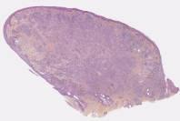
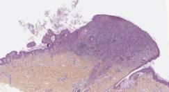
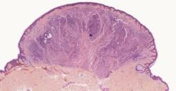
4 M15 Back (Dr de Carrère): BAP1-inactivated melanocytic tumor F34 Abdomen (Dr Bellili): Superficial Spreading Melanoma Microscopy/morphology Low magnification (Silhouette) High magnification (cytology) F6 arm (picturedr Dadban): Spitzoid melanoma Molecular Pathology of melanocytic tumors «Driver» BRAF NRAS NF1 HRAS CKIT GNAQ GNA11 «Oncogenetic» CDKN2A CDK4 MiTF BAP1 «Passenger» PTEN htert p53 Mutations 4


5 Main drivers SSM ALM Bastian B The molecular pathology of melanoma. Annu Rev Pathol Mech 2014 LMM NM Melanomaarising from blue nevi BRAF V600E Kit exon 11, 13, 17 or 18 Molecular Tools: use is guided by clinical morphological features IHC NGS FISH CNV BRAF V600K NRAS exon 2 or 3 GNAQ/11 exon 4 or 5 What is new? Realm of «Molecular Pathology» What is new? Realm of «Molecular Pathology» New concepts New concepts New techniques applied to FFPE New techniques applied to FFPE Antibodies, Next Generation Sequencing, RNA-seq 5

 D2-40 CD68 Why perform IHC?")
 Unpigmented metastases Desmoplastic melanoma 6")
6 Immunohistochemistry It s all about chosing the adapted antibody(ies) for the selected task(s) Antibodies «Melanocytic» antibodies S100 MelanA HMB45 PNL2 MiTF SOX10 «Anomaly-specific» antibodies BRAF V600E NRAS Q61R ALK ROS1 NTRK1 MET P16 BAP1 PDL1 Other antibodies D2-40 CD68 Specificity vs Sensitivity HMB45 Antibodies «Melanocytic» antibodies S100 MelanA HMB45 PNL2 MiTF SOX10 «Anomaly-specific» antibodies BRAF V600E NRAS Q61R ALK ROS1 NTRK1 MET P16 BAP1 PDL1 Other antibodies D2-40 CD68 NTRK1 Antibodies «Melanocytic» antibodies S100 MelanA HMB45 PNL2 MiTF SOX10 «Anomaly-specific» antibodies BRAF V600E NRAS Q61R ALK ROS1 NTRK1 MET P16 BAP1 PDL1 Other antibodies (DD mainly) D2-40 CD68 Why perform IHC? Confirm melanocytic lineage Visualize the melanocytes Benign vs Malignant Molecular characterization IHC to confirm melanocytic lineage Unpigmented dermal or ulcerated tumor (No recognizable junctional melanocytes) Unpigmented metastases Desmoplastic melanoma 6
")

7 IHC to confirm melanocytic lineage M69 Torso. No MM history Unpigmented dermal or ulcerated tumor (No recognizable junctional melanocytes) Unpigmented metastases Desmoplastic melanoma If usual melanocytic markers are negative Combination of SOX10 and MiTF nuclear positivity favors melanocytic origin M69 Torso. No MM history S100Protein S100Protein MelanA/Mart1/A103 7
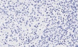
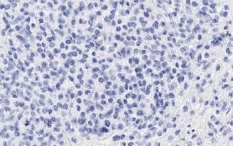
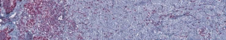
8 MelanA/Mart1/A103 HMB45 HMB45 SOX10 SOX10 MiTF 8


9 MiTF IHC for Molecular characterization Point mutations Gene fusions Loss of function (tumor suppressor genes) «Theragnostic» tools Point mutations BRAF VE1 BRAF V600E NRAS Q61R BRAF VE1 NRAS Q61R Melanoma arising from a large congenital nevus 9


")


10 Kinase Fusions IHC Screening tool Spitzoid tumors with kinase fusions ALK ROS1 NTRK1 Pan-NTRK MET Currently no reliable BRAF or NTRK3 fusion Ab Wiesner et al. pathology2015 IHC Gene Fusions Spitzoid tumors with ALK fusions NTRK1 MET ROS1 ALK Yeh et al Am J Surg Pathol 2015 Spitzoid tumors with kinase fusions Lesional spectrum of low grade tumors Kinase fusions allow a subclassification of tumors with (but not limited to) Spitzoid morphology Genotype/morphotype link? Work in progress Potential therapeutic targets Potential prognostic factors (BRAF fusion worse?) Drivers are mutually exclusive Nevoid Vs Spitzoid? BRAF V600E mutation vs Kinase fusions 10


11 «Raise your hand» vote BRAF mutation Raise Fusion 11

12 BRAF VE1 Ab IHC to explore loss of gene function (tumor suppressor genes) BAP1 p53 Usefulness of molecular pathology in cutaneous melanocytic tumors Level 1 Usefulness of molecular pathology in cutaneous melanocytic tumors Level 2 100% Morphological analysis with knowledge of essential clinical information: age of patient, size, localization and evolution of the tumor (recent modification for instance) 10% IHC screening if «benign vs malignant» doubt : 4 Ab panel (anti-melana, HMB45, anti-p16 et Ki67) with a red chromogene for thick lesions (>1 mm). Usefulness of molecular pathology in cutaneous melanocytic tumors Level 3 Usefulness of molecular pathology in cutaneous melanocytic tumors Level 4 5% Complementary IHC study searching for a specific anomaly guided by morphology and 4 Ab panel (anti-bap1, anti-alk, etc.). 1% Extensive molecular analysis in the event of a wide surgery (digital amputation, etc.), in pediatric variants of melanoma or rare melanocytic tumors (unclassified) Case specific combination of techniques : FISH : 4 color, CDKN2A (P16), fusions specific probes (ALK, ROS1, etc.) ; Gene sequencing accessing somatic mutations (BRAF, NRAS, GNAQ, GNA11, HRAS, etc.) ; CGH-array; RNA-seq analysis. 12
13 Hyperbole of uncertainty/technique Clinical evolution Uncertainty Local relapse Nodal extension Metastatic dissemination Site Biological comparison to primary tumor Technique Melanoledge project Convergence of viewpoints Embryogenesis Clinical features Microscopy/morphology Immunophenotype Genomic profile Mutation status What is new? Realm of «Molecular Pathology» New concepts New techniques applied to FFPE New entities Clinical evolution What is new? Realm of «Molecular Pathology» New concepts New techniques applied to FFPE BAP1-inactivated melanocytic tumors (BAPimt) Arnaud de la Fouchardière, MD, PhD Lyon, France New entities Kinase fusions in spitzoid tumors Melanocytic tumors with loss of BAP1 expression 13


/ melanoma(s) in the context of a")
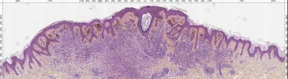
 Sporadic epidermal linked melanomas")


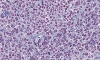
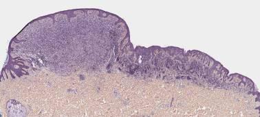
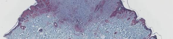
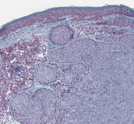
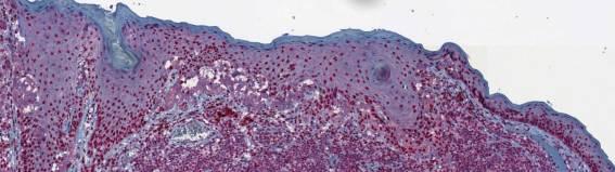
14 BAP1: BRCA1- Associated Protein 1 A gene with multiple faces BAP1 IHC Normal staining M Carbone et al Compound nevus Loss of nuclear BAP1 expression = loss of gene function Loss of BAP1 expression in melanocytic lesions of the skin Distinct scenarii Solitary BAPimt BAPimt(s)/ melanoma(s) in the context of a BAP1 cancer predisposition syndrome (germline mutation) Sporadic epidermal linked melanomas (DM) Melanomas arising from/mimicking cellular blue nevus BAPimts Somatic > germline mutation of BAP1 BAPimts Children or young adults Female>Male Sun-exposed areas Modification of a nevus Growing unpigmented nodule <1cm Inflammatory features Multiple lesions: germline Eur J Dermatol : F, 14 back M,56 Arm 14


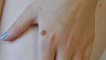
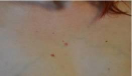
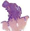

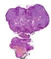
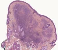
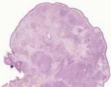
15 BAPimts M 27 with uveal melanoma F 16, cheek M 10 ; shoulder F25 multiple lesions since age 11 BAPimts keep the morphologic features of the nevi they arised from 15
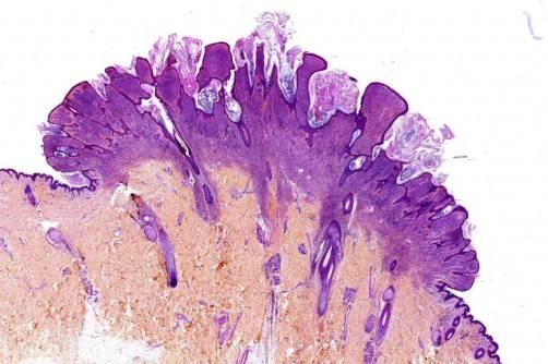
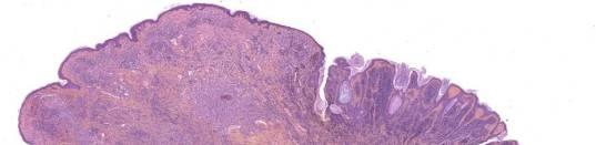
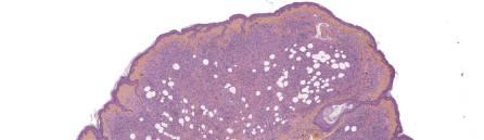
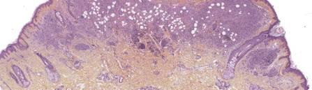
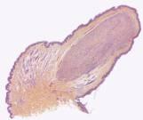
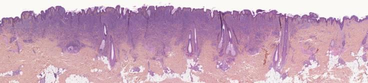
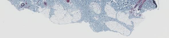
16 Congenital-like nevus with nodule Lipidic inclusions Lipidic inclusions 16


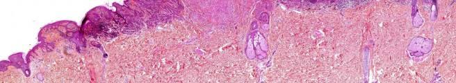
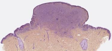
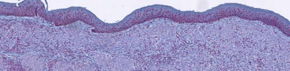
17 Lateral combined aspect commun nevus F 8: Ear; familial history of uveal melanoma M, 15 Back F, 19 Back 17


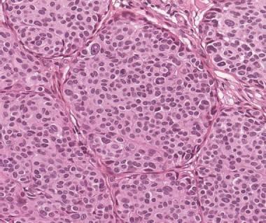
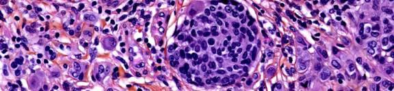
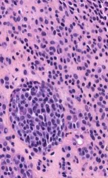
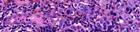
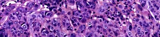
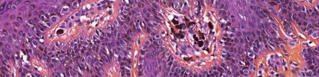
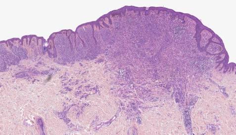
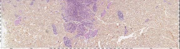
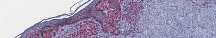
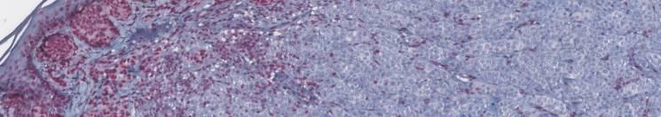
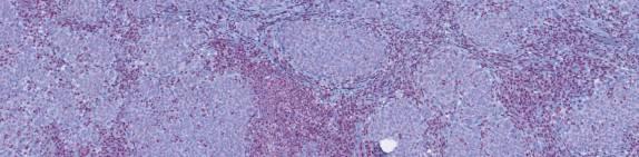
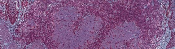
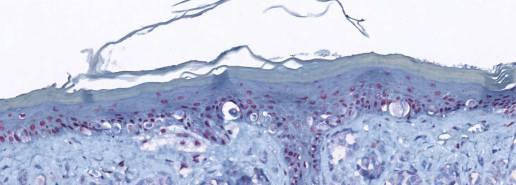
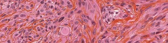
18 Nests of nevoid cells Nests of nevoid cells Large epithelioid and nevoid melanocytes +/- lymphocytes 18
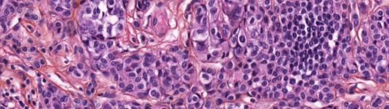
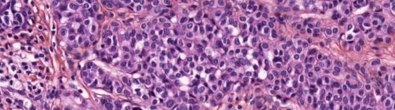
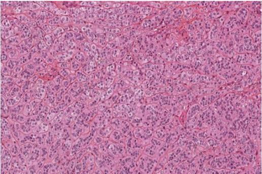
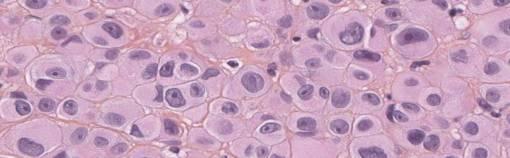
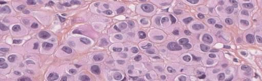
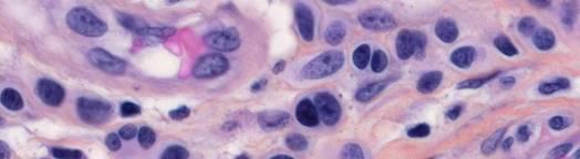
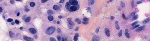
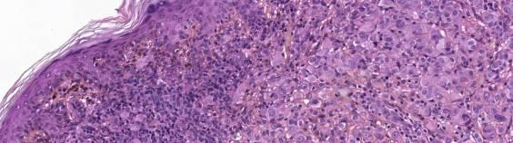
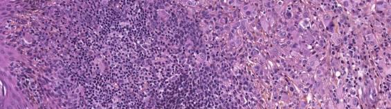
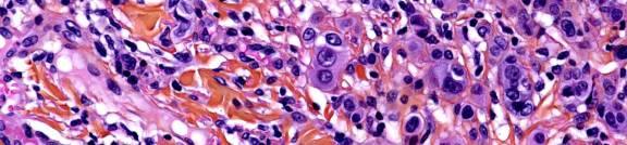
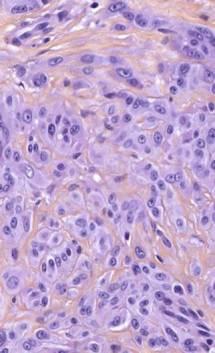
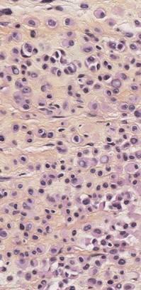
19 Large epithelioid and nevoid melanocytes +/- lymphocytes Clonal large unpigmented melanocytes Clonal large unpigmented melanocytes Multinucleations 19

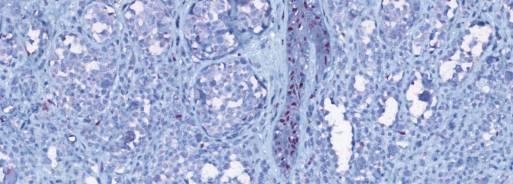
20 BAP1 IHC BAP1 IHC BAP1 IHC BAP1 IHC BAP1 IHC BAP1 IHC 20
 BAPimts No formal guidelines Unknown prognosis")

 Clear cell renal cancer Meningiomas")

21 BAP1 IHC BRAF V600E mutation association (> 80%) A C A G W G A A A c.1799t>a BRAF mutation IHC (VE1) BAPimts No formal guidelines Unknown prognosis Complete surgical removal Follow-up Identify patients needing oncogenetic counseling Tumors associated with a BAP1 germline mutation Atypical cutaneous melanocytic tumors (BAPimts ) Uveal melanoma Cutaneous melanoma Leptomeningeal melanoma Mesothelioma (pleural and abdominal) Clear cell renal cancer Meningiomas Multiple basal cell carcinomas These cancers arise at a younger age Maybe other cancers: lung, stomach, pancreas, CholangioK Probably incomplete cancer spectrum : ongoing work Frequency of germline mutations? 21


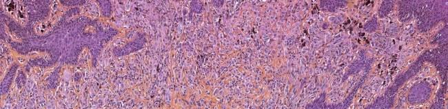
22 Estimated a <16% frequency of germline mutation We recommend, following the diagnosis of a Melanocytic tumor with loss of BAP1 expression, performing a BAP1 immunohistochemistry in all other cutaneous melanocytic tumors removed previously or simultaneously and all skin melanomas. Malignant transformation of BAPimts? M43, history in childhood of BAPimt on the neck 3cm buttocks mass that grew in a few months Malignant transformation of BAPimts? M43, history in childhood of BAPimt on the neck 3cm buttocks mass that grew in a few months Malignant transformation of BAPimts? M43, history in childhood of BAPimt on the neck 3cm buttocks mass that grew in a few months Malignant transformation of BAPimts? Malignant transformation of BAPimts? M43, history in childhood of BAPimt on the neck 3cm buttocks mass that grew in a few months BAP1 22


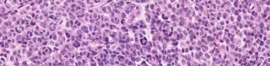
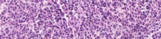
23 Malignant transformation of BAPimts? Malignant transformation of BAPimts? BAP1 Ki67 Differential Diagnosis DD: Spitz nevus Spitz nevus Nevoid melanoma Dystrophic nevus BAPimt-like melanocytic tumors DD: Spitz nevus DD:Spitz nevus 23


24 Nevoid melanoma ex-nevus Dystrophic nevus M, 64 Neck BAP1 «BAPimt-like» lesions F29, shoulder BAP1 BAP1 take home messages BAPology is a fascinating new science BAP1-inactivated melanocytic tumors can identify carriers of a germline syndrome with cancer-predisposition. Isolated cases with somatic mutations prevail. Exophytic combined architecture Large unpigmented epithelioid and nevoid dermal melanocytes +/- lymphocytes. IHC is an excellent technique to identify loss of function. Global Take home messages Extremely active field for molecular pathology Molecular techniques are strongly dependant on high quality clinical and morphological analysis Enhancing classifications improves patient care 24


25 Definition of Primary Tumor (T) - AJCC 8 th Edition Gershenwald, Scolyer, et al. Melanoma. In Amin, M.B., Edge, S.B., Greene, F.L., et al. (Eds.)AJCC Cancer Staging Manual. 8th Ed. New York: Springer;
Update on Spitzoid and Blue nevus-like melanocytic lesions Emphasis on molecular studies informing diagnosis, prognosis and therapy
 Update on Spitzoid and Blue nevus-like melanocytic lesions Emphasis on molecular studies informing diagnosis, prognosis and therapy Michael T. Tetzlaff MD, PhD Associate Professor Department of Pathology,
Molecular Aspects of Melanocytic Neoplasia. Iwei Yeh MD, PhD University of California, San Francisco
 Molecular Aspects of Melanocytic Neoplasia Iwei Yeh MD, PhD University of California, San Francisco Thanks to: Boris Bastian Timothy McCalmont Philip LeBoit Beth Ruben Jeff North Laura Pincus Thaddeus
Michael T. Tetzlaff MD, PhD
 Molecular alterations informing the diagnosis of melanocytic tumors Michael T. Tetzlaff MD, PhD Associate Professor Department of Pathology, Section of Dermatopathology Department of Translational and
Dermatopathology. Dr. Rafael Botella Estrada. Hospital La Fe de Valencia
 Dermatopathology Dr. Rafael Botella Estrada. Hospital La Fe de Valencia Melanoma and mimics Dr. Martin Mihm Malignant lesions result from the accumulation of mutations Class I lesions (benign) Class II
The Enigmatic Spitz Lesion
 The Enigmatic Spitz Lesion The Dawn of Spitz S Spitz Sophie Spitz Melanomas of Childhood ; Am J Pathol 1948 1910-1956 13 children (18 mo - 12 yrs) 12/13 had a benign clinical course Sophie Spitz Born 1910
Melanoma and the genes: Molecular alterations informing the diagnosis of melanocytic tumors
 Melanoma and the genes: Molecular alterations informing the diagnosis of melanocytic tumors Michael T. Tetzlaff MD, PhD Associate Professor Department of Pathology, Section of Dermatopathology Department
Patricia Chevez-Barrrios AAOOP-USCAP /12/2016
 Biomarkers in Ocular Melanoma Patricia Chévez-Barrios, MD Pathology and Genomic Medicine, Houston Methodist Hospital Professor of Pathology and Laboratory Medicine and Ophthalmology, Weill Cornell Medical
10/2/17. MELTUMP, SAMPUS, AST.An Algorithmic Approach to Challenging (Often Borderline) Melanocytic Tumors. An Introduction to SNP Arrays
 Melanocytic Tumors. An Introduction to SNP Arrays") MELTUMP, SAMPUS, AST.An Algorithmic Approach to Challenging (Often ) Melanocytic Tumors An Introduction to SNP Arrays Rajiv M. Patel, M.D. RCPA NZ ASM 2017 (11:45-12:30pm, Saturday, 23-09-17) Why do we
MELTUMP, SAMPUS, AST.An Algorithmic Approach to Challenging (Often ) Melanocytic Tumors An Introduction to SNP Arrays Rajiv M. Patel, M.D. RCPA NZ ASM 2017 (11:45-12:30pm, Saturday, 23-09-17) Why do we
Diagnoses of Cases 1. Lentigo, other melanosis and the acquired nevus 2. Variations on the acquired nevus 3. Dermal melanocytosis
 Diagnoses of Cases 1. Lentigo, other melanosis and the acquired nevus 1 1A. Lentigo simplex 4 1B. Psoralens and ultraviolet A (PUVA) lentigo 6 1C. Solar lentigo 8 1D. Café au lait macule 10 1E. Ink-spot
Vernon K. Sondak. Department of Cutaneous Oncology Moffitt Cancer Center Tampa, Florida
 Vernon K. Sondak Department of Cutaneous Oncology Moffitt Cancer Center Tampa, Florida Australasian Melanoma Conference 2016 Sydney, NSW, Australia October 29, 2016 Disclosures Dr. Sondak is a compensated
Genetic Testing: When should it be ordered? Julie Schloemer, MD Dermatology
 Genetic Testing: When should it be ordered? Julie Schloemer, MD Dermatology Outline Germline testing CDKN2A BRCA2 BAP1 Somatic testing Gene expression profiling (GEP) BRAF Germline vs Somatic testing
Ways to get into trouble, ideas on avoiding trouble, and diagnostic approaches to keep trouble at bay
 Pitfalls in the diagnosis of melanocytic tumors Timothy McCalmont, MD University of California, San Francisco Ways to get into trouble, ideas on avoiding trouble, and diagnostic approaches to keep trouble
MAPK Pathway. CGH Next Generation Sequencing. Molecular Tools in Care of Patients with Pigmented Lesions 7/20/2017
 Molecular Tools in Care of Patients with Pigmented Lesions Tammie Ferringer, MD Geisinger Medical Center, Danville, PA tferringer@geisinger.edu DISCLOSURE OF RELATIONSHIPS WITH INDUSTRY Tammie Ferringer,
Melanocytic Lesions: Use of Immunohistochemistry and Special Studies Napa Valley 2018
 Melanocytic Lesions: Use of Immunohistochemistry and Special Studies Napa Valley 2018 Victor G. Prieto, MD, PhD Professor Depts. of Pathology and Dermatology University of Texas - MD Anderson Cancer Center
Springer Healthcare. Staging and Diagnosing Cutaneous Melanoma. Concise Reference. Dirk Schadendorf, Corinna Kochs, Elisabeth Livingstone
 Concise Reference Staging and Diagnosing Cutaneous Melanoma Dirk Schadendorf, Corinna Kochs, Elisabeth Livingstone Extracted from Handbook of Cutaneous Melanoma: A Guide to Diagnosis and Treatment Published
There is NO single Melanoma Stain. > 6000 Mutations in Melanoma. What else can be done to discriminate atypical nevi from melanoma?
 Las Vegas Fall Clinical 2016: The Assessment and Diagnosis of Melanoma Whitney A. High, MD, JD, MEng Associate Professor, Dermatology & Pathology Director of Dermatopathology (Dermatology) University of
BAP-oma & BEYOND MICHAEL A NOWAK, MD
 BAP-oma & BEYOND MICHAEL A NOWAK, MD CONFLICTS No conflicts with the content of this lecture BAP-oma Wiesner 2011: Families with multiple tan dome-shaped papules of head, neck, trunk, and extremities.
Financial disclosures
 Mesenchymal Neoplasms with Melanocytic Differentiation By Konstantinos Linos MD, FCAP, FASDP Bone, Soft Tissue and Dermatopathology Assistant Professor of Pathology Dartmouth-Hitchcock Medical Center Geisel
MRC-Holland MLPA. Description version 06; 23 December 2016
 SALSA MLPA probemix P417-B2 BAP1 Lot B2-1216. As compared to version B1 (lot B1-0215), two reference probes have been added and two target probes have a minor change in length. The BAP1 (BRCA1 associated
The Pathology of Neoplasia Part II
 The Pathology of Neoplasia Part II February 2018 PAUL BOGNER, MD A S S O C I A T E P R O F E S S O R O F O N C O L O G Y P A T H O L O G Y A N D D E R M A T O L O G Y Clinical goals of cancer pathology
Melanocytic Tumours. Molecular Biology 02/06/2015. Cutaneous Melanocytic Tumours Introduction. Thomas Brenn. Intermediate Malignancy
 Cutaneous Melanocytic Tumours Introduction Melanocytic Tumours: Update on Epidemiology and Molecular Biology Thomas Brenn Wide clinical and morphological spectrum Ranging from benign naevi to melanoma
Case RAC7783. M46. Ear. Mole. r/o MM.?Blue naevus RAC7783
 Case RAC7783. M46. Ear. Mole. r/o MM.?Blue naevus RAC7783 Pie Chart Participants N=74 Benign: 48 N=74 Blue naevus: 38 Intradermal: 12 DPN: 10 Compound 3 Clonal: 3; Spitz 2; Special Site: 1; Congenital:
Which melanoma patients benefit from genetic testing?
 Which melanoma patients benefit from genetic testing? Michael A. Marchetti, MD Assistant Attending, Dermatology Service Memorial Sloan Kettering Cancer Center American Academy of Dermatology Annual Meeting
A PRACTICAL APPROACH TO ATYPICAL MELANOCYTIC LESIONS BIJAN HAGHIGHI M.D, DIRECTOR OF DERMATOPATHOLOGY, ST. JOSEPH HOSPITAL
 A PRACTICAL APPROACH TO ATYPICAL MELANOCYTIC LESIONS BIJAN HAGHIGHI M.D, DIRECTOR OF DERMATOPATHOLOGY, ST. JOSEPH HOSPITAL OBJECTIVES Discuss current trends and changing concepts in our understanding of
Update on Genetic Testing for Melanoma
 Update on Genetic Testing for Melanoma Emily Y. Chu, M.D., Ph.D. Assistant Professor of Dermatology & Pathology and Laboratory Medicine Hospital of the University of Pennsylvania February 18, 2018 AAD
Next Generation Sequencing in Clinical Practice: Impact on Therapeutic Decision Making
 Next Generation Sequencing in Clinical Practice: Impact on Therapeutic Decision Making November 20, 2014 Capturing Value in Next Generation Sequencing Symposium Douglas Johnson MD, MSCI Vanderbilt-Ingram
David B. Troxel, MD. Common Medicolegal Situations: Misdiagnosis of Melanoma
 Common Medicolegal Situations: Misdiagnosis of Melanoma David B. Troxel, MD Medical Director, The Doctors Company, Napa, California Clinical Professor Emeritus, University of California at Berkeley Past
Desmoplastic Melanoma R/O BCC. Clinical Information. 74 y.o. man with lesion on left side of neck r/o BCC
 R/O BCC Sabine Kohler, M.D. Professor of Pathology and Dermatology Dermatopathology Service Stanford University School of Medicine Clinical Information 74 y.o. man with lesion on left side of neck r/o
Melanocytic proliferations in sundamaged
 Atypical Spitzoid Tumor: What Does It Mean And How Should It Be Managed? Melanocytic proliferations in sundamaged skin Jane L. Messina, Jane L. Messina MD International Melanoma Pathology Working Group
Supplementary Figure 1. Spitzoid Melanoma with PPFIBP1-MET fusion. (a) Histopathology (4x) shows a domed papule with melanocytes extending into the
 Histopathology (4x) shows a domed papule with melanocytes extending into the") Supplementary Figure 1. Spitzoid Melanoma with PPFIBP1-MET fusion. (a) Histopathology (4x) shows a domed papule with melanocytes extending into the deep dermis. (b) The melanocytes demonstrate abundant
Supplementary Figure 1. Spitzoid Melanoma with PPFIBP1-MET fusion. (a) Histopathology (4x) shows a domed papule with melanocytes extending into the deep dermis. (b) The melanocytes demonstrate abundant
6/22/2015. Original Paradigm. Correlating Histology and Molecular Findings in Melanocytic Neoplasms
 6 Correlating Histology and Molecular Findings in Melanocytic Neoplasms Pedram Gerami MD, Associate Professor of Dermatology and Pediatrics at Northwestern University Disclosures: I have been a consultant
Molecular Testing in Lung Cancer
 Molecular Testing in Lung Cancer Pimpin Incharoen, M.D. Assistant Professor, Thoracic Pathology Department of Pathology, Ramathibodi Hospital Genetic alterations in lung cancer Source: Khono et al, Trans
Conflict of Interest 9/2/2014. Pathogenesis and Comparison of Atypical Spitz Nevi vs Benign Spitz, and Childhood Melanoma
 Pathogenesis and Comparison of Atypical Spitz Nevi vs Benign Spitz, and Childhood Melanoma Martin C. Mihm Jr., M.D., F.A.C.P. Harvard Medical School Brigham and Women s Hospital Dana Farber Cancer Center
Female 18. Deeply pigmented lesion on trunk.?warty naevus?seborrhoeic keratosis?malignant melanoma. The best diagnosis is:
 Female 18. Deeply pigmented lesion on trunk.?warty naevus?seborrhoeic keratosis?malignant melanoma. The best diagnosis is: A. deep penetrating naevus B. naevoid malignant melanoma C. pigment synthesising
Case 26 Male 37. Right jawline 5mm nodule?keloid. The best diagnosis is:
 Case 26 Male 37. Right jawline 5mm nodule?keloid. The best diagnosis is: A. Desmoplastic Spitz naevus B. Atypical Spitz Tumour C. Spitzoid melanoma D. Deep penetrating naevus E. Spitz naevus Case 26: M
Benign and malignant epithelial lesions: Seborrheic keratosis: A common benign pigmented epidermal tumor occur in middle-aged or older persons more
 Benign and malignant epithelial lesions: Seborrheic keratosis: A common benign pigmented epidermal tumor occur in middle-aged or older persons more common on the trunk; but extremities, head and neck are
Guy Perrot (Ги Перро)
") НАУЧНО-ПРАКТИЧЕСКАЯ КОНФЕРЕНЦИЯ (МАСТЕР-КЛАСС) «ПРАКТИЧЕСКИЕ АСПЕКТЫ ДИАГНОСТИКИ И ЛЕЧЕНИЯ МЕЛАНОМЫ КОЖИ» DIAGNOSTIC AND PITFALLS IN MELANOMA Guy Perrot (Ги Перро) MD PHD pathologist, University Hospital
НАУЧНО-ПРАКТИЧЕСКАЯ КОНФЕРЕНЦИЯ (МАСТЕР-КЛАСС) «ПРАКТИЧЕСКИЕ АСПЕКТЫ ДИАГНОСТИКИ И ЛЕЧЕНИЯ МЕЛАНОМЫ КОЖИ» DIAGNOSTIC AND PITFALLS IN MELANOMA Guy Perrot (Ги Перро) MD PHD pathologist, University Hospital
Rare melanoma: Are the options improving? Dr Neil Steven Consultant in Medical Oncology University Hospital Birmingham University of Birmingham
 Rare melanoma: Are the options improving? Dr Neil Steven Consultant in Medical Oncology University Hospital Birmingham University of Birmingham Classifying melanoma Melanoma (site of origin, thickness,
ACCME/Disclosures ALK FUSION-POSITIVE MESENCHYMAL TUMORS. Tumor types with ALK rearrangements. Anaplastic Lymphoma Kinase. Jason L.
 Companion Meeting of the International Society of Bone and Soft Tissue Pathology The Evolving Concept of Mesenchymal Tumors ALK FUSION-POSITIVE MESENCHYMAL TUMORS Jason L. Hornick, MD, PhD March 13, 2016
Melanoma Update: 8th Edition of AJCC Staging System
 Melanoma Update: 8th Edition of AJCC Staging System Rosalie Elenitsas, M.D. Professor of Dermatology Director, Dermatopathology University of Pennsylvania DISCLOSURE OF RELATIONSHIPS WITH INDUSTRY None
Predisposition of Melanoma
 Predisposition of Melanoma Nelleke Gruis Department of Dermatology Leiden University Medical Center The Netherlands OCTOBER 27TH 2017 Melanoma Risk Factors? Melanoma Predisposition 10% familial Manolio
Reviewers' comments: Reviewer #1 (Remarks to the Author):
:") Reviewers' comments: Reviewer #1 (Remarks to the Author): In this study the authors analysed 18 deep penetrating nevi for oncogenic genomic changes (single nucleotide variations, insertions/deletions,
Reviewers' comments: Reviewer #1 (Remarks to the Author): In this study the authors analysed 18 deep penetrating nevi for oncogenic genomic changes (single nucleotide variations, insertions/deletions,
Management of pediatric melanocytic lesions
 Open Journal of Clinical & Medical Case Reports Management of pediatric melanocytic lesions Volume 3 (2017) Issue 8 ISSN 2379-1039 Jin Kim, BS; Emmanuel Gabriel MD, PhD; Weiguo Liu MD, PhD; Lin Lin MD,
Cutaneous Mesenchymal Neoplasms with EWSR1 Rearrangement
 Cutaneous Mesenchymal Neoplasms with EWSR1 Rearrangement By Konstantinos Linos MD, FCAP, FASDP Bone, Soft Tissue and Dermatopathology Assistant Professor of Pathology Dartmouth-Hitchcock Medical Center
Case 231: F7. Exophytic naevus over left trapezious. Grown over a few weeks. Iniitally flat.?spitz naevus,?malignant
 Case 231: F7. Exophytic naevus over left trapezious. Grown over a few weeks. Iniitally flat.?spitz naevus,?malignant Dermoscopy: coarse vascular structures. c/o A, B, C RAC7750 Case 231: F7. Exophytic
An update on molecular alterations in melanocytic tumors with emphasis on Spitzoid lesions
 Review Article on Molecular Oncology age 1 of 17 An update on molecular alterations in melanocytic tumors with emphasis on Spitzoid lesions Emmanouil Dimonitsas 1#, Aliki Liakea 2#, Stratigoula Sakellariou
I have no relevant conflicts of interest to disclose. John T. Seykora MD PhD Departments of Dermatology & Pathology and Laboratory Medicine
 Molecular Characterization of Stage 1-3 Melanoma: Are we close to accurate prognostication and prediction? I have no relevant conflicts of interest to disclose. John T. Seykora MD PhD Departments of Dermatology
Self assessment case. Dr Saleem Taibjee Dorset County Hospital, Dorchester
 Self assessment case Dr Saleem Taibjee saleemtaibjee@gmail.com Dorset County Hospital, Dorchester Clinical details 34-year-old man: Shave excision Skin tag / papilloma left thigh The best diagnosis is:
Disclosure Information. Lecture Outline. Lecture Outline. Introduction. Molecular Pathology of Cutaneous Melanoma. Nothing to disclose
 Molecular Pathology of Cutaneous Melanoma Disclosure Information Nothing to disclose Jonathan L. Curry, MD Assistant Professor of Pathology and Dermatology University of Texas-MD Anderson Cancer Center
Index. Springer-Verlag Berlin Heidelberg 2017 J.A. Plaza, V.G. Prieto, Pathology of Pigmented Skin Lesions, DOI /
 A Acral lentiginous (mucosal lentiginous) melanoma, 483 Acral lentiginous melanoma (ALM) asymmetric and irregular lentiginous junctional growth, 431 clinical features, 427 428 differential diagnosis, 428
The Relevance of Cytologic Atypia in Cutaneous Neural Tumors
 The Relevance of Cytologic Atypia in Cutaneous Neural Tumors Recent Findings - New Developments New Problems Zsolt B. Argenyi, M.D. Professor of Pathology & Dermatology Director of Dermatopathology Department
Diploma Examination. Dermatopathology: First paper. Tuesday 20 March Candidates must answer FOUR questions. Time allowed: 3 hours
 Dermatopathology: First paper Tuesday 20 March 2018 Candidates must answer FOUR questions Time allowed: 3 hours 1. Give an account of the genetic aberrations encountered in Spitzoid neoplasms and how these
أملس عضلي غرن = Leiomyosarcoma. Leiomyosarcoma 1 / 5
 Leiomyosarcoma 1 / 5 EPIDEMIOLOGY Exact incidence is unknown, but older studies suggest that leiomyosarcomas comprise approximately 3 percent of soft-tissue sarcomas. Superficial leiomyosarcoma occurs
5 th July 2016 ACGS Dr Michelle Wood Laboratory Genetics, Cardiff
 5 th July 2016 ACGS Dr Michelle Wood Laboratory Genetics, Cardiff National molecular screening of patients with lung cancer for a national trial of multiple novel agents. 2000 NSCLC patients/year (late
MELANOMA IN ADOLESCENTS AND YOUNG ADULTS
 Cancer in Adolescents and Young Adults (AYA) Working Group MELANOMA IN ADOLESCENTS AND YOUNG ADULTS Emmanouil Saloustros MD, DSc General Hospital of Heraklion Venizelio Heraklion, Crete, Greece ESMO Preceptorship
Normal RAS-RAF (MAPK) pathway signaling
 pathway signaling") BRAF-Mutations in Melanomas L. Mazzucchelli Istituto Cantonale di Patologia, Locarno 77. Annual Meeting Swiss Society of Pathology, Lucerne 2011 Sponsored by Roche Pharma Switzerland Melanoma has increased
BRAF-Mutations in Melanomas L. Mazzucchelli Istituto Cantonale di Patologia, Locarno 77. Annual Meeting Swiss Society of Pathology, Lucerne 2011 Sponsored by Roche Pharma Switzerland Melanoma has increased
Mesothelioma: diagnostic challenges from a pathological perspective. Naseema Vorajee August 2016
 Mesothelioma: diagnostic challenges from a pathological perspective Naseema Vorajee August 2016 Naseema.vorajee@nhls.ac.za Pleural diseases (whether neoplastic, reactive or infective) may have similar
Integrating Fluorescence in situ Hybridization and Genomic Array Results into the Diagnostic Workup of Melanoma
 Integrating Fluorescence in situ Hybridization and Genomic Array Results into the Diagnostic Workup of Melanoma Association for Molecular Pathology United States and Canadian Academy of Pathology Companion
Living Beyond Cancer Skin Cancer Detection and Prevention
 Living Beyond Cancer Skin Cancer Detection and Prevention Cutaneous Skin Cancers Identification Diagnosis Treatment options Prevention What is the most common cancer in people? What is the most common
Bersagli molecolari nel melanoma
 Bersagli molecolari nel melanoma Giuseppe Palmieri - Unit of Cancer Genetics Institute of Biomolecular Chemistry, CNR, Sassari Essential alterations in malignant cells Hanahan & Weinberg, Cell 2000 Essential
2/6/2018. Original Paradigm. Clonal Chromosomal A berrations. Only 20% of Spitz Nevi 95% 6p, 7q, 17q, 20q, 4q,8q, 1q, 11q. Isolated Gain in 11p
 Molecular Diagnostics for Melanocytic Neoplasms: Moving towards a Revolution in the Management of Melanocytic Neoplasms Pedr am Gerami MD Associate Professor of Dermatology, Pathology and Pediatrics at
Pathology of the skin. 2nd Department of Pathology, Semmelweis University
 Pathology of the skin 2nd Department of Pathology, Semmelweis University Histology of the skin Epidermis: Stratum corneum Stratum granulosum Stratum spinosum Stratum basale Dermis: papillary and reticular
المركب النموذج--- سبيتز وحمة = Type Spitz's Nevus, Compound SPITZ NEVUS 1 / 7
 SPITZ NEVUS 1 / 7 Epidemiology An annual incidence rate of 1.4 cases of Spitz nevus per 100,000 individuals has been estimated in Australia, compared with 25.4 per 100,000 individuals for cutaneous melanoma
Cutaneous Melanoma: Epidemiology (USA) The Sentinel Node in Head and Neck Melanoma. Cutaneous Melanoma: Epidemiology (USA)
 The Sentinel Node in Head and Neck Melanoma. Cutaneous Melanoma: Epidemiology (USA)") The Sentinel Node in Head and Neck Melanoma Cutaneous Melanoma: Epidemiology (USA) 6 th leading cause of cancer among men and women 68,720 new cases of invasive melanoma in 2009 8,650 deaths from melanoma
The Sentinel Node in Head and Neck Melanoma Cutaneous Melanoma: Epidemiology (USA) 6 th leading cause of cancer among men and women 68,720 new cases of invasive melanoma in 2009 8,650 deaths from melanoma
Enterprise Interest Nothing to declare
 Enterprise Interest Nothing to declare Diagnoses one would not like to miss in soft tissue pathology early in your career Marta Sbaraglia, MD Department of Pathology Hospital of Treviso University of Padua
Molecular Testing Updates. Karen Rasmussen, PhD, FACMG Clinical Molecular Genetics Spectrum Medical Group, Pathology Division Portland, Maine
 Molecular Testing Updates Karen Rasmussen, PhD, FACMG Clinical Molecular Genetics Spectrum Medical Group, Pathology Division Portland, Maine Keeping Up with Predictive Molecular Testing in Oncology: Technical
Simulators of melanoma
 Simulators of melanoma Philip E. LeBoit, M.D. Depts. of Pathology and Dermatology University of California, San Francisco Simulators of melanoma Simulators of melanoma in situ Melanocytic Non-melanocytic
Racial differences in six major subtypes of melanoma: descriptive epidemiology
 Wang et al. BMC Cancer (2016) 16:691 DOI 10.1186/s12885-016-2747-6 RESEARCH ARTICLE Racial differences in six major subtypes of melanoma: descriptive epidemiology Yu Wang 1, Yinjun Zhao 2 and Shuangge
Update: Morphologic Considerations in Mesothelioma within the Pleural and Peritoneal Cavities. Douglas J. Hartman, MD June 7, 2018
 Update: Morphologic Considerations in Mesothelioma within the Pleural and Peritoneal Cavities Douglas J. Hartman, MD June 7, 2018 Objectives Review Historical Features associated with prognosis Present
1/10/2018. Soft Tissue Tumors Showing Melanocytic Differentiation. Overview. Desmoplastic/ Spindle Cell Melanoma
 2016 MFMER slide-1 2016 MFMER slide-2 2016 MFMER slide-3 Soft Tissue Tumors Showing Melanocytic Differentiation Andrew L. Folpe, M.D. Professor of Laboratory Medicine and Pathology Mayo Clinic, Rochester,
Malignant tumors of melanocytes: Part 1. Deba P Sarma, MD., Omaha
 Malignant tumors of melanocytes: Part 1 Deba P Sarma, MD., Omaha The melanocytic tumor is one of the most difficult and confusing areas in Dematopathology. It is true that most (95%) of such lesions are
Melanoma-Back to Basics I Thought I Knew Ya! Paul K. Shitabata, M.D. Dermatopathologist APMG
 Melanoma-Back to Basics I Thought I Knew Ya! Paul K. Shitabata, M.D. Dermatopathologist APMG At tumor board, a surgeon insists that all level II melanomas are invasive since they have broken through the
Interesting Case Series. Desmoplastic Melanoma
 Interesting Case Series Desmoplastic Melanoma Anthony Maurice Kordahi, MD, Joshua B. Elston, MD, Ellen M. Robertson, MD, and C. Wayne Cruse, MD Division of Plastic Surgery, Department of Surgery, University
Multiple Primary Melanoma in a Thai Male: A Case Report
 Case Report Multiple Primary Melanoma in a Thai Male: A Case Report J Med Assoc Thai 2014; 97 (Suppl. 2): S234-S238 Full text. e-journal: http://www.jmatonline.com Kittisak Payapvipapong MD*, Pinyapat
Patient age and cutaneous malignant melanoma: Elderly patients are likely to have more aggressive histological features and poorer survival
 MOLECULAR AND CLINICAL ONCOLOGY 7: 1083-1088, 2017 Patient age and cutaneous malignant melanoma: Elderly patients are likely to have more aggressive histological features and poorer survival FARUK TAS
Difficult Diagnoses and Controversial Entities in Neoplastic Lung
 Difficult Diagnoses and Controversial Entities in Neoplastic Lung Lynette M. Sholl, M.D. Associate Pathologist, Brigham and Women s Hospital Chief, Pulmonary Pathology Service Associate Professor, Harvard
From Morphology to Molecular Pathology: A Practical Approach for Cytopathologists Part 1-Cytomorphology. Songlin Zhang, MD, PhD LSUHSC-Shreveport
 From Morphology to Molecular Pathology: A Practical Approach for Cytopathologists Part 1-Cytomorphology Songlin Zhang, MD, PhD LSUHSC-Shreveport I have no Conflict of Interest. FNA on Lymphoproliferative
Prevalence of Exon 15 BRAF Mutations in Primary Melanoma of the Superficial Spreading, Nodular, Acral, and Lentigo Maligna Subtypes
 Prevalence of Exon 15 BRAF Mutations in Primary Melanoma of the Superficial Spreading, Nodular, Acral, and Lentigo Maligna Subtypes Julie Lang and Rona M. MacKie w Duncan Guthrie Institute of Medical Genetics,
Society for Pediatric Pathology Spring Meeting Joint Symposium with American Society of Dermatopathology
 Society for Pediatric Pathology 2013 Spring Meeting Joint Symposium with American Society of Dermatopathology Update on Cutaneous Melanocytic, Mesenchymal and Lymphoproliferative Lesions in Children Melanocytic
Primary Cutaneous CD30-Positive T-cell Lymphoproliferative Disorders
 Primary Cutaneous CD30-Positive T-cell Lymphoproliferative Disorders Definition A spectrum of related conditions originating from transformed or activated CD30-positive T-lymphocytes May coexist in individual
We are IntechOpen, the world s leading publisher of Open Access books Built by scientists, for scientists. International authors and editors
 We are IntechOpen, the world s leading publisher of Open Access books Built by scientists, for scientists 3,500 108,000 1.7 M Open access books available International authors and editors Downloads Our
Dilemmas in Cytopathology and Histopathology
 Dilemmas in Cytopathology and Histopathology Yuri E. Nikiforov, MD, PhD Division of Molecular & Genomic Pathology University of Pittsburgh Medical Center, USA Objectives Discuss new WHO classification
Michael T. Tetzlaff MD, PhD
 American Joint Cancer Committee (AJCC) staging system for primary cutaneous melanoma (8 th Edition) and principles of sentinel lymph node evaluation Emphasis on concise and accurate reporting of primary
Associate Clinical Professor of Dermatology MUSC
 Re-excision of Moderately Dysplastic Nevi: Should we or shouldn t we? John C. Maize, Jr, M.D. Dermatologist and Dermatopathologist Trident Dermatology, Charleston SC Associate Clinical Professor of Dermatology
Pathologic prognosticators, primary tumor
 Pathologic prognosticators, primary tumor Epidermal invasion Ulceration (AJCC2009) BRESLOW (4mm) Dermal invasion (Clark) Radial or Vertical Growth Satellite nodule= in-transit metastasis
Disclosure. Relevant Financial Relationship(s) None. Off Label Usage None MFMER slide-1
 None. Off Label Usage None MFMER slide-1") Disclosure Relevant Financial Relationship(s) None Off Label Usage None 2013 MFMER slide-1 Case Presentation A 43 year old male, with partial nephrectomy for a right kidney mass 2013 MFMER slide-2 2013
Disclosure Relevant Financial Relationship(s) None Off Label Usage None 2013 MFMER slide-1 Case Presentation A 43 year old male, with partial nephrectomy for a right kidney mass 2013 MFMER slide-2 2013
History A 89 year old gentleman presenting with a scalp/forehead nodule. Patient had squamous cell carcinoma 18 m at same site, excised. Outside diagn
 Case III History A 89 year old gentleman presenting with a scalp/forehead nodule. Patient had squamous cell carcinoma 18 m at same site, excised. Outside diagnoses: Squamous cell carcinoma. R/O: SCC, Melanoma,
The Dermal Melanocytoses. Conflicts of Interest 5/22/2018. The Nevi of Ota and Ito. Martin C. Mihm M.D.
 The Dermal Melanocytoses Martin C. Mihm M.D. Director Mihm Cutaneous Pathology Consultative Service (MCPCS) Brigham and Women s Hospital Director Melanoma Program Brigham and Women s Hospital and Harvard
K Blessing, J J H Grant, D S A Sanders, M M Kennedy, A Husain, P Coburn
 J Clin Pathol 2000;53:591 595 591 Papers Pathology, Aberdeen University, Foresterhill, Aberdeen AB25 2ZD, K Blessing Pathology, Birmingham University, Birmingham B15 2TT, D S A Sanders Pathology, Heartlands
Special slide seminar
 Special slide seminar Tomáš Rozkoš The Fingerland Department of Pathology Charles University Medical Faculty and Faculty Hospital in Hradec Králové Czech Republic Case history, 33 years old resistance
Genomic Medicine: What every pathologist needs to know
 Genomic Medicine: What every pathologist needs to know Stephen P. Ethier, Ph.D. Professor, Department of Pathology and Laboratory Medicine, MUSC Director, MUSC Center for Genomic Medicine Genomics and
Benign versus Cancerous Lesions How to tell the difference FMF 2014 Christie Freeman MD, CCFP, DipPDerm, MSc
 1 Benign versus Cancerous Lesions How to tell the difference FMF 2014 Christie Freeman MD, CCFP, DipPDerm, MSc Benign lesions Seborrheic Keratoses: Warty, stuck-on Genetics and birthdays Can start in late
Update on 8 th Edition Cutaneous AJCC Staging of Primary Cutaneous Melanoma. Michael T. Tetzlaff MD, PhD
 Update on 8 th Edition Cutaneous AJCC Staging of Primary Cutaneous Melanoma Michael T. Tetzlaff MD, PhD Associate Professor Departments of Pathology (Dermatopathology) and Translational and Molecular Pathology
patients in the era of
 Communicating with cancer patients in the era of personalized medicine September 9 th, 2017 Gerald Prager, M.D. Comprehensive Cancer Center Vienna Medical University of Vienna, Austria Gerald Prager, M.D.
Diploma examination. Dermatopathology: First paper. Tuesday 21 March Candidates must answer FOUR questions ONLY. Time allowed: Three hours
 Dermatopathology: First paper Tuesday 21 March 2017 1. Discuss the role of fluorescent in-situ hybridization (FISH) and emerging molecular techniques in the diagnosis of cutaneous melanocytic lesions,
THE SPITZ NEVUS OFTEN POSES
 OBSERVATION ONLINE FIRST Melanoma Mimic A Case of Multiple Pagetoid Spitz Nevi KaLynne Harris, MD; Scott R. Florell, MD; Jason Papenfuss, MD; Wendy Kohlmann, MS, CGC; Mona Jahromi, BS; Joshua D. Schiffman,
MECHANISMS OF HUMAN DISEASE: LABORATORY SESSION PATHOLOGY OF THE SKIN LAB. Friday, February 12, :30 am 11:00 am
 MECHANISMS OF HUMAN DISEASE: LABORATORY SESSION PATHOLOGY OF THE SKIN LAB Friday, February 12, 2012 9:30 am 11:00 am FACULTY COPY GOALS: Describe the basic clinical and morphologic features of various
I. Diagnosis of the cancer type in CUP
 Latest Research: USA I. Diagnosis of the cancer type in CUP II. Outcomes of site-specific therapy of the cancer type in CUP a. Prospective clinical trial b. Retrospective clinical trials 1 Latest Research:
Select analysis on the next pages. Sample request and sending address see last page. Institut für Pathologie und Molekularpathologie
 Diagnostic Tumor Genome Analysis Schmelzbergstrasse 12 8091 Zürich Tel.: (+41) 044 255 3929 Fax.: (+41) 044 255 4416 Client (address, telephone number): ngs.pathologie@usz.ch www.pathologie.usz.ch Sample-Nr:
Follow up of the Guidelines for Cytopathologic Diagnosis of Malignant Mesothelioma
 Follow up of the Guidelines for Cytopathologic Diagnosis of Malignant Mesothelioma Assoc. Prof. Katalin Dobra, Senior Lecturer in Molecular Pathology Karolinska University Hospital Stockholm, Sweden Disclosure