Criteria that really discriminate
|
|
|
- Alaina Mosley
- 5 years ago
- Views:
Transcription

1 Criteria that really discriminate Dirk M. Elston, MD Medical Universiity of South Carolina, Charleston 1


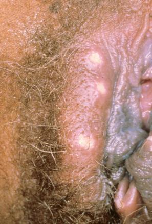
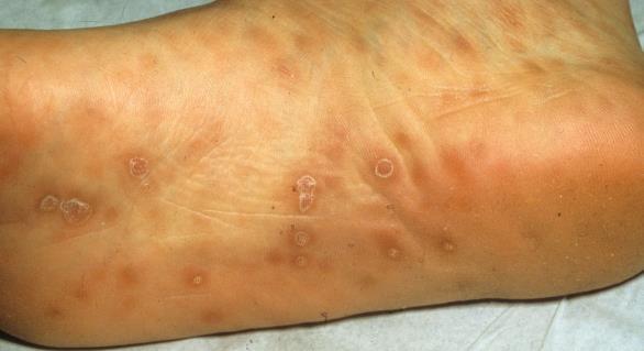
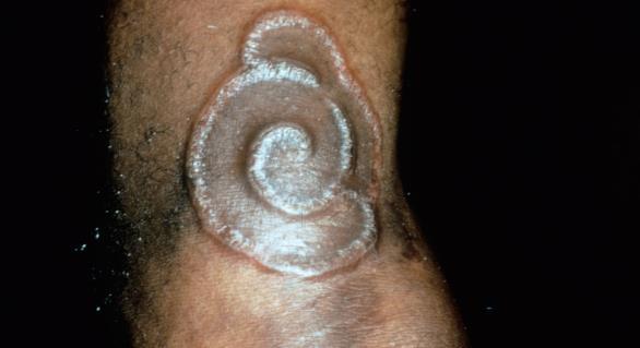
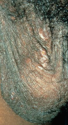
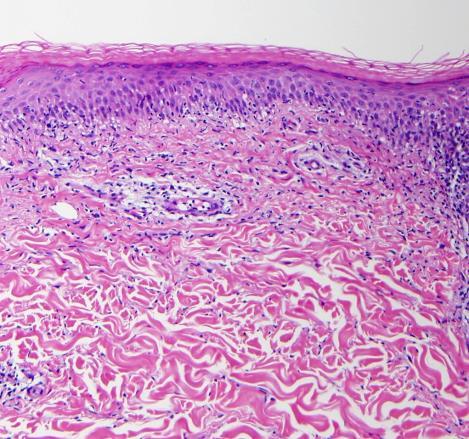
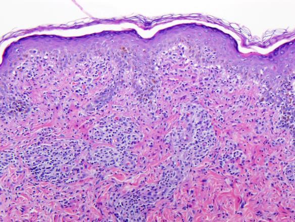
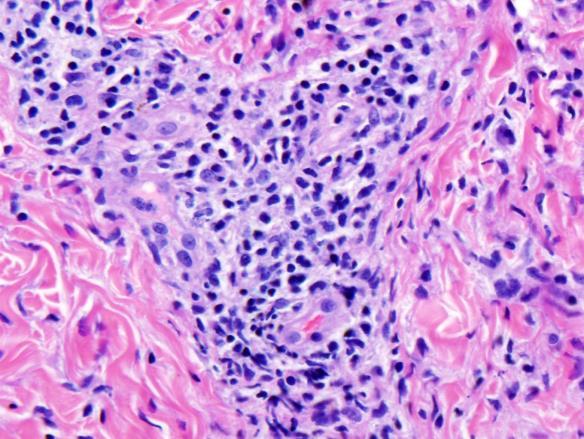
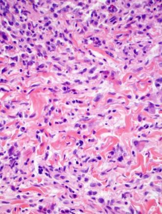
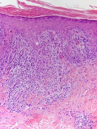
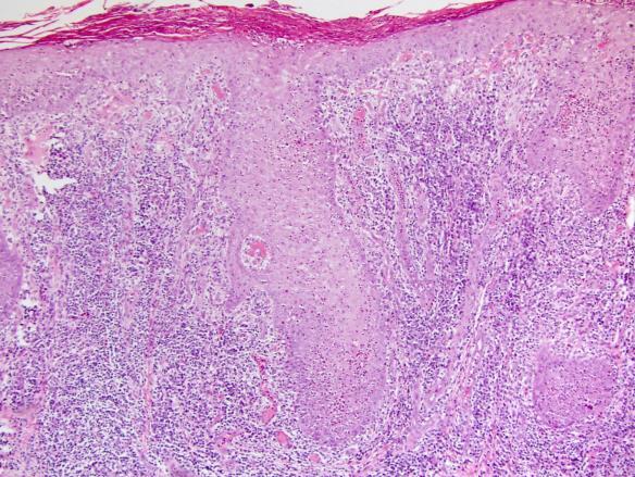
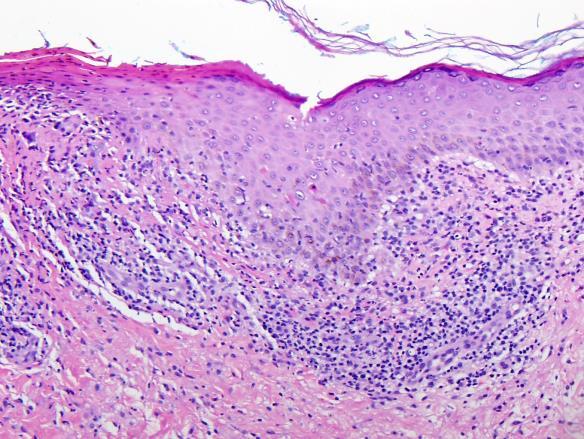
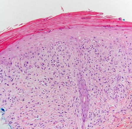
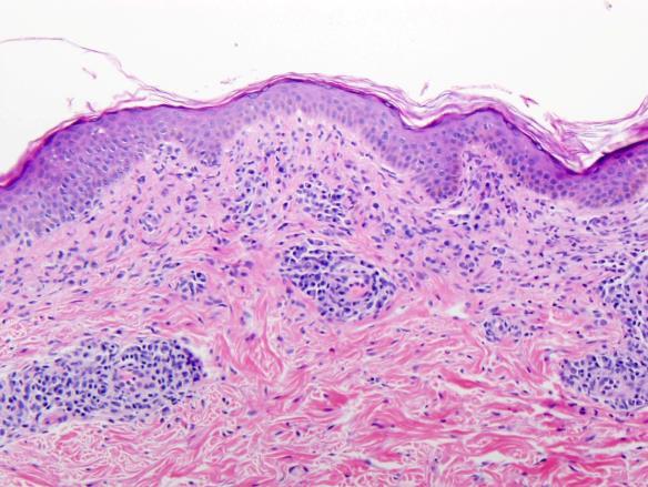
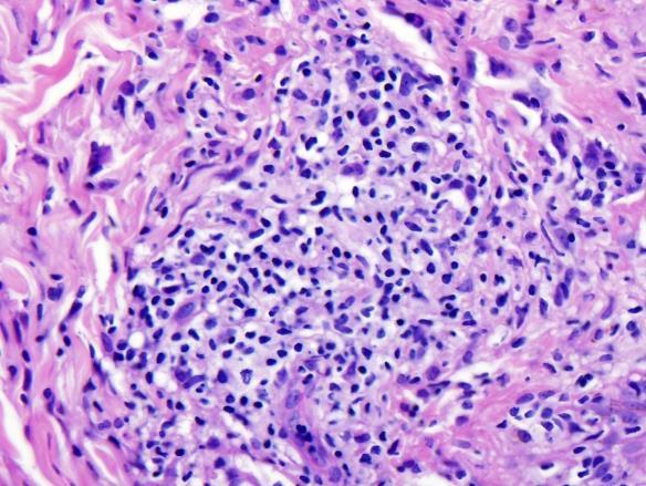
2 Secondary syphilis is protean Syphilis Often not suspected clinically Old men widowed and lonely Old women Sexual abuse Sometimes nursing home patients If you don t consider the diagnosis, you will miss it. Classic presentation Plasma Elongation of rete Endothelial swelling Scale/crust Lymphs w/ cytoplasm 2


3 Would you have ordered a T pallidum immunostain on this case? Untreated syphilis: Tabes Paresis Aortic dissection 3

4 4
5 5
 regardless of race, ethnicity, or socioeconomic status The great imitator\" Often not suspected clinically Atypical")
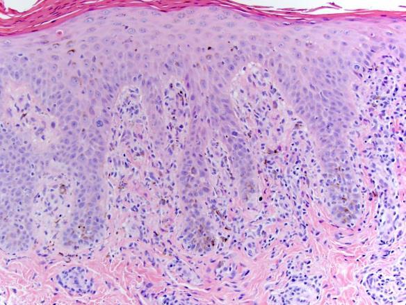
6 Incidence doubled since 2001 Incidence 5.3 cases in 100,000 Local outbreaks Majority in men who have sex with men (MSM) regardless of race, ethnicity, or socioeconomic status The great imitator" Often not suspected clinically Atypical clinical and false negative laboratory tests are more common in patients with human immunodeficiency virus (HIV) infection Prozone reactions Histology Wide spectrum, from interface dermatitis to granulomatous disease Detection rates of spirochetes Silver stain 33%-70% IHC 71% Review of 106 cases of syphilis Most common findings Interstitial inflammatory pattern Endothelial swelling Irregular acanthosis Elongated slender rete ridges Is it acceptable to miss 30% of your cases of syphilis? 6
7 Most common vs Most helpful The most helpful features are those that occur in cases with relatively few diagnostic features As few as 2 features could be present in some slides Review of 106 cases of syphilis No biopsy specimen demonstrated only a single feature... but as few as 2 or as many as 10 features could be present in a given slide Number of diagnostic features Overall Frequency Neutrophils in Stratum Corneum 34 (32.1%) Effacement 21 (19.8%) Total Cases 106 Number of Features Number of Cases (%) 1 0 (0%) 2 2 (1.9%) Irregular Acanthosis 82 (77.4%) Psoriasiform Acanthosis 3 (2.8%) Elongated Rete Ridges 80 (75.5%) Vacuolar Pattern (more holes than lymphs) 15 (14.2%) 3 5 (4.7%) 4 7 (6.6%) 5 15 (14.2%) 6 30 (28.3%) 7 28 (26.4%) 8 14 (13.2%) 9 4 (3.8%) 10 1 (1.0%) Vacuolar Pattern (equal holes and lymphs) 77 (72.6%) Lichenoid Pattern 25 (23.6%) Interstitial Inflammation 97 (91.5%) Plasma Cells 74 (69.8%) Lymphocytes with ample cytoplasm 58 (54.7%) Endothelial Swelling 93 (87.7%) Cases with 5 or fewer features Neutrophils in Stratum Corneum 2 (6.9%) Effacement 13 (44.8%) Irregular Acanthosis 7 (24.1%) Psoriasiform Acanthosis 2 (6.9%) Elongated Rete Ridges 8 (27.6%) Vacuolar Pattern (more holes than lymphs) 2 (6.9%) 4 or fewer features Neutrophils in Stratum Corneum 0 (0%) Effacement 6 (42.9%) Irregular Acanthosis 1 (7.1%) Psoriasiform Acanthosis 2 (14.3%) Elongated Rete Ridges 1 (7.1%) Vacuolar Pattern (more holes than lymphs) 2 (14.3%) Vacuolar Pattern (equal holes and lymphs) 15 (51.7%) Vacuolar Pattern (equal holes and lymphs) 4 (28.9%) Lichenoid Pattern 1 (3.4%) Interstitial Inflammation 22 (75.9%) Plasma Cells 14 (48.3%) Lymphocytes with ample cytoplasm 11 (37.9%) Endothelial Swelling 21 (72.4%) Lichenoid Pattern 1 (7.1%) Interstitial Inflammation 9 (64.3%) Plasma Cells 5 (35.7%) Lymphocytes with ample cytoplasm 4 (28.9%) Endothelial Swelling 8 (57.1%) 7
8 3 or fewer features Only 2 features Total Cases 7 Neutrophils in Stratum Corneum 0 (0%) Effacement 2 (28.6%) Irregular Acanthosis 1 (14.3%) Psoriasiform Acanthosis 0 (0%) Elongated Rete Ridges 0 (0%) Vacuolar Pattern (more holes than lymphs) 1 (14.3%) Total Cases 2 Neutrophils in Stratum Corneum 0 (0%) Effacement 0 (0%) Irregular Acanthosis 0 (0%) Psoriasiform Acanthosis 0 (0%) Elongated Rete Ridges 0 (0%) Vacuolar Pattern (more holes than lymphs) 0 (0%) Vacuolar Pattern (equal holes and lymphs) 1 (14.3%) Vacuolar Pattern (equal holes and lymphs) 0 (0%) Lichenoid Pattern 0 (0%) Interstitial Inflammation 5 (71.4%) Plasma Cells 3 (42.9%) Lymphocytes with ample cytoplasm 3 (42.9%) Endothelial Swelling 3 (42.9%) Lichenoid Pattern 0 (0%) Interstitial Inflammation 2 (100%) Plasma Cells 0 (0%) Lymphocytes with ample cytoplasm 1 (50%) Endothelial Swelling 1 (50%) Conclusions Few cases demonstrate all diagnostic features Most cases demonstrate between 5 and 7 diagnostic features (13.2%) 9 4 (3.8%) 10 1 (1.0%) Conclusions Interstitial inflammatory pattern, endothelial swelling, irregular acanthosis and elongated slender rete ridges were the most common findings overall Some more likely to be seen in specimens that demonstrated many findings, so their independent value may be overestimated. Conclusions More important is the likelihood that a given finding will be present in a slide with 5 or fewer diagnostic features these are the cases most prone to misdiagnosis. Most common Intersitial inflammatory pattern ( busy dermis ) Endothelial swelling Conclusions Other helpful findings vacuolar interface dermatitis with a lymphocyte in nearly every vacuole lymphocytes with visible cytoplasm Cases with fewer findings were more likely to demonstrate effacement of the rete pattern, making them more prone to misdiagnosis. 8
9 Take-Home Points Most cases have 5-7 features Many less frequent in cases with 5 features endothelial swelling (87.7% overall vs 72.4% of cases with 5 features) plasma cells (69.8% vs 48.3%) elongated rete ridges (75.5% vs 27.6%) Take-Home Points Specimens with 5 features more likely to be from the trunk (61.1% of cases with 5 features vs 34.4% overall) demonstrate rete ridge effacement (44.8% vs 19.8%) have pityriasis rosea (33.3% vs 17.2%) or drug eruption (33.3% vs 10.9%) in the clinical differential Take-Home Points An interstitial inflammatory pattern was the most common characteristic of specimens with 5 features (present in 75.9%). Cases with only two diagnostic features demonstrated an interstitial pattern with either endothelial swelling or lymphocytes with ample cytoplasm. Next step Determine positive and negative predictive values of each feature when syphilis is compared with other forms of interface dermatitis. Some, like a lymph in every hole pattern will help you suspect syphilis but discriminate poorly from conditions like PLC How Do We Use Diagnostic Criteria? Our diagnoses should be based on reproducible criteria Criteria Our diagnoses should be based on reproducible criteria How rigorously have our criteria been validated? 9
, renal impairment, hepatitis B infection, abnormal arteriogram, biopsy of an artery with neutrophilic inflammation 3 criteria give >82% sensitivity, >86% specificity Major and minor")
10 Polyarteritis nodosa American College of Rheumatology Criteria Weight loss >4 kg, livedo reticularis, testicular pain or tenderness, myalgia / myopathy or muscle tenderness, neuropathy, hypertension (diastolic >90), renal impairment, hepatitis B infection, abnormal arteriogram, biopsy of an artery with neutrophilic inflammation 3 criteria give >82% sensitivity, >86% specificity Major and minor criteria Give weight to individual criteria Polyarteritis nodosa American College of Rheumatology Criteria Weight loss >4 kg, livedo reticularis, testicular pain or tenderness, myalgia / myopathy or muscle tenderness, neuropathy, hypertension (diastolic >90), renal impairment, hepatitis B infection, abnormal arteriogram, biopsy of an artery with neutrophilic inflammation 3 criteria give >82% sensitivity, >86% specificity Major and minor criteria Give weight to individual criteria Dermatopathology KA vs SCC Alopecia Areata Melanoma Neoplastic vs reactive squamous atypia Keratoacanthoma Clinicopathologic correlation Rapid growth followed by termination differentiation and involution Histological Diagnosis KA vs SCC Does it matter? History of rapid growth Crateriform Glassy Neutrophilic microabcesses Eosinophilic infiltrate Central hypergranulosis Lack of acantholysis Elastic trapping 10


 Shah K, Kazlouskaya V, Lal K, et al.")
 (95%CI)")
 Lesions with elastic trapping are twice as likely to behave like KA")
11 Scheduled for amputation Eruptive but not crateriform No neutrophilic microabscesses No eosinophils Glassy Lacked acantholysis Elastic trapping Treated with IL MTX Images courtesy of Omar Noor, MD Elastic trapping Does Elastic Trapping Discriminate? Incidence 71% in KA 37% in SCC (p<0.0001) 5.9% in Hypertrophic LE (p<0.0001) Shah K, Kazlouskaya V, Lal K, et al. J Cutan Pathol 2014; 41: Does Elastic Trapping Discriminate? Alopecia areata Compared entities Sensitivity Specificity LR (+) (95%CI) LR(-) (95%CI) KA vs. SCC ( ) 0.5 ( ) Lesions with elastic trapping are twice as likely to behave like KA Good but not perfect Use in concert with other findings Shah K et al. J Cutan Pathol 2014; 41:


 Miniaturization of follicles 15/17 (88%) Melanin in fibrous tracts 12/17 (71%)")
12 What Critera Discriminate Dilated infundibulum Lymphocytes in fibrous tracts 94% Catagen follicles 93% Miniaturized follicles 90% Peribulbar lymphocytic infiltrate 84% Melanin in fibrous tracts 84% Eosinophils in fibrous tracts 44% Pigment casts 44% Peckham SJ, et al. J Am Acad Dermatol. 2011; 65: Histologic features present in cases without a peribulbar infiltrate Peckham SJ, et al. J Am Acad Dermatol. 2011; 65: HISTOLOGIC FEATURE Catagen/telogen follicles 15/17 (88%) NUMBER OF CASES (PERCENT) Miniaturization of follicles 15/17 (88%) Melanin in fibrous tracts 12/17 (71%) Pigment casts 10/17 (59%) Lymphocytes in fibrous tracts 7/17 (41%) Eosinophils in fibrous tracts or around bulbs 3/17 (18%) Trichotillomania 12
 Pigment casts Shared Lymphocytes in fibrous tracts Eosinophils in fibrous tracts or around bulbs")
13 Trichotillomania shares Alopecia Areata HISTOLOGIC FEATURE Catagen/telogen follicles Shared HISTOLOGIC FEATURE NUMBER OF CASES (PERCENT) Miniaturization of follicles Can be difficult to judge when many catagen hairs are present Melanin in fibrous tracts Melanin in fibrous tracts 12/17 (71%) Pigment casts Shared Lymphocytes in fibrous tracts Eosinophils in fibrous tracts or around bulbs Lymphocytes in fibrous tracts 7/17 (41%) Eosinophils in fibrous tracts or around bulbs 3/17 (18%) Melanoma Miniaturized dystrophic hairs: 4% but highly specific Absolute criteria are lacking in many cases Ron Rapini has said he sometimes feels like the weatherman 80% chance of rain today Melanoma Absolute criteria are lacking in many cases Ron Rapini has said he sometimes feels like the weatherman 80% chance of melanoma today Melanoma We can improve our diagnostic criteria by determining which discriminate best alone or in groups Compare with a gold standard Global assessment CGH/FISH Outcome 13


14 Computer Diagnoses Computer-aided diagnoses Based on diagnostic algorithms and weighted criteria Has been tried in clinical medicine and dermatopathology Computer Diagnoses Computer-aided diagnoses Based on diagnostic algorithms and weighted criteria Has been tried in clinical medicine and dermatopathology Not bad overall, but none of us will have to look for another line of work anytime soon Pagetoid Spitz in a child Pagetoid Intraepidermal Spitz Nevus Pagetoid scatter of epithelioid melanocytes Small Sharply circumscribed End laterally in a nest 14
 Parallel theque pattern (trabecular at base) Lack HMB-45 gradient Deep mitoses Atypical mitoses MIB + cells deep, >10%")
15 Sunburned nevus Pagetoid Scatter Melanoma Spitz Pigmented Spindle Cell Nevus Acral Nevus Trauma Sunburn Nevoid Melanoma nasal tip Helpful diagnostic features Lack of maturation and dispersion (especially spitzoid melanomas in children bulbous outline like a blue nevus) Parallel theque pattern (trabecular at base) Lack HMB-45 gradient Deep mitoses Atypical mitoses MIB + cells deep, >10% overall, > 5% deep Displace elastic Nevoid Melanoma: Parallel theque pattern 15


16 VVG - elastic As reported by Kamino et al Benign nevus MIB-1 / Ki-67 16


 Simple Techniques you may not be using Acral sites: Hyperchromatic, angulated nuclei")

17 Architecture vs Cytology Spitz We focus mostly on architecture at scan But cytology should not be ignored Melanoma: Irregular nuclear contour Clumped chromatin Irregular/stellate nucleoli Multiple/peripheral nucleoli Cytological Criteria for Melanoma Logistic regression multivariate analysis Independent diagnostic value of each feature Features found to be independently useful Pleomorphism with enlarged nuclei Notching of the nuclear envelope Mitotic figures Peppered moth chromatin pattern 1)How We make diagnoses 2) Simple Techniques you may not be using Acral sites: Hyperchromatic, angulated nuclei Irregular dendrites Dirk M. Elston, MD Medical University of South Carolina Research team: Alex Flamm, EunJi Kwon, Qiang Xie, Nathan Cleaver, Khan Thieu, Jeff Shackleton, Filamer Kabigting, Amira Elbendary, Kara Melissa Torres, Manuel Valdebran, 17


18 Bowen s Squamous Atypia Irregular nuclear contour Nuclear crowding High N/C ratio Enlarged prominent nucleoli Hyerchromasia Squamous Atypia Atypical mitotic figures Rarely seen, but highly specific Malhotra S, Kazlouskaya V, Andres C, et al. J Cutan Pathol Apr;40(4):371-8 Not Statistically Significant Multiple nucleoli Clumped chromatin Number of mitotic figures At the End of the Day Dermatopathology will remain an art Malhotra S, Kazlouskaya V, Andres C, et al. J Cutan Pathol Apr;40(4):
19 At the End of the Day Dermatopathology will remain an art Weighing histological features in context will remain a complex skill set At the End of the Day Dermatopathology will remain an art Weighing histological features in context will remain a complex skill set Skills can be learned Validated objective criteria can improve our accuracy Challenge Deconstruct each set of diagnostic criteria in dermatopathology and test the positive predictive value of individual criteria alone and in groups Challenge Deconstruct each set of diagnostic criteria in dermatopathology and test the positive predictive value of individual criteria alone and in groups We re really good, but we can always be a bit better better 19
Benign and malignant epithelial lesions: Seborrheic keratosis: A common benign pigmented epidermal tumor occur in middle-aged or older persons more
 Benign and malignant epithelial lesions: Seborrheic keratosis: A common benign pigmented epidermal tumor occur in middle-aged or older persons more common on the trunk; but extremities, head and neck are
Histopathology: skin pathology
 Histopathology: skin pathology These presentations are to help you identify, and to test yourself on identifying, basic histopathological features. They do not contain the additional factual information
Desmoplastic Melanoma R/O BCC. Clinical Information. 74 y.o. man with lesion on left side of neck r/o BCC
 R/O BCC Sabine Kohler, M.D. Professor of Pathology and Dermatology Dermatopathology Service Stanford University School of Medicine Clinical Information 74 y.o. man with lesion on left side of neck r/o
Melanocytic Lesions: Use of Immunohistochemistry and Special Studies Napa Valley 2018
 Melanocytic Lesions: Use of Immunohistochemistry and Special Studies Napa Valley 2018 Victor G. Prieto, MD, PhD Professor Depts. of Pathology and Dermatology University of Texas - MD Anderson Cancer Center
Basal cell carcinoma 5/28/2011
 Goal of this Presentation A practical approach to the diagnosis of cutaneous carcinomas and their mimics Thaddeus Mully, MD University of California San Francisco To review common non-melanoma skin cancers
Dermatopathology. Dr. Rafael Botella Estrada. Hospital La Fe de Valencia
 Dermatopathology Dr. Rafael Botella Estrada. Hospital La Fe de Valencia Melanoma and mimics Dr. Martin Mihm Malignant lesions result from the accumulation of mutations Class I lesions (benign) Class II
Histopathology of Melanoma
 THE YALE JOURNAL OF BIOLOGY AND MEDICINE 48, 409-416 (1975) Histopathology of Melanoma G. J. WALKER SMITH Department ofpathology, Yale University School ofmedicine, 333 Cedar Street, New Haven, Connecticut
Clinicopathologic Self-Assessment
 Clinicopathologic Self-Assessment Handout Symposium (S003), July 27 th 2017 Maija Kiuru MD PhD Assistant Professor, Departments of Dermatology & Pathology University of California Davis CASE 1: History
David B. Troxel, MD. Common Medicolegal Situations: Misdiagnosis of Melanoma
 Common Medicolegal Situations: Misdiagnosis of Melanoma David B. Troxel, MD Medical Director, The Doctors Company, Napa, California Clinical Professor Emeritus, University of California at Berkeley Past
Malignant tumors of melanocytes: Part 1. Deba P Sarma, MD., Omaha
 Malignant tumors of melanocytes: Part 1 Deba P Sarma, MD., Omaha The melanocytic tumor is one of the most difficult and confusing areas in Dematopathology. It is true that most (95%) of such lesions are
Female 18. Deeply pigmented lesion on trunk.?warty naevus?seborrhoeic keratosis?malignant melanoma. The best diagnosis is:
 Female 18. Deeply pigmented lesion on trunk.?warty naevus?seborrhoeic keratosis?malignant melanoma. The best diagnosis is: A. deep penetrating naevus B. naevoid malignant melanoma C. pigment synthesising
Case 26 Male 37. Right jawline 5mm nodule?keloid. The best diagnosis is:
 Case 26 Male 37. Right jawline 5mm nodule?keloid. The best diagnosis is: A. Desmoplastic Spitz naevus B. Atypical Spitz Tumour C. Spitzoid melanoma D. Deep penetrating naevus E. Spitz naevus Case 26: M
Associate Clinical Professor of Dermatology MUSC
 Re-excision of Moderately Dysplastic Nevi: Should we or shouldn t we? John C. Maize, Jr, M.D. Dermatologist and Dermatopathologist Trident Dermatology, Charleston SC Associate Clinical Professor of Dermatology
Tips on getting the most from your alopecia pathology reports. D irector, H a ir C linic, Boston Medical C e n ter
 Tips on getting the most from your alopecia pathology reports Lynne J. Goldberg, MD J a g Bhawan Professor o f Dermatology a n d Pathology & Laboratory Medicine Boston U n iversity School of Medicine D
Conflict of Interest 9/2/2014. Pathogenesis and Comparison of Atypical Spitz Nevi vs Benign Spitz, and Childhood Melanoma
 Pathogenesis and Comparison of Atypical Spitz Nevi vs Benign Spitz, and Childhood Melanoma Martin C. Mihm Jr., M.D., F.A.C.P. Harvard Medical School Brigham and Women s Hospital Dana Farber Cancer Center
Simulators of melanoma
 Simulators of melanoma Philip E. LeBoit, M.D. Depts. of Pathology and Dermatology University of California, San Francisco Simulators of melanoma Simulators of melanoma in situ Melanocytic Non-melanocytic
Pathology of the skin. 2nd Department of Pathology, Semmelweis University
 Pathology of the skin 2nd Department of Pathology, Semmelweis University Histology of the skin Epidermis: Stratum corneum Stratum granulosum Stratum spinosum Stratum basale Dermis: papillary and reticular
21/07/2017. Hobnail endothelial cells are not the same as epithelioid endothelial cells
 UPDATE IN CUTANEOUS VASCULAR S DERMATOPATHOLOGY SESSION BELFAST PATHOLOGY JUNE 21/2017 Dr E Calonje St John s Institute of Dermatology, London, United Kingdom THE FAMILY OF VASCULAR S WITH EPITHELIOID
The Enigmatic Spitz Lesion
 The Enigmatic Spitz Lesion The Dawn of Spitz S Spitz Sophie Spitz Melanomas of Childhood ; Am J Pathol 1948 1910-1956 13 children (18 mo - 12 yrs) 12/13 had a benign clinical course Sophie Spitz Born 1910
Spitz nevi in the classic histopathological pattern - lamb in wolf`s clothing *
 DERMATOPATHOLOGY 91 Spitz nevi in the classic histopathological pattern - lamb in wolf`s clothing * Gustavo Costa Verardino 1 Mayra Carrijo Rochael 1 DOI: http://dx.doi.org/10.1590/abd1806-4841.20153310
Lichenoid Tissue Reaction in Malignant Melanoma A Potential Diagnostic Pitfall
 natomic Pathology / LICHENOID TISSUE RECTION IN MLIGNNT MELNOM Lichenoid Tissue Reaction in Malignant Melanoma Potential Diagnostic Pitfall CPT Scott R. Dalton, MC, US, 1,3 Capt Matt. aptista, USF, MC,
Keratinocyte tumors. Actinic Keratosis. Squamous cell carcinoma in situ. Squamous Cell Carcinoma. (aka Bowen s disease)
") Actinic Keratosis Keratinocyte tumors Prepared by Kurt Schaberg Precancerous, risk of malignancy ~8-20% per year (progresses to SCC); Due to chronic sun exposure Rough scaly plaque; typically due to sun
Actinic Keratosis Keratinocyte tumors Prepared by Kurt Schaberg Precancerous, risk of malignancy ~8-20% per year (progresses to SCC); Due to chronic sun exposure Rough scaly plaque; typically due to sun
, , 2011 HODGKIN LYMPHOMA
 European Federation of Cytology Societies 4tu Annual Tutorial in Cytopathology Trieste, June 6-10, 2011 HODGKIN LYMPHOMA Classification The World Health Organization Classification of Lymphomas (2001)
Whitney A. High, MD, JD, MEng
 ADS Dermatopathology Meeting 2014 Selected Adnexal Tumors Whitney A. High, MD, JD, MEng Associate Professor, Dermatology & Pathology Director of Dermatopathology (Dermatology) University of Colorado School
Gastrooesophageal reflux disease. Jera Jeruc Institute of pathology, Faculty of Medicine, Ljubljana, Slovenia
 Gastrooesophageal reflux disease Jera Jeruc Institute of pathology, Faculty of Medicine, Ljubljana, Slovenia Reflux esophagitis (RE) GERD: a spectrum of clinical conditions and histologic alterations resulting
1/10/2018. Soft Tissue Tumors Showing Melanocytic Differentiation. Overview. Desmoplastic/ Spindle Cell Melanoma
 2016 MFMER slide-1 2016 MFMER slide-2 2016 MFMER slide-3 Soft Tissue Tumors Showing Melanocytic Differentiation Andrew L. Folpe, M.D. Professor of Laboratory Medicine and Pathology Mayo Clinic, Rochester,
المركب النموذج--- سبيتز وحمة = Type Spitz's Nevus, Compound SPITZ NEVUS 1 / 7
 SPITZ NEVUS 1 / 7 Epidemiology An annual incidence rate of 1.4 cases of Spitz nevus per 100,000 individuals has been estimated in Australia, compared with 25.4 per 100,000 individuals for cutaneous melanoma
Objectives. Atypical Glandular Cells. Atypical Endocervical Cells. Reactive Endocervical Cells
 2013 California Society of Pathologists 66 th Annual Meeting San Francisco, CA Atypical Glandular Cells to Early Invasive Adenocarcinoma: Cervical Cytology and Histology Christina S. Kong, MD Associate
MECHANISMS OF HUMAN DISEASE: LABORATORY SESSION PATHOLOGY OF THE SKIN LAB. Friday, February 12, :30 am 11:00 am
 MECHANISMS OF HUMAN DISEASE: LABORATORY SESSION PATHOLOGY OF THE SKIN LAB Friday, February 12, 2012 9:30 am 11:00 am FACULTY COPY GOALS: Describe the basic clinical and morphologic features of various
Special slide seminar
 Special slide seminar Tomáš Rozkoš The Fingerland Department of Pathology Charles University Medical Faculty and Faculty Hospital in Hradec Králové Czech Republic Case history, 33 years old resistance
EARLY ONLINE RELEASE
 EARLY ONLINE RELEASE Note: This article was posted on the Archives Web site as an Early Online Release. Early Online Release articles have been peer reviewed, copyedited, and reviewed by the authors. Additional
F006 Imaging in Dermatology Melanocytic Neoplasia Clinical-Confocal-Pathological-Correlations
 F006 Imaging in Dermatology Melanocytic Neoplasia Clinical-Confocal-Pathological-Correlations Melissa Gill, MD SkinMedical Research and Diagnostics Dobbs Ferry, NY, USA Department of Pathology SUNY Downstate
Chapter 6 Squamous Cell Carcinoma: Variants and Challenges
 Chapter 6 Squamous Cell Carcinoma: Variants and Challenges Michael B. Morgan EPIDEMIOLOGY: Second most common skin cancer, rare in the dark-skinned races. ETIOLOGY: Ultraviolet light, HPV infection. PATHOGENESIS:
A PRACTICAL APPROACH TO ATYPICAL MELANOCYTIC LESIONS BIJAN HAGHIGHI M.D, DIRECTOR OF DERMATOPATHOLOGY, ST. JOSEPH HOSPITAL
 A PRACTICAL APPROACH TO ATYPICAL MELANOCYTIC LESIONS BIJAN HAGHIGHI M.D, DIRECTOR OF DERMATOPATHOLOGY, ST. JOSEPH HOSPITAL OBJECTIVES Discuss current trends and changing concepts in our understanding of
Clinicopathologic Self- Assessment S003 AAD 2017
 Clinicopathologic Self- Assessment S003 AAD 2017 Clay J. Cockerell, M.D. Director, Cockerell Dermatopathology Director, Division of Dermatopathology UT Southwestern Medical Center July 2017 No relevant
Prepared By Jocelyn Palao and Layla Faqih
 Prepared By Jocelyn Palao and Layla Faqih The structure of the suspected atypical cell should always be compared to the structure of other similar, benign, cells which are present in the smears. The diagnosis
04/09/2018. Squamous Cell Neoplasia and Precursor Lesions. Agenda. Squamous Dysplasia. Squamo-proliferative lesions. Architectural features
 Squamous Cell Neoplasia and Precursor Lesions Jennifer L. Hunt, MD, MEd Aubrey J. Hough Jr, MD, Endowed Professor of Pathology Chair of Pathology and Laboratory Medicine University of Arkansas for Medical
Disorders of Cell Growth & Neoplasia. Histopathology Lab
 Disorders of Cell Growth & Neoplasia Histopathology Lab Paul Hanna April 2010 Case #84 Clinical History: 5 yr-old, West Highland White terrier. skin mass from axillary region. has been present for the
Metastatic balloon cell malignant melanoma: a case report and literature review
 Int J Clin Exp Pathol 2011;4(1):315-321 www.ijcep.com /IJCEP1102002 Metastatic balloon cell malignant melanoma: a case report and literature review Lili Lee 1*, Fang Zhou 1*, Anthony Simms 1, Rosemary
S everal morphological features are frequently used in the
 1194 ORIGINAL ARTICLE Interobserver reproducibility of histological features in cutaneous malignant melanoma C Urso, F Rongioletti, D Innocenzi, C Saieva, D Batolo, S Chimenti, R Filotico, R Gianotti,
Management of pediatric melanocytic lesions
 Open Journal of Clinical & Medical Case Reports Management of pediatric melanocytic lesions Volume 3 (2017) Issue 8 ISSN 2379-1039 Jin Kim, BS; Emmanuel Gabriel MD, PhD; Weiguo Liu MD, PhD; Lin Lin MD,
Critical Analysis of Histologic Criteria for Grading Atypical (Dysplastic) Melanocytic Nevi
 Melanocytic Nevi") Anatomic Pathology / GRADING ATYPICAL MELANOCYTIC NEVI Critical Analysis of Histologic Criteria for Grading Atypical (Dysplastic) Melanocytic Nevi Lucia Pozo, MD, Mahmoud Naase, PhD, Rino Cerio, MD,, Alfredo
Anatomic Pathology / GRADING ATYPICAL MELANOCYTIC NEVI Critical Analysis of Histologic Criteria for Grading Atypical (Dysplastic) Melanocytic Nevi Lucia Pozo, MD, Mahmoud Naase, PhD, Rino Cerio, MD,, Alfredo
Update on Spitzoid and Blue nevus-like melanocytic lesions Emphasis on molecular studies informing diagnosis, prognosis and therapy
 Update on Spitzoid and Blue nevus-like melanocytic lesions Emphasis on molecular studies informing diagnosis, prognosis and therapy Michael T. Tetzlaff MD, PhD Associate Professor Department of Pathology,
K Blessing, J J H Grant, D S A Sanders, M M Kennedy, A Husain, P Coburn
 J Clin Pathol 2000;53:591 595 591 Papers Pathology, Aberdeen University, Foresterhill, Aberdeen AB25 2ZD, K Blessing Pathology, Birmingham University, Birmingham B15 2TT, D S A Sanders Pathology, Heartlands
Guy Perrot (Ги Перро)
") НАУЧНО-ПРАКТИЧЕСКАЯ КОНФЕРЕНЦИЯ (МАСТЕР-КЛАСС) «ПРАКТИЧЕСКИЕ АСПЕКТЫ ДИАГНОСТИКИ И ЛЕЧЕНИЯ МЕЛАНОМЫ КОЖИ» DIAGNOSTIC AND PITFALLS IN MELANOMA Guy Perrot (Ги Перро) MD PHD pathologist, University Hospital
НАУЧНО-ПРАКТИЧЕСКАЯ КОНФЕРЕНЦИЯ (МАСТЕР-КЛАСС) «ПРАКТИЧЕСКИЕ АСПЕКТЫ ДИАГНОСТИКИ И ЛЕЧЕНИЯ МЕЛАНОМЫ КОЖИ» DIAGNOSTIC AND PITFALLS IN MELANOMA Guy Perrot (Ги Перро) MD PHD pathologist, University Hospital
The Relevance of Cytologic Atypia in Cutaneous Neural Tumors
 The Relevance of Cytologic Atypia in Cutaneous Neural Tumors Recent Findings - New Developments New Problems Zsolt B. Argenyi, M.D. Professor of Pathology & Dermatology Director of Dermatopathology Department
Diagnostic problems in uterine smooth muscle tumors
 Diagnostic problems in uterine smooth muscle tumors Marina Kos Ljudevit Jurak Clinical Department of Pathology, Clinical Hospital Center Sestre milosrdnice, Zagreb Institute of Pathology, University of
ARTICLE INFO ABSTRACT
 Melanocytic Pigmentation: A Single Manifestation of Myriad of Pathologies [PP: 05-09] Dr. Swapna Honwad Department of Oral Pathology dr.swapnahonwad@gmail.com Dr. Elsy P. Simon Department of Endodontics
Important Decisions in Dermatopathology: The Clinico- Pathologic Correlation. Dermatopathology Specialists Needed. Changing Trends
 Important Decisions in Dermatopathology: The Clinico- Pathologic Correlation Uma Sundram, MD, PhD Departments of Pathology and Dermatology Stanford University May 29, 2008 Dermatopathology Specialists
How to decipher a pathology report for alopecia
 How to decipher a pathology report for alopecia DISCLOSURE OF RELATIONSHIPS WITH INDUSTRY Lynne J. Goldberg, MD S063-Hair Disorders Made Easier DISCLOSURES I do not have any relationships with industry
Ways to get into trouble, ideas on avoiding trouble, and diagnostic approaches to keep trouble at bay
 Pitfalls in the diagnosis of melanocytic tumors Timothy McCalmont, MD University of California, San Francisco Ways to get into trouble, ideas on avoiding trouble, and diagnostic approaches to keep trouble
HISTOPATHOLOGY. Shannon Martinson
 HISTOPATHOLOGY Shannon Martinson March 2013 Case #1 History: 8 year old beagle Neck pain for the past couple of weeks Paresis, followed by paralysis developed over the past few days Gross Description courtesy
Malignant tumors of melanocytes : Part 3. Deba P Sarma, MD., Omaha
 Malignant tumors of melanocytes : Part 3 Deba P Sarma, MD., Omaha Let s go over one case of melanoma using the following worksheet. Of the various essential information that needs to be included in the
A cquired melanocytic naevi usually appear in the 1st
 459 ORIGINAL ARTICLE Histopathological characteristics of small diameter melanocytic naevi M Braun-Falco, R Hein, J Ring, N S McNutt... See end of article for authors affiliations... Correspondence to:
MECHANISMS OF HUMAN DISEASE: LABORATORY SESSION PATHOLOGY OF THE SKIN LAB. Friday, February 13, :30 am 11:00 am
 MECHANISMS OF HUMAN DISEASE: LABORATORY SESSION PATHOLOGY OF THE SKIN LAB Friday, February 13, 2009 9:30 am 11:00 am FACULTY COPY GOALS: Describe the basic clinical and morphologic features of various
Dermoscopy: Recognizing Top Five Common In- Office Diagnoses
 Dermoscopy: Recognizing Top Five Common In- Office Diagnoses Vu A. Ngo, DO Department of Family Medicine and Dermatology Choctaw Nation Health Services Authority Learning Objectives Introduction to dermoscopy
Respiratory Tract Cytology
 Respiratory Tract Cytology 40 th European Congress of Cytology Liverpool, UK Momin T. Siddiqui M.D. Professor of Pathology and Laboratory Medicine Director of Cytopathology Emory University Hospital, Atlanta,
Histopathology: Cell necrosis and cytoplasmic accumulations
 Histopathology: Cell necrosis and cytoplasmic accumulations These presentations are to help you identify basic histopathological features. They do not contain the additional factual information that you
Diagnostic difficulties with lesions of the oral mucosa
 BDIAP London, November 2010 School of Clinical Dentistry University of Sheffield Diagnostic difficulties with lesions of the oral mucosa Paul M Speight Dept Oral & Maxillofacial Pathology University of
Maligna Melanoma and Atypical Fibroxanthoma: An Unusual Collision Tumour G Türkcü 1, A Keleş 1, U Alabalık 1, D Uçmak 2, H Büyükbayram 1 ABSTRACT
 Maligna Melanoma and Atypical Fibroxanthoma: An Unusual Collision Tumour G Türkcü 1, A Keleş 1, U Alabalık 1, D Uçmak 2, H Büyükbayram 1 ABSTRACT Two different neoplasia in the same biopsy material called
Among the benign intraepithelial melanocytic proliferations, Inflamed Conjunctival Nevi. Histopathological Criteria. Resident Short Reviews
 Resident Short Reviews Inflamed conjunctival nevi (ICN) may suggest malignancy because of their rapid growth and atypical histology. The objective of this study was to characterize the diagnostic features
Melanoma Update: 8th Edition of AJCC Staging System
 Melanoma Update: 8th Edition of AJCC Staging System Rosalie Elenitsas, M.D. Professor of Dermatology Director, Dermatopathology University of Pennsylvania DISCLOSURE OF RELATIONSHIPS WITH INDUSTRY None
Case Report A Rare Cutaneous Adnexal Tumor: Malignant Proliferating Trichilemmal Tumor
 Case Reports in Medicine Volume 2015, Article ID 742920, 4 pages http://dx.doi.org/10.1155/2015/742920 Case Report A Rare Cutaneous Adnexal Tumor: Malignant Proliferating Trichilemmal Tumor Omer Alici,
Cellular Neurothekeoma
 Cellular Neurothekeoma Scott W Binder, MD Pritzker Professor of Pathology & Dermatology Sr. Vice Chair Director, Pathology Clinical Services Chief, Dermatopathology Geffen/UCLA School of Medicine Clinical
Almost any suspected tumor can be aspirated easily and safely. Some masses are more risky to aspirate including:
 DOES THIS PATIENT HAVE CANCER? USING IN-HOUSE CYTOLOGY TO HELP YOU MAKE THIS DIAGNOSIS. Joyce Obradovich, DVM, Diplomate, ACVIM (Oncology) Animal Cancer & Imaging Center, Canton, Michigan Almost every
Pathological Classification of Hepatocellular Carcinoma
 3 rd APASL Single Topic Conference: HCC in 3D Pathological Classification of Hepatocellular Carcinoma Glenda Lyn Y. Pua, M.D. HCC Primary liver cancer is the 2 nd most common cancer in Asia HCC is the
Review of the AP Part II Practical Examination. Dr David Clift Co Chief Examiner
 Review of the AP Part II Practical Examination Dr David Clift Co Chief Examiner General Remarks The part II practical examination involved 15 cases which were presented with sufficient clinical data to
Squamous Cell Neoplasia and Precursor Lesions
 Squamous Cell Neoplasia and Precursor Lesions Jennifer L. Hunt, MD, MEd Aubrey J. Hough Jr, MD, Endowed Professor of Pathology Chair of Pathology and Laboratory Medicine University of Arkansas for Medical
Dermatopathology Workshop Summary, Berlin 2004
 Dermatopathology Workshop Summary, Berlin 2004 David A. Whiting and Rolf Hoffmannw Baylor Hair Research and Treatment Center, Dallas, Texas, USA; wdermatology Practice, Freiburg, Germany Figure 1 Case
Case year female. Routine Pap smear
 Case 1 57 year female Routine Pap smear Diagnosis? 1. Atypical glandular cells of unknown significance (AGUS) 2. Endocervical AIS 3. Endocervical adenocarcinoma 4. Endometrial adenocarcinoma 5. Adenocarcinoma
Blue Melanocytic Proliferations
 Blue Melanocytic Proliferations Labib R. Zakka M.D., M.A. Research Fellow Melanoma Program Department of Dermatology Brigham and Women s Hospital Harvard Medical School Conflicts of Interest No conflicts
Abrupt Intralesional Color Change on Dermoscopy as a New Indicator of Early Superficial Spreading Melanoma in a Japanese Woman
 Published online: June 24, 2015 1662 6567/15/0072 0123$39.50/0 This is an Open Access article licensed under the terms of the Creative Commons Attribution-NonCommercial 3.0 Unported license (CC BY-NC)
When Immunostains Can Get You in Trouble: Gynecologic Pathology p16: Panacea or Pandora s Box?
 When Immunostains Can Get You in Trouble: Gynecologic Pathology p16: Panacea or Pandora s Box? Teri A. Longacre, MD Stanford Medicine Stanford California pi6 in Gynecologic Pathology: Panacea or Pandora
Myxo-inflammatory Fibroblastic sarcoma
 AKA Myxo-inflammatory Fibroblastic sarcoma Acral Myxoinflammatory fibroblastic sarcomaam.j.surg.path1998; 22; 911-924 Inflammatory myxoid tumour of soft parts with bizarre giant cells [Pathol.Res.Pract.
There is NO single Melanoma Stain. > 6000 Mutations in Melanoma. What else can be done to discriminate atypical nevi from melanoma?
 Las Vegas Fall Clinical 2016: The Assessment and Diagnosis of Melanoma Whitney A. High, MD, JD, MEng Associate Professor, Dermatology & Pathology Director of Dermatopathology (Dermatology) University of
Atypical Histologic Features in Melanocytic Nevi
 The American Journal of Dermatopathology 22(5): 391 396, 2000 2000 Lippincott Williams & Wilkins, Inc., Philadelphia Atypical Histologic Features in Melanocytic Nevi Carmelo Urso, M.D. The atypical histologic
أملس عضلي غرن = Leiomyosarcoma. Leiomyosarcoma 1 / 5
 Leiomyosarcoma 1 / 5 EPIDEMIOLOGY Exact incidence is unknown, but older studies suggest that leiomyosarcomas comprise approximately 3 percent of soft-tissue sarcomas. Superficial leiomyosarcoma occurs
Pathology of the skin. Dr Fónyad László, 1sz. Patológiai és Kísérleti Rákkutató Intézet, SE
 Pathology of the skin Dr Fónyad László, 1sz. Patológiai és Kísérleti Rákkutató Intézet, SE The skin Biggest organ Kb. 1.8 nm Kb. 10 kg Most frequent site for tumor development (BCC) Pathology of the skin
Diagnostic Cytology of Cancer Cases
 Diagnostic Cytology of Cancer Cases Somporn Techangamsuwan Companion Animal Cancer Research Unit (CAC-RU) Department of Pathology, Faculty of Veterinary Science, Chulalongkorn University 1 Tumor or Non-tumor
IT S FUNDAMENTAL MY DEAR WATSON! A SHERLOCKIAN APPROACH TO DERMATOLOGY
 IT S FUNDAMENTAL MY DEAR WATSON! A SHERLOCKIAN APPROACH TO DERMATOLOGY Skin, Bones, and other Private Parts Symposium Dermatology Lectures by Debra Shelby, PhD, DNP, FNP-BC, FADNP, FAANP Debra Shelby,
LGM International, Inc.
 Liqui-PREP TM Cytology Atlas Preface The following pictures are examples with descriptions of cytology slides processed with the Liqui-PREP TM System.. The descriptions are reviewed by Pathologists. It
Case 231: F7. Exophytic naevus over left trapezious. Grown over a few weeks. Iniitally flat.?spitz naevus,?malignant
 Case 231: F7. Exophytic naevus over left trapezious. Grown over a few weeks. Iniitally flat.?spitz naevus,?malignant Dermoscopy: coarse vascular structures. c/o A, B, C RAC7750 Case 231: F7. Exophytic
Benign Lichenoid Keratosis
 Benign Lichenoid Keratosis ALAN F. FRIGY, M.D. AND PHILIP H. COOPER, M.D. The microscopic spectrum of benign lichenoid keratosis (BLK) was studied by examination of 30 examples. BLK consists of a segment
Papillary Lesions of the breast
 Papillary Lesions of the breast Emad Rakha Professor of Breast Pathology The University of Nottingham Papillary lesions of the breast are a heterogeneous group of disease, which are characterised by neoplastic
Hyperchromatic Crowded Groups: What is Your Diagnosis? Session 3000
 Hyperchromatic Crowded Groups: What is Your Diagnosis? Session 3000 Thomas A. Bonfiglio, M.D. Professor Emeritus, Pathology and Laboratory Medicine University of Rochester Disclosures In the past 12 months,
Brief Report. Shivanand Gundalli 1, Smita Kadadavar 1, Somil Singhania 1, Rutuja Kolekar 2 INTRODUCTION. Melanocytic Nevus
 Our Dermatology Online Histopathological spectrum of benign melanocytic nevi our experience in a tertiary care centre Shivanand Gundalli 1, Smita Kadadavar 1, Somil Singhania 1, Rutuja Kolekar 2 1 Department
Actinic keratosis (AK): Dr Sarma s simple guide
: Dr Sarma s simple guide") Actinic keratosis (AK): Dr Sarma s simple guide Actinic keratosis is a very common lesion that you will see in your day-to-day practice. First, let me explain the name Actinic keratosis. It means keratosis
Actinic keratosis (AK): Dr Sarma s simple guide Actinic keratosis is a very common lesion that you will see in your day-to-day practice. First, let me explain the name Actinic keratosis. It means keratosis
Basics in Dermoscopy
 Basics in Dermoscopy Manal Bosseila Professor of Dermatology, Cairo University Member of European Academy Dermatology & Venereology EADV Member of International Dermoscopy Society IDS Member of Aesthetic
Diploma examination. Dermatopathology: First paper. Tuesday 21 March Candidates must answer FOUR questions ONLY. Time allowed: Three hours
 Dermatopathology: First paper Tuesday 21 March 2017 1. Discuss the role of fluorescent in-situ hybridization (FISH) and emerging molecular techniques in the diagnosis of cutaneous melanocytic lesions,
Mody. AIS vs. Invasive Adenocarcinoma of the Cervix
 Common Problems in Gynecologic Pathology Michael T. Deavers, M.D. Houston Methodist Hospital, Houston, Texas Common Problems in Gynecologic Pathology Adenocarcinoma in-situ (AIS) of the Cervix vs. Invasive
Malignant Peripheral Nerve Sheath Tumor
 C H A P T E R 120 Malignant Peripheral Nerve Sheath Tumor Currently, malignant peripheral nerve sheath tumor (MPNST) is the most commonly used generic name for the neoplasms known in the past as neurosarcoma,
Tinh hoàn
 Tinh hoàn Tinh hoàn Tinh hoàn Tiền liệt tuyến Tiền liệt tuyến Mào tinh hoàn Mào tinh hoàn Túi tinh Túi tinh Túi tinh Túi tinh So-called cystadenoma of seminal vesicle. Gross appearance of granulomatous
Cutaneous Leiomyoma: Novel Histologic Findings for Classification and Diagnosis
 Cutaneous Leiomyoma: Novel Histologic Findings for Classification and Diagnosis Alireza Ghanadan 1, Ata Abbasi 2, and Kambiz Kamyab Hesari 1 1 Department of Dermatopathology, Razi Hospital, Tehran University
DERMATOLOGY PRACTICAL & CONCEPTUAL. Introduction. Dermoscopy. Hiroshi Sakai 1, Kyoko Tonomura 1, Hirotsugu Shirabe 1, Masaru Tanaka 2
 DERMATOLOGY PRACTICAL & CONCEPTUAL www.derm101.com Assessment of the colors of melanin pigment in acral compound nevus by using a novel dermoscopy technique with surgical light illumination and saturation
Less Common Variants of Cutaneous Melanoma
 Less Common Variants of Cutaneous Melanoma Raymond L. Barnhill* 1, G. Peter Sarantopoulos 1, and Kapil Gupta 2 1 Department of Pathology and Laboratory Medicine, University of California, Los Angeles,
Pathological diagnosis of melanocytic tumours: clues and pitfalls # Richard A. Scolyer 1,2,3* and Stanley W. McCarthy 1,2,3
 Pathological diagnosis of melanocytic tumours: clues and pitfalls # Richard A. Scolyer 1,2,3* and Stanley W. McCarthy 1,2,3 1 Tissue Pathology and Diagnostic Oncology, Royal Prince Alfred Hospital, Sydney,
Dermatopathology: The tumor is composed of keratinocytes which show atypia, increase mitoses and abnormal mitoses.
 Squamous cell carcinoma (SCC): A common malignant tumor of keratinocytes arising in the epidermis, usually from a precancerous condition: 1- UV induced actinic keratosis, usually of low grade malignancy.
CHAPTER 4 PREDICTIVE VALUE OF EXFOLIATIVE CYTOL- OGY IN PIGMENTED CONJUNCTIVAL LESIONS
 CHAPTER 4 PREDICTIVE VALUE OF EXFOLIATIVE CYTOL- OGY IN PIGMENTED CONJUNCTIVAL LESIONS Acta Ophthalmologica Scandinavica 26;84:88-9. S. Keijser, C.M. van Luijk, G.S. Missotten, M. Veselic-Charvat, 2 D.
SESSION 1: GENERAL (BASIC) PATHOLOGY CONCEPTS Thursday, October 16, :30am - 11:30am FACULTY COPY
 PATHOLOGY CONCEPTS Thursday, October 16, :30am - 11:30am FACULTY COPY") SESSION 1: GENERAL (BASIC) PATHOLOGY CONCEPTS Thursday, October 16, 2008 9:30am - 11:30am FACULTY COPY GOAL: Describe the basic morphologic (structural) changes which occur in various pathologic conditions.
SESSION 1: GENERAL (BASIC) PATHOLOGY CONCEPTS Thursday, October 16, 2008 9:30am - 11:30am FACULTY COPY GOAL: Describe the basic morphologic (structural) changes which occur in various pathologic conditions.
Diploma Examination. Dermatopathology: First paper. Tuesday 20 March Candidates must answer FOUR questions. Time allowed: 3 hours
 Dermatopathology: First paper Tuesday 20 March 2018 Candidates must answer FOUR questions Time allowed: 3 hours 1. Give an account of the genetic aberrations encountered in Spitzoid neoplasms and how these