Chapter 11 Lecture Outline Part 1 of 2
|
|
|
- Camron Brown
- 5 years ago
- Views:
Transcription

1 Chapter 11 Lecture Outline Part 1 of 2 See separate PowerPoint slides for all figures and tables preinserted into PowerPoint without notes. Copyright McGraw-Hill Education. Permission required for reproduction or display. 1
2 Axial muscles Introduction: Axial and Appendicular Muscles Have both origins and insertions on axial skeleton Support and move the head and vertebral column Function in facial expression, breathing, chewing and swallowing Support and protect abdominal and pelvic organs Appendicular muscles Control movements of upper and lower limbs Control movements of pectoral and pelvic girdles Organized into groups based on locations 2

3 Body Musculature: Anterior View Figure 11.1a 3

4 Body Musculature: Posterior View Figure 11.1b 4
5 11.1a Origin and Insertion Origin Less movable attachment of a muscle Typically the more proximal attachment Insertion More movable attachment of a muscle: pulled toward origin when muscle contracts Typically the more distal attachment 5

6 11.1a Origin and Insertion Origins of biceps brachii are on scapula Insertion of biceps brachii is on radius Contraction pulls forearm toward shoulder Figure
7 11.1b Organizational Patterns of Skeletal Muscle Fibers Varied organization of fascicles (bundles of muscle fibers) Circular muscles: concentrically arranged fascicles o Create a sphincter o Control material passage through an opening Parallel muscles: fascicles run parallel to muscle s long axis o Sometimes have an expanded central belly o High endurance Convergent muscles: fascicles merge toward a common attachment site o Common site might be a tendon, sheet, or raphe (seam) o Can pull in varying directions, but not as hard as parallel muscles of same size Pennate muscles: fascicles organized as if part of a large feather o Fibers pull at an angle to the tendon o Generate more tension but don t pull their tendons as far as parallel muscles of same size o Three subtypes: unipennate, bipennate, and multipennate 7

8 11.1b Organizational Patterns of Skeletal Muscle Fibers Figure 11.3 Unipennate: All fibers on same side of tendon Bipennate: Fibers on both sides of tendon Multipennate: Tendon branches within muscle 8
9 11.1c Actions of Skeletal Muscles Muscles grouped by primary actions Agonist o Prime mover; muscle that contracts to produce a movement o E.g., triceps brachii is agonist for forearm extension Antagonist o Muscle whose contraction opposes that of the agonist This allows for smooth movement of controlled speed o E.g., biceps brachii, is antagonist for forearm extension Synergist o Muscle that assists agonist by contributing tension or stabilizing point of origin (acting as fixators) o E.g., biceps brachii and brachialis muscle work synergistically to flex elbow joint 9
10 11.2 Skeletal Muscle Naming How are muscles named? Muscle action: indicates muscle s primary function o E.g., flexor digitorum longus flexes digits Specific body regions: indicates muscle location o E.g., rectus femoris is near the femur Muscle attachments: indicates origins and/or insertions o E.g., sternocleidomastoid originates on the sternum and clavicle and inserts on the mastoid process Orientation of muscle fibers: indicates organization of muscle fascicles o E.g., rectus abdominis is composed of fibers running in vertically straight ( rectus ) orientation 10
11 11.2 Skeletal Muscle Naming How are muscles named? (continued) Muscle shape o E.g., deltoid is shaped like a triangular delta symbol o E.g., abductor pollicis longus is a long muscle Muscle size o E.g., gluteus maximus is the largest of the buttocks muscles Muscle heads/tendons of origin: indicates how many tendons are present at less movable attachment o E.g., triceps brachii has three heads attaching by tendons to the skeleton 11
12 Clinical View: Intramuscular Injections One route of medication administration May be inserted into muscle with a syringe Medication to cardiovascular system through muscle s blood vessels Allows large amount of medication given at once Ensures slower and more uniform delivery than orally or intravenously Common sites: deltoid, gluteus medius, quadriceps 12
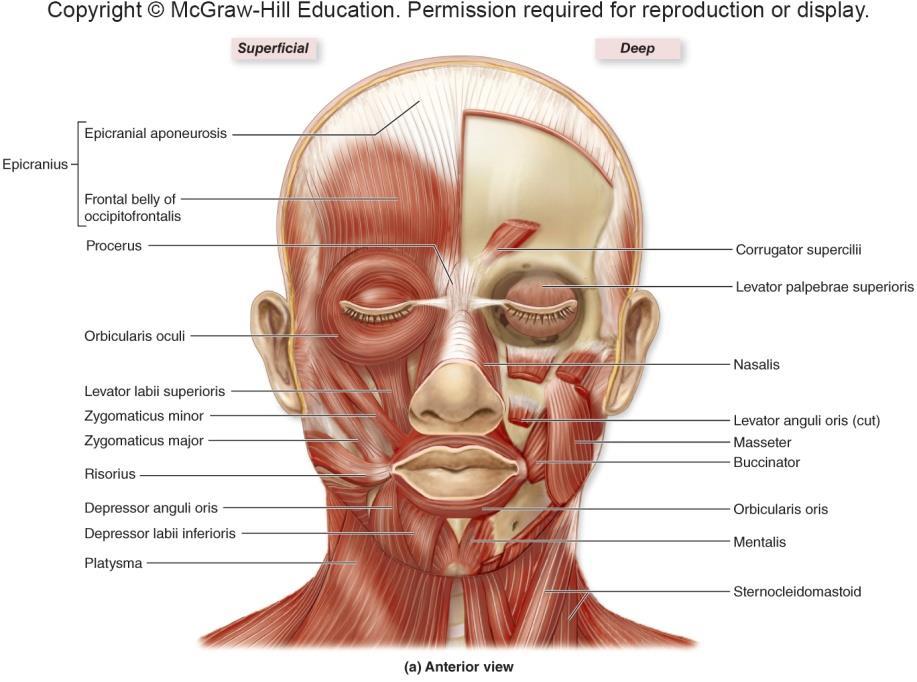
13 11.3a Muscles of Facial Expression Characteristics Insert into superficial fascia of skin of face Cause skin to move (facial expression) during contraction Most are innervated by facial nerve (CN VII) 13
14 11.3a Muscles of Facial Expression Muscles of forehead, scalp, and eyebrows Occipitofrontalis muscle o Connects with epicranial aponeurosis (galea aponeurotica) to form epicranius o Frontal belly raises eyebrows o Occipital belly retracts scalp slightly Corrugator supercilii muscle o Draws eyebrows together Orbicularis oculi o Circular muscle fibers surround orbit o Closes eye Levator palpebrae superioris o Elevates upper eyelid 14
15
16 11.3a Muscles of Facial Expression Muscles associated with the nose Nasalis o Elevates corners of the nostrils for flared nostrils Procerus o Wrinkles nose in distaste o Continuous with frontal belly of occipitofrontalis muscle 16
17 Procerus 10-17
18 11.3a Muscles of Facial Expression Muscles associated with the mouth Orbicularis oris o Closes mouth, puckers lips Depressor labii inferioris o Pulls lower lip inferiorly Depressor anguli oris o Pulls corners of the mouth inferiorly to frown Levator labii superioris o Pulls upper lip superiorly Levator anguli oris o Pulls corners of mouth superiorly and laterally o Works with zygomaticus major and minor in smiling 18
19 11.3a Muscles of Facial Expression Muscles associated with the mouth (continued) Risorius o Pulls corner of the lips laterally Mentalis o Protrudes lower lip Platysma o Tenses skin of neck and pulls lower lip inferiorly Buccinator o Compresses cheek against the teeth when chewing 19
20 Muscles of Facial Expression Figure 11.5a 20

21 Muscles of Facial Expression Figure 11.5b 21
22 Clinical View: Idiopathic Facial Nerve Paralysis (Bell Palsy) May be without known cause (idiopathic) Also known as Bell palsy Facial nerve inflamed and compressed Muscles on same side paralyzed Concern of eyes drying out and becoming damaged Prednisone often used to reduce swelling Level and timing of recovery varies 22
23 11.3b Extrinsic Eye Muscles Extrinsic eye muscles are extraocular Insert onto outer surface of the eye and move it Six muscles: four rectus and two oblique Rectus eye muscles originate from common tendinous ring Medial rectus pulls eye medially o Innervated by CN III (oculomotor) Lateral rectus pulls eye laterally o Innervated by CN VI (abducens) Inferior rectus pulls eye inferiorly and medially o Innervated by CN III (oculomotor) Superior rectus pulls eye superiorly and medially o Innervated by CN III (oculomotor) 23
24 11.3b Extrinsic Eye Muscles Oblique eye muscles Inferior oblique elevates and turns eye laterally o Innervated by CN III (oculomotor) Superior oblique depresses and turns the eye laterally o Passes through pulleylike loop, trochlea o Innervated by CN IV (trochlear) 24

25 Extrinsic Muscles of the Eye Figure 11.7a 25

26 Extrinsic Muscles of the Eye Figure 11.7b 26
27 Clinical View: Cranial III, IV, and VI Eye movements tested together with H-pattern Weakness (ptosis) of the eye in one direction can indicate cranial nerve involvement Double vision, Lazy eye, Cross eyed 27
28 11.3c Muscles of the Oral Cavity and Pharynx Muscles of mastication (chewing) Four paired muscles move mandible at temporomandibular joint (TMJ) All four are innervated by CN V (trigeminal) Temporalis o Elevates and pulls the mandible posteriorly (retracts) Masseter o Elevates and pulls the mandible anteriorly (protracts) o Most powerful and important masticatory muscle Medial and lateral pterygoid o Side to side movement of mandible o Medial pterygoid also helps with elevation of mandible o Lateral pterygoid helps with depression of the mandible 28

29 Muscles of Mastication Figure
30 11.3c Muscles of the Oral Cavity and Pharynx Muscles that move the tongue Left and right genioglossus o Protract (stick out) the tongue Left and right styloglossus o Elevate and retract the tongue Left and right hyoglossus o Depress and retract the tongue Left and right palatoglossus o Elevate posterior portion of the tongue 30

31 Muscles That Move the Tongue Figure
32 11.3d Muscles of the Anterior Neck: The Hyoid Muscles Suprahyroid muscles: above hyoid bone Elevate hyoid bone during swallowing or speaking Digastric: has two bellies, anterior and posterior o Also helps depress mandible Geniohyoid o Elevates the hyoid bone Mylohyoid o Provides muscular floor of mouth; also raises it up Stylohyoid: attaches to styloid process of temporal bone o Elongates oral cavity during swallowing 32
33 11.3d Muscles of the Anterior Neck: The Hyoid Muscles Infrahyoid muscles: inferior to hyoid bone Depress hyoid bone or thyroid cartilage of larynx as swallowing finishes Omohyoid o Depresses hyoid by pulling it toward scapula Sternohyoid o Depresses hyoid by pulling it toward sternum Sternothyroid o Depresses thyroid cartilage by pulling it toward sternum Thyrohyoid: extends from thyroid cartilage to hyoid o Depresses hyoid bone and elevates thyroid cartilage o Closes off larynx during swallowing 33

34 Muscles of the Anterior Neck Figure
35 11.3e Muscles That Move the Head and Neck Anterolateral neck muscles Generally act to flex neck Sternocleidomastoid: from sternum and clavicle to mastoid o Unilateral (one side) flexion of head to contracting muscle s side and contralateral rotation of head to opposite side Anterior, middle, and posterior scalene muscles Work with sternocleidomastoid to flex neck Pull cervical vertebrae toward 1 st and 2 nd ribs Elevate 1 st and 2 nd ribs during forced inhalation Tighten with paradoxical breathing 35

36 Muscles That Move the Head and Neck Figure
37 11.3e Muscles That Move the Head and Neck Posterior neck muscles Generally act to extend the neck Trapezius o Primary function is to help move pectoral girdle, but also helps extend neck Splenius capitis, splenius cervicis, semispinalis capitis, longissimus capitis o When contracted bilaterally, extend neck o When contracted unilaterally, turn head to same side Suboccipital muscles o Obliquus capitis superior, obliquus capitis inferior turn head to same side o Rectus capitis posterior major, rectus capitis posterior minor extend neck 37

38 Posterior Neck Muscles 38
39 Clinical View: Congenital Muscular Torticollis Newborn with shortened sternocleidomastoid May persist into childhood From birth trauma or prenatal position Head tilts to affected side and chin to unaffected side Treatments include Physical therapy Botulinum toxin, which impairs contraction of affected muscle Chiropractic care 39
40 What did you learn? Which muscles open and close the eyes? Which cranial nerve controls the lateral rectus muscle? What action does contraction of the masseter cause? What muscle is affected in Torticollis and what clinical presentation might be present? Copyright 2016 McGraw-Hill Education. All rights reserved. No reproduction or distribution without the prior written consent of McGraw-Hill Education 40
41 11.4 Muscles of the Vertebral Column Muscles of the vertebral column Complex, multiple origins and insertions, overlapping muscles Erector spinae o Maintain upright posture; if bilaterally contracted, vertebral column extends o If unilaterally contracted, lateral flexion toward that side o Three groups of erector spinae muscles from lateral to medial Iliocostalis group Longissimus group Spinalis group Quadratus lumborum muscles o Extends vertebral column when bilaterally contracted o Laterally flexes column when unilaterally contracted o Located primarily in the lumbar region 41

42 Deep Muscles of the Vertebral Column Figure
43 11.5 Muscles of Respiration Muscles of respiration Diaphragm o Internally placed dome-shaped muscle that partitions thoracic and abdominal cavities o Most important muscle associated with breathing o Muscle fibers converge toward fibrous central tendon o Openings for vena cava, aorta, and esophagus o Contracts during inspiration o Central tendon pulled inferiorly, increasing dimension of thoracic cavity 43

44 Muscles of Respiration Figure 11.15c-d 44
45 11.5 Muscles of Respiration Muscles of respiration External intercostals o Elevate ribs during inspiration, expanding cavity Internal intercostals o Deep to external intercostals o Fibers at right angles to external intercostals o Depress ribs during forced expiration Transversus thoracis o Depresses ribs during forced expiration 45

46 Muscles of Respiration Figure 11.15a 46
47 11.6 Muscles of the Abdominal Wall Muscles of the abdominal wall Compress and hold abdominal organs in place External oblique o Ipsilateral flexion and Contralateral rotation Internal oblique o Ipsilateral flexion and Ipsilateral rotation Transversus abdominis o Stabilizes the vertebral column (core muscle) Rectus abdominis o Partitioned into four segments by fibrous tendinous intersections o Enclosed within fibrous sleeve, rectus sheath Formed from aponeuroses of external and internal oblique, transversus abdominis Rectus sheaths of two sides connected by fibrous strip, linea alba 47

48 Muscles of the Abdominal Wall Figure 11.16a 48

49 Muscles of the Abdominal Wall Figure 11.16b 49
 can push intestine into canal Physicians test for it by palpating inguinal ring while patient coughs (cough raises abdominal pressure) 50")
50 Portion of viscera protrudes through weak point of abdominal wall Inguinal hernia Clinical View: Hernias Loop of small intestine protrudes through superficial inguinal ring More likely to occur in males since their inguinal canals are larger to accommodate spermatic cord High abdominal pressure (e.g., straining to lift something heavy) can push intestine into canal Physicians test for it by palpating inguinal ring while patient coughs (cough raises abdominal pressure) 50

51 Perineum Muscles of the Pelvic Floor Diamond-shaped region between lower appendages Bounded by pubic symphysis, coccyx, ischial tuberosities Contains two triangles divided by transverse line between ischial tuberosities o Urogenital triangle contains external genitalia and urethra o Anal triangle contains anus Figure 11.17b,c (left) 51
52 11.8a Muscles That Move the Pectoral Girdle Anterior thoracic muscles Pectoralis minor o Deep to pectoralis major o Helps depress and protract scapula; hunches shoulders Serratus anterior o Fan-shaped muscle between ribs and scapula o Protracts, stabilizes scapula Subclavius o Extends from clavicle to first rib o Stabilizes and depresses scapula 52

53 Muscles That Move Pectoral Girdle and Arm Figure 11.21a 53
54 11.8a Muscles That Move the Pectoral Girdle Posterior thoracic muscles Levator scapulae o Attaches to cervical vertebrae and scapula Tends to be involved in tension headaches o Elevates and inferiorly rotates the scapula Rhomboid major and minor o Runs inferolaterally from vertebrae to scapula, deep to trapezius o Helps elevate, retract, and inferiorly rotate the scapula Trapezius o Diamond-shaped muscle extending from skull and vertebral column to pectoral girdle o Can elevate, depress, retract, or rotate scapula 54

55 Muscles That Move Pectoral Girdle and Arm Figure 11.21b 55

56 Actions of Some Thoracic Muscles on the Scapula Figure 11.20a 56
57 11.8b Muscles That Move the Glenohumeral Joint/Arm Eleven muscles cross the glenohumeral joint and move the humerus Deltoid o Prime abductor of the arm o Its anterior fibers flex and medially rotate arm o Its lateral fibers abduct the arm o Its posterior fibers extend and laterally rotate arm Latissimus dorsi o Prime arm extensor; adducts, medially rotates Pectoralis major o Prime arm flexor; adducts, medially rotates Coracobrachialis o Flexes and adducts the arm 57
58 Triceps brachii 11.8b Muscles That Move the Glenohumeral Joint/Arm o Helps extend and adduct the arm Biceps brachii o Assists in flexing the arm Teres major Extends, adducts, medially rotates arm Rotator cuff muscles Subscapularis o Medially rotates arm Supraspinatus o Abducts the arm Infraspinatus and teres minor o Adduct and laterally rotate arm 58

59 Rotator Cuff Muscles Subscapularis helps in wind up for pitch Supraspinatus helps in executing pitch delivery Infraspinatus and teres minor slow the arm at end of pitch Figure

60 Clinical View: Rotator Cuff Injuries Result of trauma or disease Can be caused by repetitive use Can be caused by falling on the shoulder or lifting too heavy of an object Supraspinatus most commonly involved Symptoms are swelling, tenderness, and pain with movement Especially common in baseball players May require physical therapy or surgical repair 60
61 11.8c Arm and Forearm Muscles That Move the Elbow Joint/Forearm Muscles of the arm s anterior compartment Biceps brachii o Two-headed muscle on anterior humerus o Flexes and supinates forearm; weakly helps flex humerus Brachialis o Deep to biceps brachii o Most powerful flexor of forearm (prime mover) Brachioradialis o Located on anterolateral forearm o Synergist in elbow flexion 61

62 Anterior Muscles with Actions at Elbow Figure
63 11.8c Arm and Forearm Muscles That Move the Elbow Joint/Forearm Muscles of the arm s posterior compartment Triceps brachii o Large three-headed muscle on posterior arm o Major extensor of forearm; also helps extend humerus Anconeus o Weak elbow extensor (synergist) o Crosses posterolateral region of elbow 63

64 Posterior Muscles with Actions at Elbow Figure
65 11.8c Arm and Forearm Muscles That Move the Elbow Joint/Forearm Muscles of the forearm that act on the elbow joint Pronator teres and pronator quadratus o Rotate the radius across the ulna to pronate forearm o Located in anterior compartment of forearm Supinator o Supinates forearm o Located in posterior compartment of forearm 65

66 Forearm Muscles That Supinate or Pronate Figure
67 11.8d Forearm Muscles That Move the Wrist Joint, Hand, and Fingers Muscles of the forearm s anterior compartment Muscles originate on medial epicondyle Flexor carpi radialis o Flexes wrist and abducts hand Palmaris longus o Weakly assists in wrist flexion Flexor carpi ulnaris o Flexes wrist and adducts hand Flexor digitorum superficialis o Flexes wrist, MP, and proximal interphalangeal (PIP) joints of fingers 2 5 Flexor digitorum profundus o Flexes wrist, MP joints, PIP joints, and distal interphalangeal (DIP) joints of fingers

68 Anterior Forearm Muscles Figure 11.27a 68
69 Anterior Forearm Muscles Figure 11.27b-c 69
70 11.8d Forearm Muscles That Move the Wrist Joint, Hand, and Fingers Muscles of forearm s posterior compartment Muscles originate from lateral epicondyle Extensor carpi radialis longus/brevis o Extends wrist and abducts hand Extensor digitorum o Extends wrist, MP joints, PIP joints, and DIP joints of fingers 2 5 Extensor digit minimi o Extends the little finger Extensor carpi ulnaris o Extends wrist and adducts hand Extensor pollicis longus/brevis o Helps extend MP joint of the thumb Extensor indicis o Extends MP, PIP, and DIP joints of index finger 70

71 Posterior Forearm Muscles Figure 11.29a 71
72 Posterior Forearm Muscles Figure 11.29b 72
73 Clinical View: Lateral Epicondylitis or Medial Epicondylitis Lateral Epicondylitis Also known as tennis elbow From trauma or overuse of common extensor tendon of posterior forearm muscles Pain at lateral epicondyle of humerus often results from repeated forceful contraction of forearm extensors Medial Epicondylitis Also known as golfers elbow From trauma or overuse of common flexor tendon of anterior forearm muscles Pain at medial epicondyle of humerus often results from repeated forceful contraction of forearm flexors 73
74 11.8d Forearm Muscles That Move the Wrist Joint, Hand, and Fingers Retinacula of the forearm Fibrous bands at the wrist formed from deep fascia Hold tendons close to bone Flexor retinaculum covers palmar surface of carpal bones o Carpal tunnel: tight space between bones and flexor retinaculum through which flexor tendons pass Extensor retinaculum superficial to dorsal surface of carpal bones o Extensor tendons of wrist and digits pass under it 74
75 Clinical View: Carpal Tunnel Syndrome Carpal tunnel Space between carpal bones and flexor retinaculum Flexor tendons extending through tunnel Median nerve extending through tunnel Syndrome caused by compression of nerve Characterized by pain and pins and needles (paresthesia) 75
76 11.8e Intrinsic Muscles of the Hand Intrinsic muscles of the hand: originate and insert in hand Thenar group: form fleshy mass at base of thumb Flexor pollicis brevis: flexes thumb Abductor pollicis brevis: abducts thumb Opponens pollicis: assists in opposition of thumb Hypothenar group: smaller fleshy mass at base of little finger Flexor digiti minimi brevis: flexes little finger Abductor digiti minimi: abducts little finger Opponens digiti minimi: assists in opposition of little finger 76

77 Intrinsic Muscles of the Hand Figure 11.30a 77
78 What did you learn? Which four muscles make up the rotator cuff? In which anatomical compartment is the brachialis muscle found? What action is typically performed by muscles in the anterior compartment of the forearm? Which part of the hand makes up the thenar group of muscles? What structure of the forearm is involved in carpal tunnel syndrome? 78
79 11.9a Muscles that Move the Hip Joint/Thigh How are thigh muscles organized? They are bound by the fascia lata, deep fascia that partitions them into compartments Anterior compartment muscles o Extend the knee or flex the thigh Medial compartment muscles o Adduct the thigh Lateral compartment muscle o Abducts the thigh Posterior compartment muscles o Flex knee and extend the thigh 79
80 11.9a Muscles that Move the Hip Joint/Thigh Muscles inserting on anterior thigh Psoas major and iliacus (collectively, iliopsoas) o Run from lumbar vertebrae and ilium to femur o Flex the thigh Sartorius tailor muscle o Flex and laterally rotate the thigh and flex the knee Quadriceps femoris: composite muscle with four heads o Consists of: rectus femoris, vastus lateralis, vastus medialis, vastus intermedius o Prime mover of knee extension o Pulls on quadriceps tendon, which becomes patellar ligament to tibia 80

81 Muscles of Anterior Thigh Figure 11.32a 81
82 11.9a Muscles that Move the Hip Joint/Thigh Muscles of the medial compartment of thigh Innervated by the obturator nerve Adductor longus, adductor brevis, gracilis, pectineus Adductor magnus Muscle of lateral thigh Tensor fasciae latae o Attaches to iliotibial tract (lateral thickening of fascia lata) o Abducts and medially rotates the thigh 82

83 Muscles That Act on the Hip and Thigh Figure 11.31a 83

84 Muscles That Act on the Hip and Thigh Figure 11.31b 84
85 11.9a Muscles That Move the Hip Joint/Thigh Muscles of the posterior thigh Gluteus maximus o Chief extensor of the thigh; also laterally rotates the thigh Gluteus medius and gluteus minimus o Deep to gluteus maximus o Abduct and medially rotate the thigh Hamstrings: group of 3 muscles o Biceps femoris, semimembranosous, semitendinosus o Extend the thigh (also rotate the knee) Group of muscles deep to gluteal muscles o Piriformis, superior gemellus, obturator internus, obturator externus, inferior gemellus, and quadratus femoris o Laterally rotate the thigh 85

86 Muscles of Gluteal Region and Posterior Thigh Figure 11.33a 86

87 Muscles That Act on the Hip and Thigh Figure 11.31c 87
88 Clinical View: Iliotibial Band Syndrome and Hamstring Strain Iliotibial Band Syndrome Both the Gluteus medius and Tensor fascia latae muscles insert on the iliotibial band Band runs from the lateral hip to the knee Pain and inflammation at the lateral knee due to friction from tightening of this band Hamstring Strain Muscle overload when the hamstring muscle undergoes eccentric contraction Sprinters, basketball players, football players, soccer players 88
89 11.9c Leg Muscles That Move the Ankle, Foot, and Toes Crural muscles: muscles located in leg that move ankle, foot, toes Partitioned into anterior, lateral, and posterior compartments 89
90 11.9c Leg Muscles That Move the Ankle, Foot, and Toes Muscles of the leg s anterior compartment Extensor digitorum longus o Dorsiflexes the foot and extends toes 2 5 Extensor hallucis longus o Dorsiflexes the foot and extends the great toe Fibularis tertius o Dorsiflexes and weakly everts the foot Tibialis anterior o Primary dorsiflexor of the foot; also inverts the foot Extensor retinaculum o Thickening of fascia at ankle that holds tendons close to bones 90

91 Muscles of the Anterior Leg Figure 11.34a 91
92 11.9c Leg Muscles That Move the Ankle, Foot, and Toes Muscles of the leg s lateral compartment Both are powerful foot evertors, weak plantar flexors o Fibularis longus: inserts on plantar side of foot o Fibularis brevis: lies deep to fibularis longus, inserts onto base of 5 th metatarsal 92

93 Muscles of the Lateral Leg Figure 11.35a 93
94 11.9c Leg Muscles That Move the Ankle, Foot, and Toes Muscles of the leg s posterior compartment Superficial layer Gastrocnemius o Has two bellies; forms calf o Flexes the leg and plantar flexes the foot Soleus o Broad muscle deep to gastrocnemius o Plantar flexes the foot Triceps surae = gastrocnemius + soleus o Insert at heel with calcaneal tendon Plantaris o Weak leg flexor and plantar flexor of the foot 94
95 11.9c Leg Muscles That Move the Ankle, Foot, and Toes Muscles of the leg s posterior compartment (continued) Deep layer Flexor digitorum longus o Flexes the foot and the MP, PIP and DIP of joints 2 5 Flexor hallucis longus o Flexes the foot and great toe Tibialis posterior o Plantar flexes and inverts the foot Popliteus o Flexes the leg and medially rotates the tibia (lock the knee) 95

96 Muscles of the Posterior Leg Figure 11.36a-b 96
97 Clinical View: Shin Splints, Tarsal Tunnel Syndrome, and Achilles Tendon Rupture Shin splints Soreness along length of tibia Often occur in new poorly conditioned runners May be considered a type of compartment syndrome Tarsal Tunnel syndrome Tension of the flexor retinaculum at the medial compartment of the foot leading to posterior tibial nerve compression Pain at the medial side of the ankle and into the heel/foot Common in overpronation Achilles tendon rupture Common in sports that involve running, jumping, pivoting, or rapid push off 97
98 11.9d Intrinsic Muscles of the Foot Intrinsic muscles of the foot originate and insert within foot Support the arches, move the toes Dorsal group Extensor hallucis brevis o Extends the MP joint of the great toe Extensor digitorum brevis o Extends the MP and PIP joints of toes
99 11.9d Intrinsic Muscles of the Foot Plantar group Supported by aponeurosis formed from deep fascia o Extends between phalanges of toes and calcaneus Muscles grouped into four layers from superficial to deep 1. Flexor digitorum brevis, abductor hallucis, abductor digiti minimi 2. Quadratus plantae, lumbricals 3. Adductor hallucis, flexor hallucis brevis, flexor digiti minimi brevis 4. Dorsal interossei, plantar interossei 99

100 Plantar Intrinsic Muscles of the Foot Figure 11.37a-c 100

101 Plantar Intrinsic Muscles of the Foot Figure 11.37d-e 101

102 Clinical View: Plantar Fasciitis Inflammation of the plantar aponeurosis Associated with overexertion that stresses the fascia E.g., weight bearing activities, excessive body weight, poor shoes, poor biomechanics Pain when stepping down from bed in the morning 102
Biology 2401 Muscles List for CPC models
 Biology 2401 List for CPC models Italicized muscles are dissect and similar in the cat = Dissect and note the differences in human and cat Major of the Human Head Facial Expression Epicranius frontalis
Epicranius (frontal belly) Zygomaticus minor. Zygomaticus major Buccinator
 Zygomaticus minor. Zygomaticus major Buccinator") Epicranius (frontal belly) Zygomaticus minor Zygomaticus major Buccinator Masseter Digastric (posterior belly) Stylohyoid Sternocleidomastoid Trapezius Scalenus Omohyoid (inferior belly) Orbicularis oris
Epicranius (frontal belly) Zygomaticus minor Zygomaticus major Buccinator Masseter Digastric (posterior belly) Stylohyoid Sternocleidomastoid Trapezius Scalenus Omohyoid (inferior belly) Orbicularis oris
Due in Lab weeks because of Thanksgiving Prelab #10. Homework #8. Both sides! Both sides!
 Lab 8 MUSCLES Due in Lab 10 2 weeks because of Thanksgiving Prelab #10 Both sides! Homework #8 Both sides! Refer to Muscles 22-23 Naming of muscles Origin Site of muscle attachment that doesn t move during
Lectures Muscular System 10-1
 Lectures 12-14 Muscular System 10-1 Properties of Muscle Ability of a muscle to shorten with force Capacity of muscle to respond to a stimulus Muscle can be stretched to its normal resting length and beyond
Human Anatomy and Physiology I Laboratory
 Human Anatomy and Physiology I Laboratory Gross Anatomy of the Muscular System (Two weeks) 1 This lab involves study of the laboratory exercise Gross Anatomy of the Muscular System. Complete the Review
The Human Muscular System Required reading before beginning this lab: Saladin, KS: Human Anatomy 5th ed (2017) Chapters 10, 11, 12 INTRODUCTION
 Chapters 10, 11, 12 INTRODUCTION") Biology 322: Human Anatomy The Human Muscular System Required reading before beginning this lab: Saladin, KS: Human Anatomy 5 th ed (2017) Chapters 10, 11, 12 INTRODUCTION We will use a number of lab periods
Biology 322: Human Anatomy The Human Muscular System Required reading before beginning this lab: Saladin, KS: Human Anatomy 5 th ed (2017) Chapters 10, 11, 12 INTRODUCTION We will use a number of lab periods
Contents. Preface xv. SECTION 1: Introduction to the Bodynamic System 1. SECTION 2: The Bodynamic Psycho-Motor Anatomy 29
 Contents Preface xv SECTION 1: Introduction to the Bodynamic System 1 Definitions in the Bodynamic System 3 Ego Formation through the Coding Elements 9 Examples of Formation of Coding 17 Using This Book
11/15/2018. Temporalis Elevates & retracts mandible. Masseter = Prime mover of jaw closure. Levator scapulae Supraspinatus Clavicle.
 Due in Lab 10 Lab 8 MUSCLES 2 weeks because of Thanksgiving Prelab #10 Both sides! Homework #8 Both sides! Refer to Muscles 22-23 Examples of Origin & Insertion Naming of muscles Origin Site of muscle
Muscle Anatomy Review Chart
 Muscle Anatomy Review Chart BACK Superficial (5) Trapezius Transverse cervical a. Latissimus dorsi Thoracodorsal a. Rhomboideus major Dorsal scapular a. Rhomboideus minor Levator scapulae Intermediate
5/21/2013. Muscle Anatomy. Thursday January, 24 th, Skeletal Muscle. Smooth Muscle. Cardiac Muscle
 Muscle Anatomy Thursday January, 24 th, 2013 Skeletal Muscle Cardiac Muscle Smooth Muscle 1 Smooth Muscle 1. Found in the walls of the digestive system, bladder, uterus and blood vessels 2. Involuntary
Chapter 10: Muscular System: Gross Anatomy
 Chapter 10: Muscular System: Gross Anatomy I. General Principles A. General Terminology 1. Tendons attach 2. What is an aponeurosis? 3. The points of muscle attachment are called & 4. How is the "origin"
Synergist Muscles. Shoulder (glenohumeral joint) Flexion Deltoid (anterior fibers) Pectoralis major (upper fibers) Biceps Brachii Coracobrachialis
 Flexion Deltoid (anterior fibers) Pectoralis major (upper fibers) Biceps Brachii Coracobrachialis") Synergist Muscles Dr Gene Desepoli DrGeneLMT@gmail.com Shoulder (glenohumeral joint) Deltoid (anterior fibers) Pectoralis major (upper fibers) Biceps Brachii Coracobrachialis Deltoid (posterior fibers)
Synergist Muscles Dr Gene Desepoli DrGeneLMT@gmail.com Shoulder (glenohumeral joint) Deltoid (anterior fibers) Pectoralis major (upper fibers) Biceps Brachii Coracobrachialis Deltoid (posterior fibers)
3/27/2012. Muscle Classification: Functional Groups. Interactions of Skeletal Muscles. Naming Skeletal Muscles. Naming Skeletal Muscles
 Interactions of Skeletal Muscles Skeletal muscles work together or in opposition Muscles only pull (never push) As muscles shorten, the insertion generally moves toward the origin Whatever a muscle (or
Biology 218 Human Anatomy. Adapted from Martini Human Anatomy 7th ed. Chapter 10 The Muscular System Axial Musculature
 Adapted from Martini Human Anatomy 7th ed. Chapter 10 The Muscular System Axial Musculature Introduction The skeletal muscle of the body can be subdivided into: Axial musculature Muscles that position
The muscular system I Muscles of the head neck and trunk
 The muscular system I Muscles of the head neck and trunk Dr. Nabil Khouri Dr. Nabil Khouri MD MSc, PhD Interactions of Skeletal Muscles Skeletal muscles work together or in opposition Muscles only pull
Chapter 11: The Muscular System. Copyright 2009, John Wiley & Sons, Inc.
 Chapter 11: The Muscular System Muscle Attachment Sites: Origin & Insertion n Skeletal muscles cause movements by exerting force on tendons, which pulls on bones or other structures. n Articulating bones
The Muscular System Lab Power Point
 The Muscular System Lab Power Point Myoneural Junction Sarcoplasm Nucleus Myofibrils Sarcomere (black line to black line) Sarcolemma Myoneural space Nucleus Endomysium Motor Neuron Muscles of Facial Expression
The Muscular System PART C. PowerPoint Lecture Slide Presentation by Patty Bostwick-Taylor, Florence-Darlington Technical College
 PowerPoint Lecture Slide Presentation by Patty Bostwick-Taylor, Florence-Darlington Technical College The Muscular System 6 PART C Five Golden Rules of Skeletal Muscle Activity Table 6.2 Muscles and Body
ACTIVITIES 5 & 6: APPENDICULAR AND AXIAL MUSCLES
 ACTIVITIES 5 & 6: APPENDICULAR AND AXIAL MUSCLES Objectives: 1) How to get ready: Read Chapter 11 & 12, McKinley et al., Human Anatomy, 4e. All text references are for this textbook. Begin identifying
The Muscular System. Chapter 10 Part D. PowerPoint Lecture Slides prepared by Karen Dunbar Kareiva Ivy Tech Community College
 Chapter 10 Part D The Muscular System Annie Leibovitz/Contact Press Images PowerPoint Lecture Slides prepared by Karen Dunbar Kareiva Ivy Tech Community College Table 10.14: Muscles Crossing the Hip and
Muscle fiber (cell) Blood vessel. Perimysium. Epimysium. Fascicle (wrapped by perimysium) Endomysium (between fibers) Tendon. Bone
 Blood vessel. Perimysium. Epimysium. Fascicle (wrapped by perimysium) Endomysium (between fibers) Tendon. Bone") Figure 6.1 Connective tissue wrappings of skeletal muscle. Blood vessel Muscle fiber (cell) Perimysium Epimysium Fascicle (wrapped by perimysium) Tendon Endomysium (between fibers) Bone Figure 6.15 Superficial
Figure 6.1 Connective tissue wrappings of skeletal muscle. Blood vessel Muscle fiber (cell) Perimysium Epimysium Fascicle (wrapped by perimysium) Tendon Endomysium (between fibers) Bone Figure 6.15 Superficial
The Muscular System. PowerPoint Lecture Presentations prepared by Jason LaPres. Lone Star College North Harris Pearson Education, Inc.
 11 The Muscular System PowerPoint Lecture Presentations prepared by Jason LaPres Lone Star College North Harris An Introduction to the Muscular System The Muscular System Consists only of skeletal muscles
Prime movers provide the major force for producing a specific movement Antagonists oppose or reverse a particular movement Synergists
 Dr. Gary Mumaugh Prime movers provide the major force for producing a specific movement Antagonists oppose or reverse a particular movement Synergists Add force to a movement Reduce undesirable or unnecessary
The Muscular System. Part A
 The Muscular System Part A 10 The Muscular System Part A 10 Hold onto your glutes, this is a big one. 10 Interactions of Skeletal Muscles Skeletal muscles work together or in opposition Muscles only pull
Musculoskeletal Anatomy Coloring Book
 Musculoskeletal Anatomy Coloring Book Muscolino, Joseph E. ISBN-13: 9780323057219 Table of Contents Introduction â How to Use This Book 1. The Skeletal System Bones of the Head â Anterior View Bones of
Temporalis Elevates & retracts mandible. Masseter Elevates mandible. Sternocleidomastoid Neck flexion. Trapezius Elevates & depresses shoulders
 Anterior Posterior Temporalis Elevates & retracts mandible Masseter Elevates mandible Sternocleidomastoid Neck flexion Trapezius Elevates & depresses shoulders Masseter Elevates mandible Temporalis Elevates
SKELETAL MUSCLE ANATOMY
 SKELETAL MUSCLE ANATOMY OUTLINE I. Anatomical Terms of Motion II. Head, Face & Neck Muscles III. Anterior Torso Muscles IV. Posterior Torso Muscles V. Arm & Shoulder Muscles VI. Leg & Hip Muscles 2 ANATOMICAL
BIOH111. o Cell Module o Tissue Module o Skeletal system o Integumentary system o Muscle system o Nervous system o Endocrine system
 BIOH111 o Cell Module o Tissue Module o Skeletal system o Integumentary system o Muscle system o Nervous system o Endocrine system TEXTBOOK AND REQUIRED/RECOMMENDED READINGS o Principles of anatomy and
Chapter 9. The Muscular System
 1 Chapter 9 The Muscular System 2 Introduction Skeletal muscles: movement in environment Smooth muscles: intestines, ureters, veins and arteries Cardiac muscle: pumps blood through heart and blood vessels
Lab Exercise #5 The Muscular System Student Performance Objectives
 Student Performance Objectives The material that you are required to learn in this exercise can be found in either the lecture text or the supplemental materials provided in lab. Prior to coming to class,
A. All movements require muscle which are organs using chemical energy to contract.
 Ch 8 Muscles Introduction: A. All movements require muscle which are organs using chemical energy to contract. B. The three types of muscle in the body are skeletal, smooth, and cardiac muscle. C. This
Bone Practical. Labs Muscle Labs. Final Practical. Divisions of the Muscular System. Quiz format
 Bone Practical Labs 17 + 18 Muscles Wed 7/11 @ 8am 40 50 stations About half axial, half appendicular bones Disarticulated bones: Skulls, partial skulls, vertebrae, ribs, skeletons, arm bones, leg bones,
A&P 1 Muscle In-Lab Guide
 A&P 1 Muscle In-Lab Guide This lab guide includes a table with all the muscles you need to ID, along with their origins, insertions and actions Dashed lines means ignore. If several actions are listed,
BIOH111. o Cell Module o Tissue Module o Skeletal system o Integumentary system o Muscle system o Nervous system o Endocrine system
 BIOH111 o Cell Module o Tissue Module o Skeletal system o Integumentary system o Muscle system o Nervous system o Endocrine system TEXTBOOK AND REQUIRED/RECOMMENDED READINGS o Principles of anatomy and
Head & Neck The muscle names are followed by the chapter number
 Head & Neck The muscle names are followed by the chapter number. Splenius capitis (9) 2. Occipitalis (2) Temporalis () 3. Temporalis () 4. Semispinalis capitis (9) Facial / Scalp (2) 5. Temporalis () Facial
CHAPTER 11 LECTURE OUTLINE I. INTRODUCTION
 CHAPTER 11 LECTURE OUTLINE I. INTRODUCTION A. The muscular system specifically concerns skeletal muscles and associated connective tissue that make individual muscle organs. B. This chapter discusses how
The Muscular System. Chapter 10 Part C. PowerPoint Lecture Slides prepared by Karen Dunbar Kareiva Ivy Tech Community College
 Chapter 10 Part C The Muscular System Annie Leibovitz/Contact Press Images PowerPoint Lecture Slides prepared by Karen Dunbar Kareiva Ivy Tech Community College Table 10.9: Muscles Crossing the Shoulder
The Muscular System Part A
 10 The Muscular System Part A Lecture Presentation by Lori Garrett Section 1: Functional Organization of the Muscular System Learning Outcomes 10.1 Describe the general function of the body s axial and
List of Muscles and Function. Region View Muscle Function Facial Anterior/Oblique Occipitofrontalis front belly Raises eyebrows
 List of Muscles and Function Region View Muscle Function Facial Anterior/Oblique Occipitofrontalis front belly Raises eyebrows Orbicularis oculi Closes eye Orbicularis oris Purses lips Zygomaticus minor/major
The Muscular System Outline 10.1 For any movement, muscles can act in one of three ways (pp ) A. Muscles only pull; they never push, and as a
 A. Muscles only pull; they never push, and as a") The Muscular System Outline 10.1 For any movement, muscles can act in one of three ways (pp. 321 322) A. Muscles only pull; they never push, and as a muscle shortens, the insertion is pulled toward the
The Muscular System Outline 10.1 For any movement, muscles can act in one of three ways (pp. 321 322) A. Muscles only pull; they never push, and as a muscle shortens, the insertion is pulled toward the
Cadaver Muscular System Practice Practical
 Cadaver Muscular System Practice Practical Station 1 Station 1 1. Specific structure 1. Rectus sheath 2. Red line 2. Linea alba Station 2 Station 2 3. Red muscle 1. Rectus abdominis 4. Red muscle actions
Human Anatomy Lab #7: Muscles of the Cadaver
 Human Anatomy Lab #7: Muscles of the Cadaver Table of Contents: Expected Learning Outcomes.... 1 Introduction...... 1 Identifying Muscles on Yourself.... 2 Muscles of the Anterior Trunk and Arm.. 2 Muscles
Scapula Spine Lateral edge of clavicle. Medial border Scapula. Medial border of Scapula, between superior angle and root of spine. Scapula.
 Muscle attachments and actions answer sheet Muscle Origins insertions Movements Joints crossed Trapezius Base of skull Spinous process of C7 Thoracic Spine Lateral edge of clavicle Elevation Retraction
In-Depth Foundations: Anatomy Terms to Know
 Be familiar with / able to identify and define all the following parts. The Spine Cranium Vertebrae Cervical, Thoracic, Lumbar Sacrum Coccyx Bones of Upper Body Cranium Mastoid process; Occipital condyle,
lesser trochanter of femur lesser trochanter of femur iliotibial tract (connective tissue) medial surface of proximal tibia
 medial surface of proximal tibia") LOWER LIMB MUSCLES OF THE APPENDICULAR SKELETON The muscles that act on the lower limb fall into three groups: those that move the thigh, those that move the lower leg, and those that move the ankle, foot,
LOWER LIMB MUSCLES OF THE APPENDICULAR SKELETON The muscles that act on the lower limb fall into three groups: those that move the thigh, those that move the lower leg, and those that move the ankle, foot,
Chapter 10. The Muscular System. AP1-Lecture Goodwin College. Introduction. In this chapter we will cover: 4/27/2015
 Chapter 10 The Muscular System AP1-Lecture Goodwin College Introduction In this chapter we will cover: Structural and functional organization of muscles Muscles of the head and neck Muscles of the trunk
Muscles of the lower extremities. Dr. Nabil khouri MD, MSc, Ph.D
 Muscles of the lower extremities Dr. Nabil khouri MD, MSc, Ph.D Posterior leg Popliteal fossa Boundaries Biceps femoris (superior-lateral) Semitendinosis and semimembranosis (superior-medial) Gastrocnemius
Name this muscle. Name this muscle
 this muscle this muscle Pectoralis Major Pectoralis Minor Serratus anterior Pectoralis minor Serratus anterior this muscle Deltoid: The major abductor of the upper limb this muscle this muscle this muscle
Muscles of the Cat. N Deltoid MUSCLES OF THE CHEST. Pectoralis major. (This muscle is superior to Pectoralis minor) MUSCLES OF THE CHEST
 MUSCLES OF THE CHEST") MUSCLES OF THE CHEST Pectoralis major (This muscle is superior to Pectoralis minor) 1. MUSCLES OF THE CHEST Pectoralis minor (This muscle is inferior to Pectoralis major) 2. MUSCLES OF THE ARM Deltoid
MUSCLES OF THE CHEST Pectoralis major (This muscle is superior to Pectoralis minor) 1. MUSCLES OF THE CHEST Pectoralis minor (This muscle is inferior to Pectoralis major) 2. MUSCLES OF THE ARM Deltoid
Biol 353 Pre-Professional Human Anatomy Exam III Fall 2017 page 1 of 8
 Biol 353 Pre-Professional Human Anatomy Exam III Fall 2017 page 1 of 8 IMPORTANT INSTRUCTIONS: ANSWER ONLY 50 QUESTIONS. Do not answer more than 50 questions. If you answer more than 50 questions, then
Muscular System. Dr. Gary Mumaugh
 Muscular System Dr. Gary Mumaugh The Muscular System. Superficial Deep Structural and functional organization of muscles Platysma Frontalis Orbicularis oculi Zygomaticus major Masseter Orbicularis oris
Exercise Science Section 3: The Muscular System
 Exercise Science Section 3: The Muscular System An Introduction to Health and Physical Education Ted Temertzoglou Paul Challen ISBN 1-55077-132-9 Major Functions of Muscles Movement Includes: breathing,
Biology 353 Pre-Professional Human Anatomy Exam III Fall 2016 page 1 of 8
 Biology 353 Pre-Professional Human Anatomy Exam III Fall 2016 page 1 of 8 IMPORTANT INSTRUCTIONS: ANSWER ONLY 50 QUESTIONS. Do not answer more than 50 questions. If you answer more than 50 questions, then
Cat Muscles Flashcards Mt SAC
 1. MUSCLES OF THE CHEST Pectoralis major (This muscle is superior to Pectoralis minor) 2. MUSCLES OF THE CHEST Pectoralis minor (This muscle is inferior to Pectoralis major) 3. MUSCLES OF THE ARM AD CHEST
Chapter 10. An Overview of the Muscle System
 Chapter 10 An Overview of the Muscle System The Muscular System Superficial Deep Deep Superficial Frontalis Platysma Deltoid Pectoralis major Biceps brachii Brachioradialis Flexor carpi radialis External
Bio 113 Anatomy and Physiology The Muscles. Muscles of the Head and Neck. Masseter. Orbicularis occuli. Orbicularis oris. Sternocleidomastoid
 Bio 113 Anatomy and Physiology The Muscles Muscles of the Head and Neck Masseter Orbicularis occuli Orbicularis oris Sternocleidomastoid Temporalis BIO 113 Fall 2011 Muscles Page 1 of 5 Muscles of the
In which arm muscle are intramuscular injections most often given? (not in text)
") AP1 Lab 9 - Muscles of the Arms and Legs Locate the following muscles on the models and on yourself. Recall anatomical position. Directional terms such as anterior, posterior, lateral, etc. all assume
AP1 Lab 9 - Muscles of the Arms and Legs Locate the following muscles on the models and on yourself. Recall anatomical position. Directional terms such as anterior, posterior, lateral, etc. all assume
Naming Skeletal Muscles
 Naming Skeletal Muscles Direction of Muscle Fibers Action Location Origin & Insertion Skeletal Muscle Size Shape Number Of Origins Direction of Muscle Fibers Relative to the Midline RECTUS = parallel to
Muscles of Lesson Five. Muscular Nomenclature and Kinesiology - Two. Muscles of Lesson Five, cont. Chapter 16
 Chapter 16 Muscular Nomenclature and Kinesiology - Two Lessons 5-6 Muscles of Lesson Five Iliopsoas (psoas major, iliacus) Hip outward rotators (piriformis, gemellus superior, gemellus inferior, obturator
Monday, November 13, 2017 A & P 2401
 Monday, November 13, 2017 A & P 2401 Today you will complete the following handouts. Study the last part of the handout for this will be on your quiz, which will be on Wednesday. It is titled steps of
Lab Activity 11: Group I
 Lab Activity 11: Group I Muscles Martini Chapter 11 Portland Community College BI 231 Origin and Insertion Origin: The place where the fixed end attaches to a bone, cartilage, or connective tissue. Insertion:
2/4/2018. Identify the two reasons why muscle cells may go through muscle fatigue. Ch.7 Review. Sternocleidomastoid.
 Ch.7 Review Identify the two reasons why muscle cells may go through muscle fatigue Temporalis Depressor anguli oris Sternocleidomastoid Tibialis anterior 1 Gluteus medius Deltoid Adducts & rotates scapula
Chapter 11 Anatomy and Physiology Lecture
 1 THE MUSCULAR SYSTEM Chapter 11 Anatomy and Physiology Lecture THE MUSCULAR SYSTEM 2 Muscle Tissue - refers to all the contractile tissues of the body; skeletal, cardiac, and smooth muscle. Muscular System
Muscular System Dr. Gary Mumaugh
 Muscular System Dr. Gary Mumaugh 1 Organization of Muscles about 600 human skeletal muscles constitute about half of our body weight three kinds of muscle tissue o skeletal, cardiac, smooth specialized
Muscles of the Gluteal Region
 Muscles of the Gluteal Region 1 Some of the most powerful in the body Extend the thigh during forceful extension Stabilize the iliotibial band and thoracolumbar fascia Related to shoulders and arms because
Module 6 - The Muscular System Introduction to the Muscular System and Muscles of the Head, Neck and Shoulder
 Module 6 - The Muscular System Introduction to the Muscular System and Muscles of the Head, Neck and Shoulder There will be three modules to cover the muscle anatomy of the body. The first module will
MUSCLES OF THE LOWER LIMBS
 MUSCLES OF THE LOWER LIMBS Naming, location and general function Dr. Nabil khouri ROLES THAT SHOULD NOT BE FORGOTTEN Most anterior compartment muscles of the hip and thigh Flexor of the femur at the hip
Muscles of the Hip 1. Tensor Fasciae Latae O: iliac crest I: lateral femoral condyle Action: abducts the thigh Nerve: gluteal nerve
 Muscles of the Hip 1. Tensor Fasciae Latae O: iliac crest I: lateral femoral condyle Action: abducts the thigh Nerve: gluteal nerve 2. Gluteus Maximus O: ilium I: femur Action: abduct the thigh Nerve:
Masseter- in front of ear Temporalis Mandible
 Frontal Belly (Epicranius) Occipital Belly (Epicranius) Orbicularis Oculi Orbicularis Oris Zygomaticus minor Zygomaticus major Buccinator Facial Expression Origin- stays still Raises eyebrows Galea aponeurotica
Match the types of muscle tissues with the words and phrases. 1) Skeletal 2) Smooth 3) Cardiac 2 Walls of blood vessels. 2 Walls of digestive tract
 Skeletal 2) Smooth 3) Cardiac 2 Walls of blood vessels. 2 Walls of digestive tract") S T U D Y G U I D E. Types of Muscle Tissues Match the types of muscle tissues with the words and phrases. ) Skeletal ) Smooth ) Cardiac, Striated Walls of blood vessels, Single nucleus Heart muscle, Involuntary
S T U D Y G U I D E. Types of Muscle Tissues Match the types of muscle tissues with the words and phrases. ) Skeletal ) Smooth ) Cardiac, Striated Walls of blood vessels, Single nucleus Heart muscle, Involuntary
Biology 218 Human Anatomy. Adapted from Martini Human Anatomy 7th ed. Chapter 12 Surface Anatomy and Cross-Sectional Anatomy
 Adapted from Martini Human Anatomy 7th ed. Chapter 12 Surface Anatomy and Introduction Surface anatomy is the study of anatomical landmarks on the exterior of the human body Knowledge of surface anatomy
TABLES OF MUSCLE ACTIONS, INNERVATIONS, AND ATTACHMENTS
 TABLES OF MUSCLE ACTIONS, INNERVATIONS, AND ATTACHMENTS Table 1-1 ERECTOR SPINAE MUSCLES Intrinsic muscles producing extension and/or lateral of the spine Muscle Joint and Action Innervation Inferior Attachment
Figure 11-1: The lever-fulcrum principle is illustrated by flexion of the forearm.
 Chapter 11: The Muscular System Read pages 325 to 399 NAME Topic Outline And Objectives: A. How skeletal muscles produce movement, and naming muscles 1. Describe the relationship between bones and skeletal
C.F.A. Christian Fitness Association
 C.F.A. Christian Fitness Association Personal Trainer/Advanced Personal Trainer/ Master Trainer/ Sports Nutritionist/ Martial Arts Strength Trainer/Step & Kickbox Aerobics Instructor/ Health Club Manager/
ANATOMY PHYSIOLOGY WORKSHEET Gross Anatomy of the Muscular System Background
 ANATOMY PHYSIOLOGY WORKSHEET Gross Anatomy of the Muscular System Background Most skeletal muscles are attached to bones by dense regular connective tissue in the form of cord-like tendons or membranous
Lab 9: Learn origin and insertion for each of the listed muscles. For Exercise 15, do Activities 1-6 in 9 th edition, Activities 1-4 in 10 th edition
 The Muscular System Exercises 14, 15, and 16 (begins: page 187 in 9 th and 10 th editions) Exercises 12, 13, and 14 (begins: page 185 in 11 th edition, page 189 in 12 th edition) Lab 8 and 9 Objectives
Muscles Built on the Maniken
 Muscles Built on the Maniken Facial Muscle Group 1. Temporalis O temporal fossa I anterior border of the ramus of the mandible A elevates the mandible (bite muscle) and holds jaw while at rest 2. Procerus
Human Anatomy Biology 351
 Human Anatomy Biology 351 Lower Limb Please place your name on the back of the last page of this exam. You must answer all questions on this exam. Because statistics demonstrate that, on average, between
BIOL 4260 Human Evolu3onary Anatomy Lecture 12: Limb Development. Lecture 2: Fossil Record
 BIOL 4260 Human Evolu3onary Anatomy Lecture 12: Limb Development Lecture 2: Fossil Record Outline Limb Evolution Limb Development Limb Function A Few Definitions Appendicular skeleton girdles & limbs Pectoral
Kaan Yücel M.D., Ph.D. 4.October.2013 Friday
 Kaan Yücel M.D., Ph.D. 4.October.2013 Friday The muscular system consists of all the muscles of the body. The discipline related to the study of muscles is myology. Musculus (muscle) is derived from the
Exercise Science Section 3: The Muscular System
 Exercise Science Section 3: The Muscular System An Introduction to Health and Physical Education Ted Temertzoglou Paul Challen ISBN 1-55077-132-9 Major Functions of Muscles Movement v Includes: breathing,
Take your baby to the gym. Which gym? BabyGym!
 Take your baby to the gym. Which gym? BabyGym! The word FOMO (short for fear of missing out) crops up everywhere, even in the nursery where parents and caregivers race from the one stimulation class to
Axial Muscles of the Head, Neck, and Back
 OpenStax-CNX module: m46484 1 Axial Muscles of the Head, Neck, and Back OpenStax College This work is produced by OpenStax-CNX and licensed under the Creative Commons Attribution License 4.0 By the end
Copy Right- Hongqi ZHANG-Department of Anatomy-Fudan University. Systematic Anatomy. Locomotor system - Part 6
 Systematic Anatomy Locomotor system - Part 6 Muscles of abdomen Muscles of the upper limb Dr.Hongqi Zhang ( 张红旗 ) Email: zhanghq58@126.com 1 Muscles of abdomen Muscles of the upper limb Muscles of abdomen
The Clavicle Right clavicle Deltoid tubercle: Conoid tubercle, conoid ligamen Impression for the
 The Clavicle Muscle Attachment Sites in the Upper Limb Pectoralis major Right clavicle Smooth superior surface of the shaft, under the platysma muscle tubercle: attachment of the deltoid Acromial facet
Muscular Nomenclature and Kinesiology - One
 Chapter 16 Muscular Nomenclature and Kinesiology - One Lessons 1-3 (with lesson 4) 1 Introduction 122 major muscles covered in this chapter Chapter divided into nine lessons Kinesiology study of human
Skeleton. The left forearm is in the position of supination, the right in pronation.
 S ystemic review A Skeleton A from the front B B from behind The left forearm is in the position of supination, the right in pronation. Skull Mandible Hyoid bone Cervical vertebrae Clavicle Sternum Costal
Anatomy and Physiology 1 Chapter 11 self quiz Pro, Dima Darwish,MD.
 Anatomy and Physiology 1 Chapter 11 self quiz Pro, Dima Darwish,MD. 1) The attachment of a muscle s tendon to the stationary bone is called the ; the attachment of the muscle s other tendon to the movable
Interactions of Skeletal Muscles, Their Fascicle Arrangement, and Their Lever Systems
 Interactions of Skeletal Muscles, Their Fascicle Arrangement, and Their Lever Systems Bởi: OpenStaxCollege To move the skeleton, the tension created by the contraction of the fibers in most skeletal muscles
Chiropractic Technician Class
 Chiropractic Technician Class Presentation By: Dr. Kay Miller. The Role of Exercise as it Relates to Our Musculoskeletal System Introduction to the topic and Preliminary Physical exam Musculoskeletal anatomy:
medial half of clavicle; Sternum; upper six costal cartilages External surfaces of ribs 3-5
 MUSCLE ORIGIN INSERTION ACTION NERVE Pectoralis Major medial half of clavicle; Sternum; upper six costal cartilages Lateral lip of intertubercular groove of horizontal adduction Medial and lateral pectoral
Anatomy & Physiology. Muscles of the Lower Limbs.
 Anatomy & Physiology Muscles of the Lower Limbs http://www.ishapeup.com/musclecharts.html Muscles of the Lower Limbs Among the strongest muscles in the body. Because pelvic girdle is composed of heavy,
Module 7 - The Muscular System Muscles of the Arm and Trunk
 Module 7 - The Muscular System Muscles of the Arm and Trunk This Module will cover the muscle anatomy of the arms and trunk. We have already seen the muscles that move the humerus, so this module will
Types of Muscle: Skeletal- muscle involved in movement of the skeleton. Striated, has alternating bands of light and dark due to overlapping
 Types of Muscle: Skeletal- muscle involved in movement of the skeleton. Striated, has alternating bands of light and dark due to overlapping filaments within the muscle cell. Skeletal muscle can be consciously
Anatomy and Physiology 141 Exam II November 6, Name Student Number
 Anatomy and Physiology 141 Exam II November 6, 2014 Name Student Number 1. In regards to the gross anatomy of muscle, which of the following is NOT TRUE? a. Perimysium is more superficial than the epimysium
Test Bank for The Human Body in Health and Illness 4th Edition by Herlihy
 Test Bank for The Human Body in Health and Illness 4th Edition by Herlihy Chapter 9: Muscular System Test Bank MULTIPLE CHOICE 1. Which of the following muscles is described as striated and involuntary?
BLUE SKY SCHOOL OF PROFESSIONAL MASSAGE AND THERAPEUTIC BODYWORK. Musculoskeletal Anatomy & Kinesiology II REVIEW
 BLUE SKY SCHOOL OF PROFESSIONAL MASSAGE AND THERAPEUTIC BODYWORK Musculoskeletal Anatomy & Kinesiology II REVIEW MSAK101-II Session 4 LEARNING OBJECTIVES: By the end of this session, the student will be
Chapter 3: Applied Kinesiology. ACE Personal Trainer Manual Third Edition
 Chapter 3: Applied Kinesiology ACE Personal Trainer Manual Third Edition Introduction Kinesiology is the study of the body s infinite number of movements, positions, and postures and is grounded in the
Certified Personal Trainer Re-Certification Manual
 Certified Personal Trainer Re-Certification Manual Section II 1 Anatomy & Physiology Terms Anatomy and physiology are closely related fields of study: anatomy is the study of form, and physiology is the
DISSECTION 1: SKELETAL MUSCLES
 8546d_c01_1-42 6/21/02 1:34 PM Page 4 mac62 mac62:1253_ge: 4 Cat Dissection DISSECTION 1: SKELETAL MUSCLES Many skeletal muscles of the cat are similar to human muscles. This dissection will reinforce