D120 and K152 within the PH domain of T cell adapter SKAP55 regulate plasma. membrane targeting of SKAP55 and LFA-1 affinity modulation in human T
|
|
|
- Willa Farmer
- 6 years ago
- Views:
Transcription
1 MCB Accepted Manuscript Posted Online 4 January 2017 Mol. Cell. Biol. doi: /mcb Copyright 2017 American Society for Microbiology. All Rights Reserved D120 and K152 within the PH domain of T cell adapter SKAP55 regulate plasma membrane targeting of SKAP55 and LFA-1 affinity modulation in human T lymphocytes Amelie Witte a, Bernhard Meineke b, Jana Sticht b, Lars Philipsen a, Benno Kuropka b, Andreas J. Müller a,c, Christian Freund b, Burkhart Schraven a,c,d, and Stefanie Kliche a,d a Otto-von-Guericke-University, Institute of Molecular and Clinical Immunology, Health Campus Immunology, Infectiology and Inflammation, Leipziger Strasse 44, Magdeburg, Germany b Freie Universität Berlin, Institut für Chemie und Biochemie, Protein Biochemistry group, Thielallee 63, Berlin, Germany c Helmholtz Center for Infection Research (HZI), Department of Immune Control, Inhoffenstrasse 7, Braunschweig, Germany d Corresponding authors contact: Dr. Stefanie Kliche or Prof. Dr. Burkhart Schraven, Ottovon-Guericke-University, Institute of Molecular and Clinical Immunology, Magdeburg, Germany, stefanie.kliche@med.ovgu.de or (burkhart.schraven@med.ovgu.de) Running title: The PH domain of SKAP
2 Abstract The β2-integrin lymphocyte function-associated antigen-1 (LFA-1) is needed for T cell receptor (TCR) induced activation of LFA-1 to promote T cell adhesion and interaction with antigen presenting cells (APCs). LFA-1-mediated cell-cell interactions are critical for proper T cell differentiation and proliferation. The Src Kinase-Associated Phosphoprotein of 55 kda (SKAP55) is a key regulator of TCR-mediated LFA-1 signaling (inside-out/outside-in signaling). To gain understanding of how SKAP55 controls TCR-mediated LFA-1 activation, we assessed the functional role of its Pleckstrin Homology (PH) domain. We identified two critical amino acid residues within the PH domain of SKAP55, aspartic acid 120 (D120) and lysine 152 (K152). D120 facilitates retention of SKAP55 in the cytoplasm of non-stimulated T cells while K152 promotes SKAP55 membrane recruitment via Actin binding upon TCRtriggering. Importantly, the K152-dependent interaction of the PH domain with Actin promotes the binding of Talin to LFA-1 thus facilitating LFA-1 activation. These data suggest that K152 and D120 within the PH domain of SKAP55 regulate plasma membrane targeting and TCR-mediated activation of LFA
3 Introduction For proper lymphocyte function, T cells interact with antigen presenting cells (APCs) that display foreign peptide antigen/major histocompatibility complexes (Ag/MHC-complexes) that are recognized by the clonotypic T cell receptor (TCR). The individual interactions between T cells with APCs strongly depend on the activation of the β2-integrin lymphocyte function-associated antigen-1 (LFA-1). On non-activated T cells, LFA-1 is expressed in an inactive conformation and exhibits a low affinity for its ligand Intercellular Adhesion Molecule-1 (ICAM-1). Triggering of the TCR by Ag/MHC-complexes or monoclonal antibodies induces a conformational change of LFA-1 that increases its affinity for ICAM-1 (affinity modulation) and facilitates clustering of LFA-1 (avidity regulation), a process termed inside-out signaling. Subsequently, the interaction between ICAM-1 and LFA-1 transmits a co-stimulatory signal into T cells, thereby driving their activation, differentiation, and proliferation; a process termed outside-in signaling (1, 2) Several molecules have been shown to play critical roles for TCR-mediated activation of LFA-1. Among these are the LFA-1-associated Talin, the GTPase Rap1, and its binding partners Regulator for cell Adhesion and Polarization enriched in Lymphoid tissues (RAPL) and Rap1 Interacting Adapter Molecule (RIAM). Loss of these molecules attenuates TCRmediated adhesion, interaction with APCs and affinity/avidity regulation of LFA-1 (2-7). Besides these molecules, additional adapter proteins are involved in TCR-mediated LFA-1 activation. These include the transmembrane adapter protein Linker of Activation of T cells (LAT) and cytosolic adapter proteins such as SH2 domain containing Leukocyte Phosphoprotein of 76 kda (SLP-76), Adhesion and Degranulation promoting Adapter Protein (ADAP), and Src Kinase-Associated Phosphoprotein of 55 kda (SKAP55) (8) 77 3
4 Analysis of ADAP -/- T cells revealed defects in LFA-1-mediated adhesion, avidity modulation, and consequently an impaired interaction of T cells with APCs in vitro and in vivo (9-13). ADAP possesses a central proline rich region, two helical Src homology 3 (SH3) domains, and one Ena/VASP homology 1 (EVH1) binding domain (8). Via its proline-rich region, ADAP directly and constitutively interacts with another adapter protein, SKAP55 (Marie- Cardine et al., 1997). This constitutive interaction with ADAP protects SKAP55 from degradation (14, 15). Consequently, ADAP -/- T cells are also deficient for SKAP55 (11, 15). Similar to ADAP, loss of SKAP55 in T cells leads to defective TCR-mediated LFA-1 function and attenuated T cell/apc interactions and is termed here the ADAP/SKAP55- module (11, 12, 15, 16) SKAP55 possesses a Dimerization (DM) domain followed by a Pleckstrin Homology (PH) domain and a C-terminal SH3 domain (interaction site with ADAP) (17, 18). Via the DM domain SKAP55 constitutively interacts with RAPL and RIAM (18-21). Loss of SKAP55 or deletion of the DM domain abrogates membrane targeting of RAPL, RIAM, and Talin and also their interaction with LFA-1 (18-22). This indicates that the interaction of RAPL and RIAM with the DM domain of SKAP55 is crucial for TCR-mediated LFA-1 activation In contrast to the DM domain, the role of the PH domain of SKAP55 for TCR-mediated LFA- 1 activation is still controversial. Two studies reported that deletion of the PH domain or mutation of arginine 131 (R131) within the PH domain of SKAP55 impairs adhesion and conjugate formation of T cells with APCs (12, 21). In contrast, two other reports showed that neither deletion of the PH domain within full-length SKAP55 nor overexpression of the isolated PH domain of SKAP55 alters TCR-mediated adhesion (15, 18)
5 We here investigated the functional role of the PH domain within SKAP55 for TCR-mediated LFA-1 activation. We show that the isolated PH domain of SKAP55 has a preference for PI(3,4,5)P 3 over PI(4,5)P 2 binding in vitro. Interestingly, however in T cells, plasma membrane (PM) recruitment of the PH domain does not depend on PI(3,4,5)P 3 formation. Rather the isolated PH domain of SKAP55 interacts with Actin via lysine 152 (K152). We show here that this interaction is critically involved in TCR-mediated adhesion to ICAM-1 and affinity modulation of LFA-1. Besides K152, we identified aspartic acid 120 (D120) within the PH domain of SKAP55 to be a critical negative regulator of SKAP55 function. Mutation of D120 to lysine (D120K mutant) induces constitutive membrane association of SKAP55 and LFA-1 activation in the absence of TCR-mediated stimuli. Importantly however, additional mutation of K152 to glutamic acid (K152E) overrides the functional properties of the D120K mutant and completely blocks inside-out-signaling leading to LFA-1 activation by attenuating the interaction of the ADAP/SKAP55-module with Actin/Talin to LFA-1. Thus, our data suggest that two critical amino acid residues within the phosphoinositide phosphate (PIP) binding site of the PH domain of SKAP55 regulate PM targeting of the ADAP/SKAP55-module and TCR mediated activation of LFA
6 Material and Methods Cell culture and transfection The Jurkat T cell line (JE6.1; ATCC) was cultured in RPMI 1640 medium (Biochrom AG) supplemented with 10% fetal calf serum (FCS; PAN) and stable L-glutamine at 37 C with 5% CO 2. Transient transfections of Jurkat T cells were performed as previously described (15, 23) and cultured for 24h (for cdna constructs encoding GFP-tagged PH domains or SKAP55 wild type (including its mutants)) or for 48h (upon transfection with suppression/reexpression plasmids or along with sirnas) before use. sirnas (sic, sirap1 and sitalin) were purchased from Santa Cruz. Primary human T cells were isolated from healthy donors by standard separation methods using AutoMACS (Miltenyi Biotech) maintained in RPMI 1640 medium containing 10% FCS, stable L-glutamine, and 2µg/ml Ciprofloxacin (Roche). Approval for these studies was obtained from the Ethics Committee of the Medical Faculty (89/13) at the Otto-von-Guericke University, Magdeburg, Germany. Informed consent was obtained in accordance with the Declaration of Helsinki. T cells (5-8 x 10 6 ) were transfected with 5µg DNA using the Amaxa human T cell Nucleofector Kit (Lonza) according to manufacture s instructions. Transfected human primary T cells were cultured for 24h before use. The anti-cd3 monoclonal antibody (mab) or in combination with anti-cd28 mab (clones OKT3 and CD28.2; both from ebioscience) were used for stimulation of T cells. Viability of Jurkat T cells and human primary T cells upon treatment with Wortmannin (Calbiochem) were assessed by trypan blue exclusion (Sigma-Aldrich) Flow cytometry and mab24 binding assay For flow cytometry analysis of LFA-1 (clone MEM48; provided by V. Horejsi (Institute of Molecular Genetics, Academy of Sciences, Prague, Czech Republic)) or CD3 (clone OKT3) were used in combination with APC-conjugated anti-mouse IgG (Dianova) and samples were analyzed using a FACSCalibur flow cytometer and CellquestPro software (BD Bioscience). 6
7 mab24 binding was assessed as previously reported (24, 25). Briefly, cells were left untreated or stimulated for 30min with plate-immobilized anti-cd3 antibody (clone OKT3) in the presence of plate-bound human Fc-tagged ICAM-1 (10μg/ml; R&D system). Cells were harvested and incubated with mab24 antibody (10μg/ml, provided by N. Hogg; Cancer Research U.K. London Research Institute, London, U.K.). Bound mab24 antibody was detected using APC-conjugated anti-mouse IgG1 (Dianova) and analyzed by flow cytometry cdna constructs and generation of plasmids The isolated PH domain of SKAP55 cloned into the pegfp-n1 vector (Clontech) has been described (15). The pef-bos expression vectors encoding N-terminal FLAG-tagged SKAP55 or deletion of the PH domain within SKAP55 have been described (26). The isolated PH domain of PLCδ was cloned into the pegfp-c1 vector (a gift from Jacob Rullo; Toronto General Research Institute, University Health Network, Toronto, Canada). The isolated PH domain of AKT cloned into the pegfp-c1 vector was provided by D. Cantrell (Department of Cell Signalling & Immunology, College of Life Sciences University of Dundee, Dundee, UK). The isolated PH domain (residues ) or the DM-PH domain (residues 7-222) of SKAP55 was sub-cloned into pet28a to generate a recombinant N-terminally His-tagged (MGSSHHHHHHSSGLVPRGSHMASMTGGQQMGRGSEF) fusion protein. Full-length human SKAP55 cdna was cloned into the pegp-n1 vector. The QuikChange site-directed mutagenesis kit II from Agilent Technologies was used to create point mutations within the full-length SKAP55 or the isolated PH domain of SKAP55 using the indicated primers: for K116M (K116M SKAP55-for/ K116M SKAP55-rev), R131M (R131M SKAP55-for/ R131M SKAP55-rev), K152E (K152E SKAP55-for/K152E SKAP55-rev), K152M (K152M SKAP55-for/K152M SKAP55-rev) and D120K (D120K SKAP55-for/D120K SKAP55-rev). The gene coding for N-terminally His-tagged PH domain containing the mutation K152E, that was used for NMR measurements, was obtained by gene synthesis (Invitrogen) and 7
8 additionally contained the S161G polymorphism. For the construction of the suppression/reexpression vector of SKAP55 and its mutants the following 19-nucleotide sequence GAAAGAATCCTGCTTTGAA was used to target SKAP55 and cloned into the pcms4 vector (a gift from D. Billadeau; Department of Biochemistry and Molecular Biology, Division of Oncology Research, Mayo Clinic, Rochester, MN, USA) using the primer pair SK55-shRNA-for/SK55-shRNA-rev. cdna sequences of full-length SKAP55 and mutants were amplified to introduce the two restriction sites Mlu1 and Not1 needed for cloning into the pcms4 vector (MluISKAP55-for/Not1SKAP55-rev). A shrna-resistant form of SKAP55 was created by mutagenesis in which the targeting sequence within the DNA was changed to AAgGAgTCgTGtTTcGAg using SKAP55 sh-res-for and SKAP55 sh-res-rev. All amplified PCR products were cloned into the pjet1.2 vector (Thermo Fischer) and sequenced prior to sub-cloning in the designated vectors or after mutagenesis. Please refer to supplemental Table 1 for sequences of primers used for cloning, PCR and mutagenesis Adhesion assay Adhesion assays were performed using a 96-well plate pre-coated with 0.5μg ICAM-1/well (R&D Systems). Transfected Jurkat T cells were left untreated or stimulated with anti-cd3 mab (clone OKT3; 1 or 5μg/ml) for 30min at 37 C prior to the adhesion assay. Cells were then allowed to adhere for 30min at 37 C and unbound cells were carefully washed off with Hanks buffered saline (HBSS, Biochrom AG). Bound cells were counted and calculated as 201 %input (2x10 5 cells) in triplicates. Conjugate formation was performed as previously 202 described (19, 23) Western Blot, immunoprecipitation and plasma membrane fractions Cell lysis and immunoprecipitations were performed as previously described (26, 27). Equivalent amounts of protein were used in precipitation experiments and Western blots. Cell 8
9 lysates or immune complexes were separated by sodium dodecyl sulfate-polyacrylamide (SDS) gel electrophoresis and transferred to nitrocellulose. Western blots were conducted with the indicated antibodies (see below), developed with the appropriate horseradish peroxidase-conjugated secondary antibodies (Dianova), and the Luminol detection system (Carl Roth). Isolation of plasma membrane fractions has been described elsewhere (15). Anti- FLAG antibodies (mouse mab clone M2 or rabbit, both from Sigma-Aldrich), anti-cd11a mab (clone 38; Calbiochem), anti-gfp mab (or conjugated to agarose (both from Santa Cruz)), anti-skap55 rat mab (clone 13B6F2; (15)), anti-rapl rat mab (clone 104B4G12 (28)); anti-riam rat mab (clone 15B7E8; (23)), phospho-akt-s473 rabbit mab, antiphospho-erk1/2 rabbit serum (both from Cell Signaling), anti-pan-actin mab AC40 (antikoerper-online), anti-talin mab (clone 8D4, Sigma-Aldrich), anti-adap mouse mab, anti-human ADAP sheep serum (29), anti-mouse mab Rap1 (both from BD Bioscience) and anti-mouse mab for His (Santa Cruz) were used for Western blotting and/or immunoprecipitation Immunofluorescence and confocal microscopy Transfected Jurkat T cells or primary human T cells (rested or stimulated with 10μg/ml of anti-cd3/cd28 mabs for 15min at 37 C) were plated onto 12-well slides coated with poly- L-lysine (Marienfeld KG) for 15min at 4 C and fixed with 3.5% PFA in PBS for 10min. Cells were permeabilized with 0.1% Triton X-100 in PBS, blocked with 5% horse serum (Biochrom AG) in PBS, and incubated with TRITC-labeled phalloidin (Sigma-Aldrich). Coverslips were mounted in Mowiol 488 and imaged with a LEICA TCS SP2 laser-scanning confocal system (Leica Microsystems) using an apochromatic oil emerging 63 objective (NA 1.4) Quantification of images 9
10 Image analysis for the cellular localization of GFP-tagged proteins was done using Adobe Photoshop CS3. The fluorescence signals near the plasma membrane were calculated and a curve (fluorescence intensity versus length of the measured area (µm)) was generated using a selfmade MATLAB (Mathworks Inc.) tool. The fluorescence signals were measured at 4 different positions (at position 3, 6, 9, and 12 o clock) of each cell. The generated curves were analyzed by (i) determining the middle of the F-Actin curve (red) and dropping a perpendicular that divides the green or yellow curve (GFP-tagged proteins) in two halves (see supplemental material (Fig. S1). The areas A (close to the plasma membrane) and B (cytoplasm) were determined using Adobe Photoshop CS3. The obtained values for A and B were used to calculate the ratio of fluorescence intensity at the plasma membrane. The calculation was done as follows: (i) calculation of the ratio: (A)/(A+B) for each curve, (ii) average of all four positions within one cell, (iii) average of the calculated values for cells. The mean average of all GFP alone expressing cells for each experiment was subtracted from the average of individual GFP-tagged fusion protein expressing cells Protein purification, NMR spectroscopy and titration experiments N-terminally His-tagged PH and DM-PH domain constructs as well as mutant variants were expressed in BL21-DE3 cells and purified via Ni 2+ -NTA affinity chromatography followed by size exclusion chromatography. NMR spectra were acquired on a Bruker AV700 MHz spectrometer equipped with a 5 mm triple-resonance cryoprobe, processed with Topspin 253 (Bruker) and analysed with CcpNMR Analysis ((30). Backbone assignments of approximately 83% of residues were obtained by standard 3D experiments in combination with 15 N amino acid selective labeling. Subsequently, assignments were transferred to the nearest neighbor peak in DM-PH or mutant spectra. For ligand titration experiments HSQC spectra of 150µM of the isolated SKAP55 PH domain were recorded in the presence of ligand concentrations of 0, 50, 150, 300, 500, 750, 1050, 1500, 2000 and 10
11 µM of IP 4, IP 3, PIP 2 -C4 and PIP 3 -C4 (all purchased from Echelon Bioscience). For the weakly binding IP 3 two additional concentrations of 3200 und 3740µM were considered. The HSQC titration with IP 4 was repeated with 270µM of the PH domain of SKAP55 (0, 50, 150, 300, 600, 1130 and 2466µM IP 4 ) in order to improve spectral quality and compared to spectra 263 of 270µM R131M-PH and K152E-PH in the presence of no or 2466µM. For K D determinations, the titration curves of the most strongly shifting resonances were individually fit to a two-state binding model and the K D determined by linear regression analysis. Structural representations are generated with PyMOL (The PyMOL Molecular Graphics System, Version 1.8 Schrödinger, LLC) Actin binding assays The non-muscle Actin binding protein spin-down assay biochem kit (Cytoskeleton Inc.) was used to assess polymer (F)-Actin binding and to sequester monomeric Actin (G-Actin) for polymerization to F-actin by ultracentrifugation to separate F-actin from G-Actin according to the manufacture s instructions. Samples were analyzed by SDS-PAGE and Coomassie blue staining (Roth) Statistical analysis Statistical differences were analyzed using Student`s t test. A p 0.05 was considered statistically significant
12 Results In vitro lipid-binding properties of the isolated PH domain of SKAP55 We used a purified recombinant His-tagged fusion protein to assess the lipid headgroup specificity of the isolated PH domain of SKAP55 by NMR (Nuclear Magnetic Resonance) spectroscopy. Because PH domains typically, recognize PI(3,4,5)P 3 or PI(4,5)P 2 we investigated the interaction of the isolated PH domain of SKAP55 with the corresponding headgroups inositol 1,3,4,5-tetrakisphosphate (IP 4 ) and inositol 1,4,5-triphosphate (IP 3 ), respectively. Exemplarily, the HSQC spectra of the 15 N-labeled PH domain in the presence of increasing amounts of IP 4 are shown in Fig. 1A. Amide group resonances of residues that experience large changes in their chemical shifts are indicated in the spectra and the titration curves for some of these resonances are shown in Fig. 1B. A mean value for the K D of 119 +/- 59µM was determined for IP 4, while IP 3 displayed a K D of 641 +/- 276µM. Comparable results were obtained when we used the short lipid-chain variants C4-PI(3,4,5)P 3 and C4- PI(4,5)P 2 as ligands (74+/- 12µM versus 604+/- 202µM). Several charged residues in the vicinity of the anticipated IP 4 binding pocket (Fig. 1C) display significant chemical shift changes and were therefore mutated in order to obtain potential non-lipid binding variants of the domain For the mutant R131M we observed that IP 4 binding was significantly reduced in NMR experiments while no binding was observed for the K152E variant. Based on these results mutants R131M, K152E and additionally K116M were generated for cellular experiments PI(3,4,5)P 3 -independent recruitment of the isolated PH domain of SKAP55 to the PM Given its PI(3,4,5)P 3 binding properties, we next investigated whether the PH domain of SKAP55 is recruited to the PM in T cells. Confocal microscopy analysis was performed using non-stimulated Jurkat T cells expressing either GFP alone or a GFP-tagged PH domain of 12
13 SKAP55 (PH-SKAP55-GFP) to determine the ratio of fluorescence intensity at the PM of GFP-fusion proteins (Fig. S1). The isolated GFP-tagged PH domain of PLCδ (phospholipase C delta; GFP-PH-PLCδ) which possesses a high preference for PI(4,5)P 2 binding (31) and of AKT (Protein kinase B; GFP-PH-AKT) displaying a high preference for PI(3,4,5)P 3 binding (32) served as controls Similar to GFP-PH-AKT and GFP-PH-PLCδ, PH-SKAP55-GFP was targeted to the PM in Jurkat T cells (Fig. 2A and Fig. S1). To assess whether binding to PI(3,4,5)P 3 is responsible for constitutive PM localization of PH-SKAP55-GFP, Jurkat T cells were treated with the PI3K-inhibtor Wortmannin, which induced a clear re-localization of GFP-PH-AKT to the cytoplasm (Fig. 2A). In contrast and as expected PM localization of GFP-PH-PLCδ was not influenced by Wortmannin treatment. Unexpectedly, the same was true for PH-SKAP55-GFP whose PM localization was not affected by Wortmannin treatment (Fig. 2A) We next investigated the sub-cellular localization of PH-SKAP55-GFP in resting and CD3/CD28-stimulated primary human T cells. PI(3,4,5)P 3 levels are low in resting primary T cells and consequently, GFP-PH-AKT mostly localizes in the cytosol (Fig. 2B). CD3/CD28 stimulation enhances PI3K activity, thereby inducing membrane recruitment of GFP-PH- AKT, a process that is counteracted by Wortmannin treatment (Fig. 2B). In marked contrast to GFP-PH-AKT, both PH domains of PLCδ and SKAP55 constitutively localize at the PM in non-activated primary human T cells. Moreover, neither CD3/CD28 stimulation nor Wortmannin-treatment affected the ratio of fluorescence intensity of both PH domains at the PM (Fig. 2B). These data collectively suggest that in contrast to GFP-PH-AKT, PM localization of PH-SKAP55-GFP does not depend on PI(3,4,5)P 3 binding
14 Lysine 152 is essential for PM targeting through Actin binding of the isolated PH domain of SKAP55 Besides PIP-binding, particular PH domains are capable to mediate protein-protein interactions with Actin through positively charged amino acids like lysine and arginine (33, 34). We therefore sought to test whether the PH domain of SKAP55 might interact with Actin. To assess this point, we performed co-immunoprecipitation studies in Jurkat T cells expressing GFP, PH-SKAP55-GFP or GFP-PH-PLCδ. Here we observed that PH-SKAP55- GFP co-precipitated Actin while (as previously reported (33, 34)), GFP-PH-PLCδ did not (Fig. 3A). This result suggests that the PH domain of SKAP55 interacts with Actin Given that Actin is a highly acidic protein itself, we asked whether the residues important for interaction with the negatively charged phosphoinositides would also affect Actin binding. As shown in Fig. 3B, the K152E (or K152M) mutant(s) completely abolished Actin binding of the SKAP55 PH domain whereas K116M and R131M mutants retained their Actin binding capability We performed co-sedimentation and co-precipitation assays employing purified G and F- Actin to assess the question whether the PH-domain of SKAP55 would directly interact with Actin. The data shown in Fig. S2 demonstrate that this is not the case. Indeed, neither the wild type PH domain nor the K152E mutant co-sedimented with F-Actin (Fig. S2A). Moreover, neither the wild type PH domain of SKAP55 nor the K152E mutant sequestered G-Actin for F-actin polymerization (Fig. S2B). Finally, anti-his precipitates of the wild type PH domain did not co-precitpitate either F- or G-Actin (Fig. S2C). Hence, the data shown in Figures 3 and S2 show that the isolated PH domain of SKAP55 i) interacts with actin, that ii) this interaction is mediated via K152 and iii) that the interaction between the two molecules is indirect. 14
15 We next asked whether the K152E (or K152M) mutant(s) would interfere with PM targeting of PH-SKAP55-GFP. As shown in Fig. 3C and D, Jurkat T cells and primary human T cells expressing PH-SKAP55 (K152E/M)-GFP displayed strongly attenuated ratios of fluorescence intensity at the PM. In contrast to the PH-SKAP55 (K152E/M)-GFP, mutation of K116M and R131M showed only a slight reduction in PM localization. These data strongly suggest that K152 is a critical residue for membrane targeting of the isolated PH domain of SKAP55 probably its ability to indirectly interact with Actin rather than to impacting PIP-binding K152 of SKAP55 is crucial for TCR-mediated adhesion In order to determine the contribution of the K116, R131, and K152 residues of the PH domain of SKAP55 to regulate TCR-mediated T-cell adhesion, we generated suppression/reexpression plasmids that simultaneously encode both a specific shrna to knockdown endogenous SKAP55 and a cdna that allows re-expression of an shrna-insensitive FLAGtagged SKAP55 molecule (Fig. 4A). As shown in Fig. 4B, the suppression/re-expression plasmids are capable of suppressing endogenous SKAP55 and allow comparable levels of reexpression of either wild type SKAP55 or variants of SKAP55 carrying K116M, R131M, or K152E point mutations, respectively. Fig. 4C further shows that Jurkat T cells in which SKAP55 was down-regulated by the shrna-approach are inhibited in their ability to adhere to ICAM-1. Re-expression of shrna-resistant wild type SKAP55 rescued TCR-mediated adherence to ICAM-1. In contrast to wild type SKAP55 (and in line with previously published data (21)), re-expression of a SKAP55-R131M mutant moderately impaired adhesion while re-expression of the SKAP55-K116M mutant showed a similar behavior as reexpression of wild type SKAP55 (Fig. 4C). However differently from the R131M and K116M mutants, cells expressing the SKAP55-K152E mutant showed strongly impaired adhesion similar to SKAP55 knockdown cells (Fig. 4C). Notably, that functional effects exerted by 15
16 individual SKAP55 mutants on adhesion were not due to an altered expression of the TCR or LFA-1, respectively (Fig. S3A) PM fractions from non-activated or TCR-stimulated Jurkat T cells re-expressing wild type SKAP55 or the above mutants were prepared to biochemically investigate PM recruitment of the individual SKAP55 mutants. Fig. 5A shows that wild type SKAP55 as well as all SKAP55 mutants tested (K116M, R131M, and K152E) were properly recruited to the PM upon TCR-triggering. The same was true for the constitutive interaction partners of SKAP55 (RAPL, RIAM, and ADAP) or the inducible binding partners Rap1 (inducibly binding to RAPL and RIAM), SLP-76 (inducibly binding to ADAP) or Talin (inducibly binding to RIAM and LFA-1, Fig. 5A). Thus, none of the mutations within the PH domain, including K152E, has an impact on TCR-mediated recruitment of SKAP55 and its associated signaling complex to the PM We next asked whether any of the mutations would affect the TCR-mediated interaction between the ADAP/SKAP55-module with Actin, Talin and/or LFA-1. To test this, FLAG- (SKAP55) immunoprecipitates were obtained from postnuclear lysates of non-stimulated or TCR-activated Jurkat T cells re-expressing wild type FLAG-SKAP55 or the corresponding K116M, R131M, or K152E mutants, respectively. Fig. 5B shows, as reported previously by us (19) and others (18, 20, 21), that ADAP, RIAM and RAPL constitutively interact with wild type SKAP55 and with all three SKAP55 mutants whereas Rap1 and SLP-76 associate with the whole complex only after TCR stimulation. However, in marked contrast to wild type SKAP55 and the K116M and R131M mutants of SKAP55, the TCR mediated association between the ADAP/SKAP55-complex with either LFA-1, Talin or Actin was strongly reduced in cells re-expressing the K152E mutant of SKAP55 (Fig. 5B). Reciprocal immunopreciptation studies for LFA-1 showed that the TCR-mediated association of LFA-1 16
17 with the ADAP/SKAP55-module, Actin and Talin was strongly attenuated in cells reexpressing the K152E mutant of SKAP55 (Fig. 5C). Taken together these data indicate that expression of the K152E mutant affects the inducible association between the ADAP/SKAP55-complex with LFA-1 via the Actin/Talin axis. The failure of LFA-1 to interact with its targets abrogates the capability of the K152E mutant to mediate adhesion upon T cell activation D120 of SKAP55 is essential for TCR independent PM targeting, adhesion and LFA-1 activation In contrast to the isolated PH domain of SKAP55 (Fig. 2A) full-length SKAP55 primarily localizes in the cytosol in non-stimulated Jurkat T cells and only re-localizes to the PM after CD3 stimulation (Fig. 6A). A previous study had shown that in non-activated macrophages the N-terminal DM domain of the SKAP55 homologue SKAP-HOM prevents membrane targeting of SKAP-HOM by blocking the PH domain via an auto-inhibitory mechanism: The SKAP-HOM PH domain β1-β2-loop forms a small interface with the DM-domain and is predicted to thereby hinder PI(3,4,5)P 3 binding by formation of a short helix (35). Assuming a similar structure of the DM-PH domain tandem in SKAP55 compared to SKAP-HOM (PDB code: 2OTX) one would expect chemical shift changes in the IP 4 /IP 3 binding pocket when comparing the isolated PH domain of SKAP55 with the PH domain as part of the DM-PH tandem construct of SKAP55. This is indeed observed by NMR (Fig. S4B) and shows that the interface between the PH and the DM domain is localized in vicinity to the critical residues K152 (see the model in Fig. S4C) and aspartate 120 (D120), homologous to D129 in SKAP- HOM (Fig. S4A) where it regulates the intramolecular switch by charge inversion in the critical interface
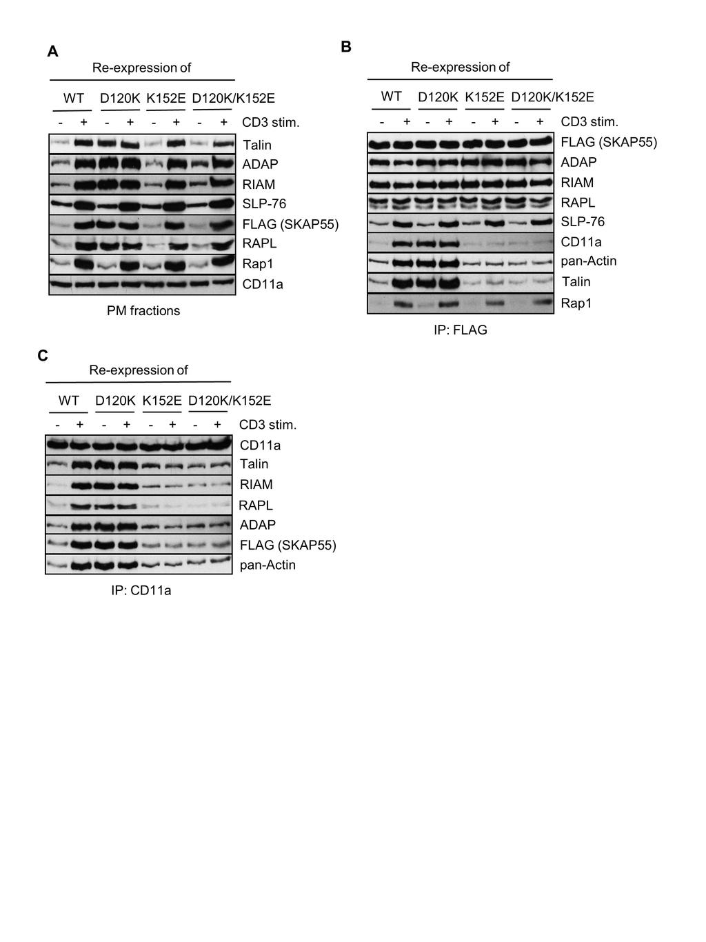
18 We generated a plasmid encoding a D120K mutant of SKAP55, which we expressed in Jurkat T cells to study the localization of this mutant. As shown in Fig. 6A, the D120K mutant displayed constitutive PM localization, comparable to the level that was found in TCRstimulated cells expressing wild type SKAP55 or the D120K/K152E double-mutant. Of note, TCR stimulation did not further increase the ratio of fluorescence intensity of the D120K mutant at the PM (Fig. 6A). Thus, similar to D129 within SKAP-HOM, D120 within the PH domain is a critical residue that regulates PM recruitment of SKAP Previous results from us and others had shown that constitutive PM targeting of SKAP55 (either using a LAT/SKAP55 chimera protein or by introducing a N-terminal myristoylation tag) induces T cell adhesion even in the absence of TCR-mediated stimuli (15, 21). Given the fact that the D120K mutant of SKAP55 displayed constitutive PM association, we hypothesized that this mutant would also induce TCR independent activation of LFA-1. Indeed, re-expression of SKAP55-D120K (Fig. 6B) induced basal adhesion to ICAM-1 (Fig. 6C) and LFA-1 affinity modulation (Fig. 6D). Note, that functional effects exerted by the D120K mutant of SKAP55 on adhesion were not due to an increased expression of the TCR or LFA-1 (Fig. S3B). In summary, D120K mediated PM targeting of SKAP55 induces spontaneous and constitutive activation of LFA To provide a biochemical basis for the functional behavior of D120K we prepared PM fractions (Fig. 7A), anti-flag- (SKAP55) immunoprecipitates (Fig. 7B) or anti-lfa-1 immunoprecipitates (Fig. 7C), respectively, from non-stimulated or TCR stimulated Jurkattransfectants expressing the SKAP55 D120K mutant. In line with the functional data, the D120K mutant as well as the known SKAP55 interaction partners ADAP, RIAM and RAPL localize to the PM even in the absence of TCR stimulation (Fig. 7A). Similarly, anti-flag (SKAP55) immunoprecipitates obtained from lysates of non-stimulated D120K transfectants 18
19 showed a constitutive association of the SKAP55 D120K mutant with Actin, Talin and LFA-1 (Fig. 7B). LFA-1-immunoprecipitates prepared from D120K expressing cells showed TCRindependent associations between LFA-1, Actin, RIAM, RAPL, ADAP, SKAP55 and Talin. Hence, mutation of D120 to K fully activates the ADAP/SKAP55-module at the PM Next we were interested to determine whether the D120K mutant would dominate/override the negative regulatory effect of the K152E mutant (or vice versa). To assess this question we generated a SKAP55 mutant carrying a double D120K/K152E mutation within the PH domain. Fig 7A shows that the D120K/K152E double mutant no longer localizes to the PM in non-stimulated cells and that cells expressing this mutant had lost their capability to adherence and to activate LFA-1 (Fig. 6C and D). Hence, the constitutive PM targeting of SKAP55 as well as the spontaneous integrin activation induced by D120K depends on K In addition to abrogating constitutive PM association of SKAP55, the D120K/K152E-doublemutant completely abrogated the TCR-induced binding of T cells to ICAM-1 and LFA-1 affinity modulation (Fig. 6C and D). Moreover TCR mediated association of the ADAP/SKAP55-module with LFA-1, Talin and Actin (Fig. 7B) as well as the TCR-mediated interaction of LFA-1 with the ADAP/SKAP55-module, Talin, RIAM and RAPL was severely attenuated (Fig. 7C). Hence, all functional effects exerted by the D120K single mutant were counteracted by additional mutation of K152 to E. Thus, K152E is dominant over D120K The constitutive adhesion of the D120K mutant of SKAP55 requires Talin but not Rap1. Unexpectedly, the experiments shown in Fig. 7A and B revealed that despite constitutive PM targeting of the ADAP/SKAP55-module and despite constitutive activation of LFA-1, Rap1 was not constitutively targeted to the PM in cells expressing D120K (Fig. 7A) and did not coprecipitate with SKAP55 in non-stimulated D120K Jurkat transfectants (Fig. 7B). Rather, 19
20 even in D120K expressing cells, membrane targeting of Rap1 as well as its interaction with the ADAP/SKAP55-module required stimulation of the TCR (Fig. 7A and B). This suggested that the constitutive activation of LFA-1 in cells expressing D120K occurs independently of Rap To address the role of Rap1 in more detail, we re-expressed the D120K mutant in Jurkat T cells in which Rap1 had been down-regulated by sirna. Fig. 8A shows that in cells reexpressing either wild type SKAP55 or the D120K mutant, knockdown of Rap1 did not affect basal adhesion, whereas TCR-mediated adhesion was almost completely blocked. This confirms that Rap1 is dispensable for steady state adhesion, while it is necessary for TCRmediated LFA-1 activation even in cells re-expressing D120K. In marked contrast to Rap1, knock-down of Talin reverted the spontaneous and the TCR-mediated integrin activation of cells expressing D120K (Fig. 8B). Hence, spontaneous adhesion induced by D120K-mutant depends on Talin
21 Discussion PH domains are generally known to bind lipids and to facilitate the translocation of proteins to the PM (36). In this study we characterized the lipid binding properties as well as the functional properties of the PH domain of SKAP55 with a focus on TCR mediated activation of the β2-integrin LFA-1. We identified two critical amino acids within the PH domain of SKAP55, D120 and K152 that appear to regulate membrane recruitment of the ADAP/SKAP55-module and T cell adhesion Structural inspection of the isolated PH domain of SKAP55 revealed that this domain contains a classical lipid-binding pocket that is present in many PH domains. It is build up by the variable β1-β2-, β3-β4- and β6-β7-loops (36). The PH domain of SKAP55 contains all amino acids (except serine 118 in the β1 strand) at homologous positions that have been predicted for the lipid binding of its homologue SKAP-HOM (35). By using NMR spectroscopy we showed that the PH domain of SKAP55 has a preference for PI(3,4,5)P 3 over PI(4,5)P 2 binding. However, in comparison to its homologue SKAP-HOM, the isolated PH domain of SKAP55 shows a much lower affinity for PI(3,4,5)P 3 binding (74±12μM for SKAP55 versus 8μM for SKAP-HOM; (35)). In part this difference could result from the different methods used in these two studies, namely NMR (this study) and fluorescence polarization (35). However, SKAP-HOM contains an arginine instead of serine (SKAP55) at position 118, a residue that is localized not far from the lipid-binding site. Thus SKAP55 likely harbours a non-optimal PIP-binding pocket Studies employing PI3-kinase inhibitor showed that both in Jurkat T cells as well as in primary human T lymphocytes the constitutive PM recruitment of the isolated PH domain of SKAP55 likely does not require PI(3,4,5)P 3 binding. In line with this finding, individual mutation of the putative PI(3,4,5)P 3 interacting residue, R131 and K116, only moderately 21
22 affected PM targeting of the isolated PH domain of SKAP55 in either Jurkat or human primary T cells. Functionally, using a genetic suppression/re-expression system, we could show that a K116M mutant of SKAP55 behaved almost as wild type SKAP55 while cells reexpressing a R131M mutant only showed slightly impaired adhesion to ICAM-1, a finding that is in line with previously published data (21). Hence, in our experimental system using suppression/re-expression plasmids for SKAP55 residue R131 appears to be of moderate importance for SKAP55 function In marked contrast to R131 and K116, mutation of K152 to E almost completely abolished PM localization of the PH domain of SKAP55. Previous studies had shown that positively charged amino acids (e.g. lysine residues such as K152) within particular PH domains allow these domains to interact with Actin (33, 34). Immunoprecipitation experiments revealed that this appears to be also true for the PH domain of SKAP55. Indeed, while the wild type SKAP55 PH domain co-precipitated Actin the corresponding K152E(M) mutants completely failed to do so. These findings strongly suggest that the isolated PH domain of SKAP55 is primarily targeted to the PM via a K152 mediated interaction with Actin. The identification of the PM-cytoskeleton linker molecules (e.g. Ezrin/radixin/moesin (ERM) proteins) that mediate the link between the isolated PH domain of SKAP55 and Actin requires further investigations Suppression of endogenous SKAP55 and re-expression of a SKAP55 K152E mutant in Jurkat T cells strongly impaired TCR induced adhesion and activation of LFA-1. Surprisingly however, biochemical analysis of PM fractions revealed that, in contrast to the isolated PH domain the K152 mutation did not alter TCR induced recruitment of SKAP55 and its association partners to the PM in the context of the full length SKAP55 molecule. Indeed, all components known to interact with SKAP55 in T cells either constitutively (ADAP, RIAM, 22
23 RAPL) or inducibly (SLP-76, Rap1) were found in membrane preparations of TCRstimulated cells expressing the K152E-mutant. This indicates that redundant pathways for membrane targeting of the whole complex exit that rescues the K152E mutant of full-length SKAP55, as found for the K152E mutant of the isolated PH domain. However, further analysis of anti-flag/skap55 and anti-lfa-1 immunoprecipitates showed that, despite normal PM targeting, the K152E mutation completely abrogated the capability of SKAP55 and its associated signalling complex to interact with Actin, Talin and LFA-1. Hence, K152 within the PH domain of SKAP55 is mandatory for the activation of LFA-1 by generating a molecular bridge between the ADAP/SKAP55-module, Actin and Talin As noted above, an important difference between the isolated PH domain of SKAP55 versus the full length SKAP55 molecule is that mutation of K152 in the isolated PH domain displaces the latter from the PM whereas mutation of K152 in the context of the full length SKAP55 molecule does not. One possibility to explain this difference would be that in contrast to the isolated PH domain, the full length SKAP55 could use two independent (or complementary) routes for PM targeting. One route would involve Actin/Talin binding via the PH domain whereas the second route would encompass an inducible association of the ADAP/SKAP55-complex with SLP-76 via ADAP (16). SLP-76 binds via Gads to LAT following phosphorylation of LAT by TCR/Lck-activated ZAP70. It is important to note that in T cells chemokine-mediated membrane-association of the ADAP/SKAP55-complex as well as chemokine induced activation of LFA-1 occurs in the absence of detectable LATphosphorylation and does not depend on an inducible association between SLP-76 and ADAP (23). Thus, it appears that SKAP55 mediated integrin activation does not necessarily depend on the ADAP/SLP76/Gads/pLAT-signaling pathway
24 Still, the fact that SKAP55 can activate integrins independently of membrane recruitment via the ADAP/SLP76/Gads/pLAT route raises the important question of an inducible (TCR or chemokine) membrane recruitment mechanism of SKAP55 that involves the PH domain. Our approach to elucidate this question was based on a previous publication by Swanson et al. in which the authors had investigated the functional properties of the PH domain of the SKAP55 homologue, SKAP-HOM (35). In this publication the colleagues had proposed a model in which an intramolecular switch mechanism occurring between the N-terminal DM domain and the PH domain of SKAP-HOM dynamically regulates PM targeting of the protein (35). In the closed conformation (i.e. in the absence of external stimuli) an interface between the DM domain and the PH domain supports a short helical segment in the β1-β2-loop incompatible with lipid binding. The authors identified D129 within the PH domain of SKAP-HOM to be critically involved in the DM-PH-switch mechanism. Indeed, mutation of D129 to K strongly increased the affinity of the PH domain of SKAP-HOM for PI(3,4,5)P 3. Consequently, expression of a D129K mutant of SKAP-HOM in macrophages induced spontaneous localization of the mutated molecule to Actin membrane ruffels (35). We identified D120 within the PH domain of SKAP55 representing the equivalent of D129 in SKAP-HOM. Reexpression of a SKAP55-D120K mutant in Jurkat T cells induced constitutive PM targeting of this mutant (as well as the constitutive binding partners of SKAP55, ADAP, RIAM and RAPL). Moreover, Jurkat T cells re-expressing the SKAP55-D120K mutant adhered to ICAM-1 in the absence of TCR mediated stimuli (Fig. 6). These findings corroborate previous reports, which had shown that forced PM recruitment of SKAP55 (by fusion with a myristoylation- or a LAT-tag) bypasses the need for TCR stimulation to induce adhesion (15, 21) Using the D120K mutant we made three important observations. First we noted that the constitutive activation of LFA-1 induced by D120K did not require membrane recruitment of 24
25 Rap1 or binding of Rap1 to either RIAM or RAPL. Indeed, knockdown of Rap1 in D120K expressing cells did not affect the spontaneously adhesion that was induced by D120K. Hence, it appears as if Rap1 is not required for this basal induced adhesion. Still, TCRmediated augmentation of adhesion was found to be depend on Rap1, both in cells expressing wild type SKAP55 as well as in cells expressing D120K, which is in line with previous published data showing that Rap1 deficiency reduces TCR-mediated adhesion but not adhesion in non-stimulated T cells (5) Second, in contrast to knockdown of Rap1, down-regulation of Talin partly blocked basal adhesion of cells expressing D120K and enhanced TCR induced adhesion by D120K was also attenuated. We have previously identified two independent ADAP/SKAP55-modules either interacting with RIAM or RAPL (28). RIAM has been reported to inducibly associate with Talin. This interaction promotes PM targeting of Talin, releases Talin from its auto-inhibition and allows binding to the cytoplasmic tail of the β-chain of LFA-1 (7, 37). Beside RIAM and Talin, RAPL interacts with the cytoplasmic tail of the α-chain of LFA-1. To our knowledge it is not known whether RAPL deficiency would affect inside-out signaling of LFA-1 upon TCR-stimulation. Independent of the exact mechanism, our experiments clearly show that Actin binding by SKAP55 crucially connects the cytoskeleton to LFA Third, we found that additional mutation of K152 (SKAP55-D120K/K152E double mutant) completely abrogated both the constitutive membrane localization of the D120K mutant as well as the constitutive activation of LFA-1 induced by D120K. Biochemical analysis of the SKAP55 and LFA-1 associated signaling complexes revealed that this was due to a failure of the double mutant to interact with Actin and Talin and, hence on a failure to interact with and to activate LFA-1. In summary, D120K can only activate integrins in the presence of K152, a residue that is crucial for both, lipid and Actin binding. 25
26 Our data suggest a model where in non-stimulated T cells spontaneous membrane recruitment of SKAP55 and its associated signaling complex is prevented by an intramolecular switch mechanism. Here the N-terminal DM domain of SKAP55 folds back on the PH domain thereby blocking the interaction of the SKAP55 associated signaling complex with Actin and Talin. Upon TCR stimulation the DM-PH domain switch undergoes a conformational change that releases the PH domain from auto-inhibition and allows the signaling complex to interact with Actin/Talin thereby activating LFA-1. In parallel (or subsequently), PM localization of the ADAP/SKAP55-module is stabilized by the SLP-76/Gads signaling complex that binds to phosphorylated LAT. Together the two membrane targeting mechanisms facilitate stable T cell adhesion (Fig. S5) The obvious question emerging from the proposed model is how the DM-PH-domain switch is released from auto-inhibition following TCR stimulation. Dimerization domains are often involved in intra- or intermolecular protein-protein interactions, which can be used for autoinhibition of functional domains (38). Moreover, Ophir and colleagues have recently shown that SKAP55 forms homo-dimers (and hetero-dimers with SKAP-HOM) and that this dimerization is mediated by the DM domain of SKAP55 (18). TCR-induced dimerization (or de-dimerization) of SKAP55 upon T cell stimulation could therefore induce conformational changes within the DM domain that could release the PH domain from auto-inhibition and thus allows PM targeting of SKAP55. An alternative scenario would be that local production of PI(3,4,5)P 3 upon T cell stimulation shifts the equilibrium towards the open, Actin binding conformation of SKAP Another puzzling question that needs clarification is why single point mutations within the PH domain of SKAP55 (or SKAP-HOM) produce dramatic functional effects in terms of integrin activation (15, 35), whereas deletion of the whole PH domain of SKAP55 (or of 26
27 SKAP-HOM) does not alter integrin functions (see also Figure S6) or subcellular localization as shown in various model systems (15, 18, 35). In striking contrast to cells expressing the D120K/K152E-double mutant of SKAP55 (in which binding to of the ADAP/SKAP55 module to LFA-1, Talin and Actin was reduced, Figure 6), we did not find a significant alteration of the LFA-1-associated signaling complex in cells expressing a SKAP55-variant where the whole PH domain was deleted (Figure S6C). Therefore, we currently cannot provide a model that would allow us to explain the contradicting and reported results that have been obtained using deletion mutants vs. single point mutations. As indicated by the structure (35) the interactions of the PH and the DM domain are intertwined, and a released DM domain will certainly be able to exert certain functions of the molecule. Furthermore, within the context of the larger complex of the SKAP55/ADAP-module redundant Actin and lipid binding properties might render the PH domain function seemingly dispensable. However, the tuning of the integrin response as shown here to be strongly modulated by the PH domain of SKAP55, might become visible only at certain relative concentrations of SKAP55 and its direct binding partners, including PIP s and Actin We here have identified two critical residues within the PH domain of SKAP55, D120 and K152 that regulate SKAP55 functions with regard to PM targeting. D120 appears to mediate the intra-molecular release of the DM-PH-switch. However, K152 overrides this feature and shows that release of auto-inhibition has to be followed by productive Actin binding in order to allow SKAP55 and its associated signalling complex to promote functional activation of LFA
28 Funding Information This work was supported by the Medical faculty of the Otto-von-Guericke University (stipend for AW) and DFG grants SFB 854 (B10, B12, B19 and Z01 for CF, SK, BS, AM and/or LP) and SFB 958/765 (A7 or C4 for CF). The authors declare no competing financial interest Acknowledgment We thank Anke Ramonat for excellent technical assistance and Nirdosh Dadwal for purification of proteins
29 References 1. Abram CL, Lowell CA The ins and outs of leukocyte integrin signaling. Annu Rev Immunol 27: doi: /annurev.immunol Hogg N, Patzak I, Willenbrock F The insider's guide to leukocyte integrin signalling and function. Nat Rev Immunol 11: doi: /nri Kinashi T Intracellular signalling controlling integrin activation in lymphocytes. Nat Rev Immunol 5: doi: /nri Moser M, Legate KR, Zent R, Fassler R The tail of integrins, talin, and kindlins. Science 324: doi: /science Su W, Wynne J, Pinheiro EM, Strazza M, Mor A, Montenont E, Berger J, Paul DS, Bergmeier W, Gertler FB, Philips MR Rap1 and its effector RIAM are required for lymphocyte trafficking. Blood 126: doi: /blood Klapproth S, Sperandio M, Pinheiro EM, Prunster M, Soehnlein O, Gertler FB, Fassler R, Moser M Loss of the Rap1 effector RIAM results in leukocyte adhesion deficiency due to impaired beta2 integrin function in mice. Blood 126: doi: /blood Calderwood DA The Rap1-RIAM pathway prefers beta2 integrins. Blood 126: doi: /blood Witte A, Degen J, Baumgart K, Waldt N, Kuropka B, Freund C, Schraven B, Kliche S Emerging roles of ADAP, SKAP55, and SKAP-HOM for integrin and NF-kB signaling in T cells. Clinical & Cellular Immunology S12. doi: / s Griffiths EK, Krawczyk C, Kong YY, Raab M, Hyduk SJ, Bouchard D, Chan VS, Kozieradzki I, Oliveira-Dos-Santos AJ, Wakeham A, Ohashi PS, Cybulsky MI, Rudd CE, Penninger JM Positive regulation of T cell activation and integrin adhesion by the adapter Fyb/Slap. Science 293: doi: /science Peterson EJ, Woods ML, Dmowski SA, Derimanov G, Jordan MS, Wu JN, Myung PS, Liu QH, Pribila JT, Freedman BD, Shimizu Y, Koretzky GA Coupling of the TCR to integrin activation by Slap-130/Fyb. Science 293: doi: /science Wang H, Liu H, Lu Y, Lovatt M, Wei B, Rudd CE Functional defects of SKAP-55-deficient T cells identify a regulatory role for the adaptor in LFA-1 adhesion. Mol Cell Biol 27: doi: /mcb
30 Burbach BJ, Srivastava R, Ingram MA, Mitchell JS, Shimizu Y The pleckstrin homology domain in the SKAP55 adapter protein defines the ability of the adapter protein ADAP to regulate integrin function and NF-kappaB activation. J Immunol 186: doi: /jimmunol Mitchell JS, Burbach BJ, Srivastava R, Fife BT, Shimizu Y Multistage T cell-dendritic cell interactions control optimal CD4 T cell activation through the ADAP-SKAP55-signaling module. J Immunol 191: doi: /jimmunol Huang Y, Norton DD, Precht P, Martindale JL, Burkhardt JK, Wange RL Deficiency of ADAP/Fyb/SLAP-130 destabilizes SKAP55 in Jurkat T cells. J Biol Chem 280: doi: /jbc.m Kliche S, Breitling D, Togni M, Pusch R, Heuer K, Wang X, Freund C, Kasirer-Friede A, Menasche G, Koretzky GA, Schraven B The ADAP/SKAP55 signaling module regulates T- cell receptor-mediated integrin activation through plasma membrane targeting of Rap1. Mol Cell Biol 26: doi: /mcb Wang H, McCann FE, Gordan JD, Wu X, Raab M, Malik TH, Davis DM, Rudd CE ADAP-SLP-76 binding differentially regulates supramolecular activation cluster (SMAC) formation relative to T cell-apc conjugation. J Exp Med 200: doi: /jem Marie-Cardine A, Bruyns E, Eckerskorn C, Kirchgessner H, Meuer SC, Schraven B Molecular cloning of SKAP55, a novel protein that associates with the protein tyrosine kinase p59fyn in human T-lymphocytes. J Biol Chem 272: Ophir MJ, Liu BC, Bunnell SC The N terminus of SKAP55 enables T cell adhesion to TCR and integrin ligands via distinct mechanisms. J Cell Biol 203: doi: /jcb Menasche G, Kliche S, Chen EJ, Stradal TE, Schraven B, Koretzky G RIAM links the ADAP/SKAP-55 signaling module to Rap1, facilitating T-cell-receptor-mediated integrin activation. Mol Cell Biol 27: doi: /mcb Raab M, Wang H, Lu Y, Smith X, Wu Z, Strebhardt K, Ladbury JE, Rudd CE T cell receptor "inside-out" pathway via signaling module SKAP1-RapL regulates T cell motility and interactions in lymph nodes. Immunity 32: doi: /j.immuni Raab M, Smith X, Matthess Y, Strebhardt K, Rudd CE SKAP1 protein PH domain determines RapL membrane localization and Rap1 protein complex formation for T cell receptor (TCR) activation of LFA-1. J Biol Chem 286: doi: /jbc.m
31 Lim D, Lu Y, Rudd CE Non-cleavable talin rescues defect in the T-cell conjugation of T-cells deficient in the immune adaptor SKAP1. Immunol Lett 172: doi: /j.imlet Horn J, Wang X, Reichardt P, Stradal TE, Warnecke N, Simeoni L, Gunzer M, Yablonski D, Schraven B, Kliche S Src homology 2-domain containing leukocyte-specific phosphoprotein of 76 kda is mandatory for TCR-mediated inside-out signaling, but dispensable for CXCR4-mediated LFA-1 activation, adhesion, and migration of T cells. J Immunol 183: doi: /jimmunol Cabanas C, Hogg N Ligand intercellular adhesion molecule 1 has a necessary role in activation of integrin lymphocyte function-associated molecule 1. Proc Natl Acad Sci U S A 90: Reedquist KA, Ross E, Koop EA, Wolthuis RM, Zwartkruis FJ, van Kooyk Y, Salmon M, Buckley CD, Bos JL The small GTPase, Rap1, mediates CD31-induced integrin adhesion. J Cell Biol 148: Marie-Cardine A, Hendricks-Taylor LR, Boerth NJ, Zhao H, Schraven B, Koretzky GA Molecular interaction between the Fyn-associated protein SKAP55 and the SLP-76-associated phosphoprotein SLAP-130. J Biol Chem 273: Togni M, Swanson KD, Reimann S, Kliche S, Pearce AC, Simeoni L, Reinhold D, Wienands J, Neel BG, Schraven B, Gerber A Regulation of in vitro and in vivo immune functions by the cytosolic adaptor protein SKAP-HOM. Mol Cell Biol 25: doi: /mcb Kliche S, Worbs T, Wang X, Degen J, Patzak I, Meineke B, Togni M, Moser M, Reinhold A, Kiefer F, Freund C, Forster R, Schraven B CCR7-mediated LFA-1 functions in T cells are regulated by 2 independent ADAP/SKAP55 modules. Blood 119: doi: /blood Musci MA, Hendricks-Taylor LR, Motto DG, Paskind M, Kamens J, Turck CW, Koretzky GA Molecular cloning of SLAP-130, an SLP-76-associated substrate of the T cell antigen receptorstimulated protein tyrosine kinases. J Biol Chem 272: Vranken WF, Boucher W, Stevens TJ, Fogh RH, Pajon A, Llinas M, Ulrich EL, Markley JL, Ionides J, Laue ED The CCPN data model for NMR spectroscopy: development of a software pipeline. Proteins 59: doi: /prot Ferguson KM, Lemmon MA, Schlessinger J, Sigler PB Structure of the high affinity complex of inositol trisphosphate with a phospholipase C pleckstrin homology domain. Cell 83:
32 Costello PS, Gallagher M, Cantrell DA Sustained and dynamic inositol lipid metabolism inside and outside the immunological synapse. Nat Immunol 3: doi: /ni Yao L, Janmey P, Frigeri LG, Han W, Fujita J, Kawakami Y, Apgar JR, Kawakami T Pleckstrin homology domains interact with filamentous actin. J Biol Chem 274: Macia E, Partisani M, Favard C, Mortier E, Zimmermann P, Carlier MF, Gounon P, Luton F, Franco M The pleckstrin homology domain of the Arf6-specific exchange factor EFA6 localizes to the plasma membrane by interacting with phosphatidylinositol 4,5-bisphosphate and F-actin. J Biol Chem 283: doi: /jbc.m Swanson KD, Tang Y, Ceccarelli DF, Poy F, Sliwa JP, Neel BG, Eck MJ The Skap-hom dimerization and PH domains comprise a 3'-phosphoinositide-gated molecular switch. Mol Cell 32: doi: /j.molcel Lemmon MA Pleckstrin homology (PH) domains and phosphoinositides. Biochem Soc Symp doi: /bss : doi: /bss Yang J, Zhu L, Zhang H, Hirbawi J, Fukuda K, Dwivedi P, Liu J, Byzova T, Plow EF, Wu J, Qin J Conformational activation of talin by RIAM triggers integrin-mediated cell adhesion. Nat Commun 5:5880. doi: /ncomms Mason JM, Arndt KM Coiled coil domains: stability, specificity, and biological implications. Chembiochem 5: doi: /cbic
33 Figure legends Figure 1. In vitro lipid binding properties of the isolated PH domain of SKAP55 (A) 1 H- 15 N HSQC titration of 270µM wild type SKAP55 PH domain with increasing concentrations (50, 150, 300, 600, 1130 and 2466µM) of IP 4, the head group of PI(3,4,5)P 3. Some of the most shifts are indicated. (B) Shown are curve fits of combined 1 H- 15 N HSQC chemical shift changes with increasing IP 4 concentrations for significantly shifting residues E115, K117, R131, Y142, T154, R185. The individual K D values derived from the fits are given. (C) Significant chemical shift differences in the isolated PH domain of wild type SKAP55 upon addition of a 10fold molar excess of IP 4 are highlighted on the structure (PDB code: 1u5d) overlayed with IP4 co-crystallized with the PH domain of AKT (PDB code: 1unq). Residues K116, R131, and K152 involved in IP 4 binding and suggested for mutation are drawn as sticks. (D) 270µM wild type (WT), R131M and K152E mutants of the PH domain of SKAP55 upon incubation with 2466µM IP 4. While large chemical shifts are seen for certain NH resonances of the wild type protein, shifts are very small for the R131M variant and are totally abolished for the K152E mutant protein Figure 2. PI(3,4,5)P 3 -independent membrane targeting of the isolated PH domain of SKAP55 (A) Jurkat T cells were transfected with plasmids encoding GFP, GFP-PH-PLCδ, GFP-PH- AKT, or PH-SKAP55-GFP. 24h after transfection, cells were treated with DMSO or Wortmannin for 1h at 37 C. Cells were fixed, permeabilized, stained with TRITC-phalloidin to visualize F-Actin and then imaged by confocal laser scanning microscopy. A histogram tool was used to determine the fluorescence intensity at the PM of individual cells (see Fig. S1) to calculate the ratio of fluorescence intensity after subtraction of GFP-background levels (n=3-4). In the right panel lysates were prepared from these transfectants to assess the inhibition of the PI3 kinase as monitored by Western blotting for pakt S473 (right panel) 33
34 (n=3). (B) Primary human T cells were transfected with plasmids as described in A. 24h after transfection, cells were pretreated with DMSO or Wortmannin for 1h at 37 C and subsequently stimulated with anti-cd3/cd28 antibodies. Cells were analyzed as described in A. In the lower panel CD3/CD28-induced activation of the PI3 kinase in the presence or absence of Wortmannin was monitored by Western blot analysis for pakt S473 (n=3). (mean ± SD; ** p 0.01, *** p 0.001) Figure 3. PM targeting of the PH domain of SKAP55 is mediated by interaction with Actin and depends on lysine 152. (A) Jurkat T cells were transfected with plasmids encoding GFP, GFP-PH-PLCδ, GFP-PH- AKT, or PH-SKAP55-GFP. 24h after transfection, the expression of the GFP and GFP fusion proteins was assessed by Western blot analysis with the indicated antibodies (left panel). Lysates from these transfectants were used for anti-gfp immunoprecipitation. Precipitates were analyzed by Western blotting for pan-actin and GFP (right panel). Lysate of Jurkat T cells (JE6) served as positive control for the detection of pan-actin (n=3). (B) Jurkat T cells were transfected with plasmids encoding GFP, PH-SKAP55-GFP wild type (WT) or its mutants (K116, R131M, K152E, and K152M), respectively. 24h after transfection, the expression of GFP and GFP-fusion proteins was assessed by Western blot analysis with the indicated antibodies (left panel). Lysates of these transfectants were used for anti-gfp immunoprecipitation. Precipitates were analyzed by Western blotting for pan-actin and GFP (right panel). Lysate of Jurkat T cells (JE6) served as positive control for detection of pan- Actin (n=2). Jurkat T cells (C) or primary human T cells (D) were transfected with plasmids encoding GFP, PH-SKAP55-GFP wild type (WT) or its mutants (K116M, R131M, K152E, and K152M), respectively. 24h after transfection, cells were fixed, permeabilized, stained with TRITC-phalloidin and then imaged by confocal laser scanning microscopy. The ratio of 34
35 fluorescence intensity after subtraction of GFP-background levels was calculated as described in Figure 2A (n=3-4). (mean ± SD; ** p 0.01) Figure 4. The K152 mutant of SKAP55 impairs TCR-mediated adhesion. (A) Schematic representation of the suppression/re-expression vector used in this study. (B) Jurkat T cells were transfected with suppression/re-expression constructs which do not suppress endogenous SKAP55 (shc), or reduce the endogenous protein level of SKAP55 (shskap55) or re-express a FLAG-tagged shrna-resistant wild type SKAP55 (WT) or its mutants (K116M, R131M, and K152E). 48h after transfection, lysates were analyzed by Western blotting with the indicated antibodies. (C) Transfected Jurkat T cells as described in B were analyzed for their ability to adhere to ICAM-1-coated wells in a resting state or upon stimulation with two different concentrations of CD3 antibodies (1μg/ml and 5μg/ml), respectively. Adherent cells were counted and calculated as percentage of input (n=3). (mean ± SD; *p 0.05; **p 0.01) Figure 5. K152E mutation abolishes the TCR-mediated interaction of LFA-1 with Actin and Talin. (A) Jurkat T cells were transfected with suppression/re-expression constructs which reexpress a FLAG-tagged shrna-resistant SKAP55 wild type (WT) or its mutants (K116M, R131M, and K152E). After 48h, cells were left untreated or stimulated with CD3 antibodies (CD3). PM fractions were isolated and analyzed by Western blotting with the indicated antibodies. Detection of CD11a served as control for fractionation of PM (n=2). (B and C) Jurkat T cells transfected as described in (A) were left untreated or stimulated with CD3 antibodies (CD3). Lysates were used for immunoprecipitation of either SKAP55 using anti- FLAG antibodies or LFA-1 using anti-cd11a antibodies. Precipitates were analyzed by Western blotting using the indicated antibodies (n=2). 35
36 Figure 6. The D120K mutant of SKAP55 induces TCR-independent adhesion and LFA- 1 activation (A) Jurkat T cells were transfected with plasmids encoding GFP, full length wild type SKAP55-GFP (WT), or its mutants (D120K and D120K/K152E). 24h after transfection, cells were left untreated or stimulated with CD3 antibodies. Subsequently cells were fixed, permeabilized, probed with TRITC-phalloidin, and imaged by confocal laser scanning microscopy. The ratio of fluorescence intensity after substraction of GFP-background levels was calculated as described in Fig. 2A (n=3). TCR stimulation was assessed by analyzing perk1/2 using Western blotting (right panel). (B) Jurkat T cells were transfected with suppression/re-expression constructs which do not suppress endogenous SKAP55 (shc), or reduce the endogenous protein level of SKAP55 (shskap55) and re-express a FLAG-tagged shrna-resistant wild type SKAP55 (WT) or its mutants (D120K, K152E or D120K/K152E). 48h after transfection, lysates were analyzed by Western blotting using the indicated antibodies. (C) Jurkat T cells were transfected as described in B. Cells were analyzed for their ability to adhere to ICAM-1-coated wells in a resting state or upon stimulation with two different concentrations of CD3 antibodies (1μg/ml and 5μg/ml), respectively. Adherent cells were counted and calculated as a percentage of input (n=3). (D) Jurkat T cells transfected as described in B were left untreated (ICAM-1) or stimulated with CD3 antibodies (ICAM- 1/CD3), followed by staining with the anti-lfa-1 antibody mab24. mab24 epitope expression was assessed by flow cytometry within the GFP gate and the mean fluorescence intensity of untreated shc-transfected Jurkat T cells was set to 1 to calculate the fold induction (n=3). (mean ± SD; *p 0.05; **p 0.01) Figure 7. The K152E mutant of SKAP55 overwrites LFA-1 activation by the D120K mutant (A) Jurkat T cells were transfected with suppression/re-expression constructs which re- 36
37 express a FLAG-tagged shrna-resistant SKAP55 wild type (WT) or its mutants (D120K, K152E, or D120K/K152E). 48h after transfection, cells were left untreated or stimulated with CD3 antibodies. PM fractions were isolated and analyzed by Western blotting using the indicated antibodies. Detection of CD11a served as control for fractionation of PMs (n=2). (B and C) Jurkat T cells transfected as described in A were left untreated or stimulated with CD3 antibodies (CD3). Lysates were used for immunoprecipitation of SKAP55 (using anti-flag antibodies) or (LFA-1 using anti-cd11a antibodies). Precipitates were analyzed by Western blotting using the indicated antibodies (n=2) Figure 8. TCR-independent adhesion induced by the D120K mutant of SKAP55 is dependent on Talin but not on Rap1. (A) Jurkat T cells were co-transfected with suppression/re-expression constructs which reexpress a FLAG-tagged shrna-resistant wild type (WT) SKAP55 or the D120K mutant in combination with either control sirna (sic) or sirna against Rap1 (sirap1). 48h after transfection, cells were left untreated or stimulated CD3 antibodies (1μg/ml). Cells were analyzed for their ability to adhere to ICAM-1-coated wells in a resting state or upon stimulation with CD3 antibodies. Adherent cells were counted and calculated as percentage of input. Lysates were prepared from transfectants and analyzed by Western blotting for FLAG, Rap1 and β-actin (right panel) (n=3). (B) Jurkat T cells were co-transfected with suppression/re-expression constructs as described in A in combination with either control sirna (sic) or sirna against Talin (sitalin). 48h after transfection, cells were left untreated or stimulated CD3 antibodies (1μg/ml). Cells were analyzed for their ability to adhere to ICAM-1-coated wells in a resting state or upon stimulation with CD3 antibodies. Adherent cells were counted and calculated as percentage of input. Lysates were prepared from transfected cells and analyzed by Western blotting using the indicated antibodies (right panel) (n=3). (mean ± SD; *p 0.05; **p 0.01) 37
38
39
40
41
42
43
44
SUPPLEMENTARY INFORMATION
 Supplementary Figures Supplementary Figure S1. Binding of full-length OGT and deletion mutants to PIP strips (Echelon Biosciences). Supplementary Figure S2. Binding of the OGT (919-1036) fragments with
Nature Immunology: doi: /ni.3631
 Supplementary Figure 1 SPT analyses of Zap70 at the T cell plasma membrane. (a) Total internal reflection fluorescent (TIRF) excitation at 64-68 degrees limits single molecule detection to 100-150 nm above
Supplementary Figure 1 CD4 + T cells from PKC-θ null mice are defective in NF-κB activation during T cell receptor signaling. CD4 + T cells were
 Supplementary Figure 1 CD4 + T cells from PKC-θ null mice are defective in NF-κB activation during T cell receptor signaling. CD4 + T cells were isolated from wild type (PKC-θ- WT) or PKC-θ null (PKC-θ-KO)
SUPPLEMENTARY INFORMATION. Supplementary Figures S1-S9. Supplementary Methods
 SUPPLEMENTARY INFORMATION SUMO1 modification of PTEN regulates tumorigenesis by controlling its association with the plasma membrane Jian Huang 1,2#, Jie Yan 1,2#, Jian Zhang 3#, Shiguo Zhu 1, Yanli Wang
Supplementary Figure 1. Normal T lymphocyte populations in Dapk -/- mice. (a) Normal thymic development in Dapk -/- mice. Thymocytes from WT and Dapk
 Normal thymic development in Dapk -/- mice. Thymocytes from WT and Dapk") Supplementary Figure 1. Normal T lymphocyte populations in Dapk -/- mice. (a) Normal thymic development in Dapk -/- mice. Thymocytes from WT and Dapk -/- mice were stained for expression of CD4 and CD8.
Supplementary Figure 1. Normal T lymphocyte populations in Dapk -/- mice. (a) Normal thymic development in Dapk -/- mice. Thymocytes from WT and Dapk -/- mice were stained for expression of CD4 and CD8.
Supplements. Figure S1. B Phalloidin Alexa488
 Supplements A, DMSO, PP2, PP3 Crk-myc Figure S1. (A) Src kinase activity is necessary for recruitment of Crk to Nephrin cytoplasmic domain. Human podocytes expressing /7-NephrinCD () were treated with
Nature Biotechnology: doi: /nbt.3828
 Supplementary Figure 1 Development of a FRET-based MCS. (a) Linker and MA2 modification are indicated by single letter amino acid code. indicates deletion of amino acids and N or C indicate the terminus
Figure S1. Generation of inducible PTEN deficient mice and the BMMCs (A) B6.129 Pten loxp/loxp mice were mated with B6.
 B6.129 Pten loxp/loxp mice were mated with B6.") Figure S1. Generation of inducible PTEN deficient mice and the BMMCs (A) B6.129 Pten loxp/loxp mice were mated with B6.129-Gt(ROSA)26Sor tm1(cre/ert2)tyj /J mice. To induce deletion of the Pten locus,
Figure S1. Generation of inducible PTEN deficient mice and the BMMCs (A) B6.129 Pten loxp/loxp mice were mated with B6.129-Gt(ROSA)26Sor tm1(cre/ert2)tyj /J mice. To induce deletion of the Pten locus,
Supplementary Figure S1. Venn diagram analysis of mrna microarray data and mirna target analysis. (a) Western blot analysis of T lymphoblasts (CLS)
 Western blot analysis of T lymphoblasts (CLS)") Supplementary Figure S1. Venn diagram analysis of mrna microarray data and mirna target analysis. (a) Western blot analysis of T lymphoblasts (CLS) and their exosomes (EXO) in resting (REST) and activated
Supplementary Figure S1. Venn diagram analysis of mrna microarray data and mirna target analysis. (a) Western blot analysis of T lymphoblasts (CLS) and their exosomes (EXO) in resting (REST) and activated
Enzyme-coupled Receptors. Cell-surface receptors 1. Ion-channel-coupled receptors 2. G-protein-coupled receptors 3. Enzyme-coupled receptors
 Enzyme-coupled Receptors Cell-surface receptors 1. Ion-channel-coupled receptors 2. G-protein-coupled receptors 3. Enzyme-coupled receptors Cell-surface receptors allow a flow of ions across the plasma
LFA-1 activates focal adhesion kinases FAK1/PYK2 to generate LAT-GRB2-SKAP1 complexes that terminate T-cell conjugate formation
 Received 27 Sep 216 Accepted 23 May 217 Published 12 Jul 217 DOI: 1.138/ncomms161 OPEN LFA-1 activates focal adhesion kinases /PYK2 to generate -GRB2- complexes that terminate T-cell conjugate formation
Supplementary Figure 1 Role of Raf-1 in TLR2-Dectin-1-mediated cytokine expression
 Supplementary Figure 1 Supplementary Figure 1 Role of Raf-1 in TLR2-Dectin-1-mediated cytokine expression. Quantitative real-time PCR of indicated mrnas in DCs stimulated with TLR2-Dectin-1 agonist zymosan
Supplementary Figure 1.TRIM33 binds β-catenin in the nucleus. a & b, Co-IP of endogenous TRIM33 with β-catenin in HT-29 cells (a) and HEK 293T cells
 and HEK 293T cells") Supplementary Figure 1.TRIM33 binds β-catenin in the nucleus. a & b, Co-IP of endogenous TRIM33 with β-catenin in HT-29 cells (a) and HEK 293T cells (b). TRIM33 was immunoprecipitated, and the amount of
Supplementary Figure 1.TRIM33 binds β-catenin in the nucleus. a & b, Co-IP of endogenous TRIM33 with β-catenin in HT-29 cells (a) and HEK 293T cells (b). TRIM33 was immunoprecipitated, and the amount of
a b G75 G60 Sw-2 Sw-1 Supplementary Figure 1. Structure predictions by I-TASSER Server.
 a b G75 2 2 G60 Sw-2 Sw-1 Supplementary Figure 1. Structure predictions by I-TASSER Server. a. Overlay of top 10 models generated by I-TASSER illustrates the potential effect of 7 amino acid insertion
Chapter 8. Interaction between the phosphatidylinositol 3- kinase SH3 domain and a photocleavable cyclic peptide
 Interaction between the phosphatidylinositol 3- kinase SH3 domain and a photocleavable cyclic peptide 129 Abstract The interaction of the PI3K SH3 domain with a cyclic photocleavable peptide and the linear
(a) Significant biological processes (upper panel) and disease biomarkers (lower panel)
 Significant biological processes (upper panel) and disease biomarkers (lower panel)") Supplementary Figure 1. Functional enrichment analyses of secretomic proteins. (a) Significant biological processes (upper panel) and disease biomarkers (lower panel) 2 involved by hrab37-mediated secretory
Supplementary Figure 1. Functional enrichment analyses of secretomic proteins. (a) Significant biological processes (upper panel) and disease biomarkers (lower panel) 2 involved by hrab37-mediated secretory
SUPPLEMENTARY INFORMATION
 SUPPLEMENTARY INFORMATION doi:1.138/nature9814 a A SHARPIN FL B SHARPIN ΔNZF C SHARPIN T38L, F39V b His-SHARPIN FL -1xUb -2xUb -4xUb α-his c Linear 4xUb -SHARPIN FL -SHARPIN TF_LV -SHARPINΔNZF -SHARPIN
Cell Biology Lecture 9 Notes Basic Principles of cell signaling and GPCR system
 Cell Biology Lecture 9 Notes Basic Principles of cell signaling and GPCR system Basic Elements of cell signaling: Signal or signaling molecule (ligand, first messenger) o Small molecules (epinephrine,
Supplementary Material and Methods
 Online Supplement Kockx et al, Secretion of Apolipoprotein E from Macrophages 1 Supplementary Material and Methods Cloning of ApoE-GFP Full-length human apoe3 cdna (pcdna3.1/zeo + -apoe) was kindly provided
Supplementary Information
 Supplementary Information Supplementary Figure 1. CD4 + T cell activation and lack of apoptosis after crosslinking with anti-cd3 + anti-cd28 + anti-cd160. (a) Flow cytometry of anti-cd160 (5D.10A11) binding
Data Sheet TIGIT / NFAT Reporter - Jurkat Cell Line Catalog #60538
 Data Sheet TIGIT / NFAT Reporter - Jurkat Cell Line Catalog #60538 Background: TIGIT is a co-inhibitory receptor that is highly expressed in Natural Killer (NK) cells, activated CD4+, CD8+ and regulatory
Supplemental information
 Carcinoemryonic antigen-related cell adhesion molecule 6 (CEACAM6) promotes EGF receptor signaling of oral squamous cell carcinoma metastasis via the complex N-glycosylation y Chiang et al. Supplemental
Supplementary Figure 1
 Supplementary Figure 1 Supplementary Figure 1. Neither the activation nor suppression of the MAPK pathway affects the ASK1/Vif interaction. (a, b) HEK293 cells were cotransfected with plasmids encoding
Signal Transduction Cascades
 Signal Transduction Cascades Contents of this page: Kinases & phosphatases Protein Kinase A (camp-dependent protein kinase) G-protein signal cascade Structure of G-proteins Small GTP-binding proteins,
TFEB-mediated increase in peripheral lysosomes regulates. Store Operated Calcium Entry
 TFEB-mediated increase in peripheral lysosomes regulates Store Operated Calcium Entry Luigi Sbano, Massimo Bonora, Saverio Marchi, Federica Baldassari, Diego L. Medina, Andrea Ballabio, Carlotta Giorgi
Signaling. Dr. Sujata Persad Katz Group Centre for Pharmacy & Health research
 Signaling Dr. Sujata Persad 3-020 Katz Group Centre for Pharmacy & Health research E-mail:sujata.persad@ualberta.ca 1 Growth Factor Receptors and Other Signaling Pathways What we will cover today: How
Nature Immunology: doi: /ni.3866
 Nature Immunology: doi:10.1038/ni.3866 Supplementary Figure 1 The effect of TIPE2 on chemotaxis. a, The expression of TIPE2 in dhl-60c, dhl-60t, TIPE2-expressing and 15/16Q-expressing dhl-60t neutrophils
Supplemental Information
 Supplemental Information Tobacco-specific Carcinogen Induces DNA Methyltransferases 1 Accumulation through AKT/GSK3β/βTrCP/hnRNP-U in Mice and Lung Cancer patients Ruo-Kai Lin, 1 Yi-Shuan Hsieh, 2 Pinpin
Incorporation of photo-caged lysine (pc-lys) at K273 of human LCK allows specific control of the enzyme activity.
 at K273 of human LCK allows specific control of the enzyme activity.") Supplementary Figure 1 Incorporation of photo-caged lysine (pc-lys) at K273 of human LCK allows specific control of the enzyme activity. (a) Modeling of the kinase domain of LCK with ATP (left) or pc-lys
Supplementary Figure 1 Incorporation of photo-caged lysine (pc-lys) at K273 of human LCK allows specific control of the enzyme activity. (a) Modeling of the kinase domain of LCK with ATP (left) or pc-lys
Supplementary Fig. 1. GPRC5A post-transcriptionally down-regulates EGFR expression. (a) Plot of the changes in steady state mrna levels versus
 Plot of the changes in steady state mrna levels versus") Supplementary Fig. 1. GPRC5A post-transcriptionally down-regulates EGFR expression. (a) Plot of the changes in steady state mrna levels versus changes in corresponding proteins between wild type and Gprc5a-/-
Supplementary Fig. 1. GPRC5A post-transcriptionally down-regulates EGFR expression. (a) Plot of the changes in steady state mrna levels versus changes in corresponding proteins between wild type and Gprc5a-/-
SUPPLEMENTARY INFORMATION
 Supplementary Table 1. Cell sphingolipids and S1P bound to endogenous TRAF2. Sphingolipid Cell pmol/mg TRAF2 immunoprecipitate pmol/mg Sphingomyelin 4200 ± 250 Not detected Monohexosylceramide 311 ± 18
Supplementary data Supplementary Figure 1 Supplementary Figure 2
 Supplementary data Supplementary Figure 1 SPHK1 sirna increases RANKL-induced osteoclastogenesis in RAW264.7 cell culture. (A) RAW264.7 cells were transfected with oligocassettes containing SPHK1 sirna
William C. Comb, Jessica E. Hutti, Patricia Cogswell, Lewis C. Cantley, and Albert S. Baldwin
 Molecular Cell, Volume 45 Supplemental Information p85 SH2 Domain Phosphorylation by IKK Promotes Feedback Inhibition of PI3K and Akt in Response to Cellular Starvation William C. Comb, Jessica E. Hutti,
Signaling Through Immune System Receptors (Ch. 7)
") Signaling Through Immune System Receptors (Ch. 7) 1. General principles of signal transduction and propagation. 2. Antigen receptor signaling and lymphocyte activation. 3. Other receptors and signaling
Signaling Through Immune System Receptors (Ch. 7) 1. General principles of signal transduction and propagation. 2. Antigen receptor signaling and lymphocyte activation. 3. Other receptors and signaling
Supplemental Materials. STK16 regulates actin dynamics to control Golgi organization and cell cycle
 Supplemental Materials STK16 regulates actin dynamics to control Golgi organization and cell cycle Juanjuan Liu 1,2,3, Xingxing Yang 1,3, Binhua Li 1, Junjun Wang 1,2, Wenchao Wang 1, Jing Liu 1, Qingsong
Supplemental Figure 1 ELISA scheme to measure plasma total, mature and furin-cleaved
 1 Supplemental Figure Legends Supplemental Figure 1 ELISA scheme to measure plasma total, mature and furin-cleaved PCSK9 concentrations. 4 Plasma mature and furin-cleaved PCSK9s were measured by a sandwich
SUPPLEMENTARY INFORMATION
 Complete but curtailed T-cell response to very-low-affinity antigen Dietmar Zehn, Sarah Y. Lee & Michael J. Bevan Supp. Fig. 1: TCR chain usage among endogenous K b /Ova reactive T cells. C57BL/6 mice
Bioluminescence Resonance Energy Transfer (BRET)-based studies of receptor dynamics in living cells with Berthold s Mithras
-based studies of receptor dynamics in living cells with Berthold s Mithras") Bioluminescence Resonance Energy Transfer (BRET)-based studies of receptor dynamics in living cells with Berthold s Mithras Tarik Issad, Ralf Jockers and Stefano Marullo 1 Because they play a pivotal role
Bioluminescence Resonance Energy Transfer (BRET)-based studies of receptor dynamics in living cells with Berthold s Mithras Tarik Issad, Ralf Jockers and Stefano Marullo 1 Because they play a pivotal role
SUPPLEMENTARY INFORMATION
 doi:10.1038/nature11700 Figure 1: RIP3 as a potential Sirt2 interacting protein. Transfected Flag-tagged Sirt2 was immunoprecipitated from cells and eluted from the Sepharose beads using Flag peptide.
RAW264.7 cells stably expressing control shrna (Con) or GSK3b-specific shrna (sh-
 or GSK3b-specific shrna (sh-") 1 a b Supplementary Figure 1. Effects of GSK3b knockdown on poly I:C-induced cytokine production. RAW264.7 cells stably expressing control shrna (Con) or GSK3b-specific shrna (sh- GSK3b) were stimulated
1 a b Supplementary Figure 1. Effects of GSK3b knockdown on poly I:C-induced cytokine production. RAW264.7 cells stably expressing control shrna (Con) or GSK3b-specific shrna (sh- GSK3b) were stimulated
Supplementary Materials and Methods
 Supplementary Materials and Methods Immunoblotting Immunoblot analysis was performed as described previously (1). Due to high-molecular weight of MUC4 (~ 950 kda) and MUC1 (~ 250 kda) proteins, electrophoresis
SUPPLEMENTAL INFORMATION
 SUPPLEMENTAL INFORMATION EXPERIMENTAL PROCEDURES Tryptic digestion protection experiments - PCSK9 with Ab-3D5 (1:1 molar ratio) in 50 mm Tris, ph 8.0, 150 mm NaCl was incubated overnight at 4 o C. The
SUPPLEMENTAL FIGURE LEGENDS
 SUPPLEMENTAL FIGURE LEGENDS Supplemental Figure S1: Endogenous interaction between RNF2 and H2AX: Whole cell extracts from 293T were subjected to immunoprecipitation with anti-rnf2 or anti-γ-h2ax antibodies
Generation of the Immune Response
 Generation of the Immune Response Sheet 18 immunity I only added extra notes that were explained in the lecture, refer back to the slides. SLIDE 3: In the generation of Immune response whether by B or
Intracellular MHC class II molecules promote TLR-triggered innate. immune responses by maintaining Btk activation
 Intracellular MHC class II molecules promote TLR-triggered innate immune responses by maintaining Btk activation Xingguang Liu, Zhenzhen Zhan, Dong Li, Li Xu, Feng Ma, Peng Zhang, Hangping Yao and Xuetao
Supplementary Material
 Supplementary Material Nuclear import of purified HIV-1 Integrase. Integrase remains associated to the RTC throughout the infection process until provirus integration occurs and is therefore one likely
Supplementary Data Table of Contents:
 Supplementary Data Table of Contents: - Supplementary Methods - Supplementary Figures S1(A-B) - Supplementary Figures S2 (A-B) - Supplementary Figures S3 - Supplementary Figures S4(A-B) - Supplementary
T cell maturation. T-cell Maturation. What allows T cell maturation?
 T-cell Maturation What allows T cell maturation? Direct contact with thymic epithelial cells Influence of thymic hormones Growth factors (cytokines, CSF) T cell maturation T cell progenitor DN DP SP 2ry
Chapter 3. Expression of α5-megfp in Mouse Cortical Neurons. on the β subunit. Signal sequences in the M3-M4 loop of β nachrs bind protein factors to
 22 Chapter 3 Expression of α5-megfp in Mouse Cortical Neurons Subcellular localization of the neuronal nachr subtypes α4β2 and α4β4 depends on the β subunit. Signal sequences in the M3-M4 loop of β nachrs
SUPPLEMENT. Materials and methods
 SUPPLEMENT Materials and methods Cell culture and reagents Cell media and reagents were from Invitrogen unless otherwise indicated. Antibiotics and Tet-certified serum were from Clontech. In experiments
TSH Receptor Monoclonal Antibody (49) Catalog Number MA3-218 Product data sheet
 Catalog Number MA3-218 Product data sheet") Website: thermofisher.com Customer Service (US): 1 800 955 6288 ext. 1 Technical Support (US): 1 800 955 6288 ext. 441 TSH Receptor Monoclonal Antibody (49) Catalog Number MA3-218 Product data sheet Details
Website: thermofisher.com Customer Service (US): 1 800 955 6288 ext. 1 Technical Support (US): 1 800 955 6288 ext. 441 TSH Receptor Monoclonal Antibody (49) Catalog Number MA3-218 Product data sheet Details
Supplementary Figure 1. PD-L1 is glycosylated in cancer cells. (a) Western blot analysis of PD-L1 in breast cancer cells. (b) Western blot analysis
 Western blot analysis of PD-L1 in breast cancer cells. (b) Western blot analysis") Supplementary Figure 1. PD-L1 is glycosylated in cancer cells. (a) Western blot analysis of PD-L1 in breast cancer cells. (b) Western blot analysis of PD-L1 in ovarian cancer cells. (c) Western blot analysis
Supplementary Figure 1. PD-L1 is glycosylated in cancer cells. (a) Western blot analysis of PD-L1 in breast cancer cells. (b) Western blot analysis of PD-L1 in ovarian cancer cells. (c) Western blot analysis
SUPPLEMENTARY INFORMATION
 SUPPLEMENTARY INFORMATION doi:10.1038/nature11429 S1a 6 7 8 9 Nlrc4 allele S1b Nlrc4 +/+ Nlrc4 +/F Nlrc4 F/F 9 Targeting construct 422 bp 273 bp FRT-neo-gb-PGK-FRT 3x.STOP S1c Nlrc4 +/+ Nlrc4 F/F casp1
Supplementary Table; Supplementary Figures and legends S1-S21; Supplementary Materials and Methods
 Silva et al. PTEN posttranslational inactivation and hyperactivation of the PI3K/Akt pathway sustain primary T cell leukemia viability Supplementary Table; Supplementary Figures and legends S1-S21; Supplementary
Follicular Lymphoma. ced3 APOPTOSIS. *In the nematode Caenorhabditis elegans 131 of the organism's 1031 cells die during development.
 Harvard-MIT Division of Health Sciences and Technology HST.176: Cellular and Molecular Immunology Course Director: Dr. Shiv Pillai Follicular Lymphoma 1. Characterized by t(14:18) translocation 2. Ig heavy
Lecture 7: Signaling Through Lymphocyte Receptors
 Lecture 7: Signaling Through Lymphocyte Receptors Questions to Consider After recognition of its cognate MHC:peptide, how does the T cell receptor activate immune response genes? What are the structural
Supplemental Information. Autophagy in Oncogenic K-Ras. Promotes Basal Extrusion. of Epithelial Cells by Degrading S1P. Current Biology, Volume 24
 Current Biology, Volume 24 Supplemental Information Autophagy in Oncogenic K-Ras Promotes Basal Extrusion of Epithelial Cells by Degrading S1P Gloria Slattum, Yapeng Gu, Roger Sabbadini, and Jody Rosenblatt
p47 negatively regulates IKK activation by inducing the lysosomal degradation of polyubiquitinated NEMO
 Supplementary Information p47 negatively regulates IKK activation by inducing the lysosomal degradation of polyubiquitinated NEMO Yuri Shibata, Masaaki Oyama, Hiroko Kozuka-Hata, Xiao Han, Yuetsu Tanaka,
Immune adaptor protein SKAP1 (SKAP 55) forms homodimers as mediated by the N terminal region
 forms homodimers as mediated by the N terminal region") https://doi.org/10.1186/s13104-018-3976-3 BMC Research Notes RESEARCH NOTE Immune adaptor protein SKAP1 (SKAP 55) forms homodimers as mediated by the N terminal region Monika Raab 1, Klaus Strebhardt 1,2
https://doi.org/10.1186/s13104-018-3976-3 BMC Research Notes RESEARCH NOTE Immune adaptor protein SKAP1 (SKAP 55) forms homodimers as mediated by the N terminal region Monika Raab 1, Klaus Strebhardt 1,2
SUPPLEMENTARY INFORMATION
 doi:10.1038/nature12652 Supplementary Figure 1. PRDM16 interacts with endogenous EHMT1 in brown adipocytes. Immunoprecipitation of PRDM16 complex by flag antibody (M2) followed by Western blot analysis
The clathrin adaptor Numb regulates intestinal cholesterol. absorption through dynamic interaction with NPC1L1
 The clathrin adaptor Numb regulates intestinal cholesterol absorption through dynamic interaction with NPC1L1 Pei-Shan Li 1, Zhen-Yan Fu 1,2, Ying-Yu Zhang 1, Jin-Hui Zhang 1, Chen-Qi Xu 1, Yi-Tong Ma
Figure S1. (A) SDS-PAGE separation of GST-fusion proteins purified from E.coli BL21 strain is shown. An equal amount of GST-tag control, LRRK2 LRR
 SDS-PAGE separation of GST-fusion proteins purified from E.coli BL21 strain is shown. An equal amount of GST-tag control, LRRK2 LRR") Figure S1. (A) SDS-PAGE separation of GST-fusion proteins purified from E.coli BL21 strain is shown. An equal amount of GST-tag control, LRRK2 LRR and LRRK2 WD40 GST fusion proteins (5 µg) were loaded
Figure S1. (A) SDS-PAGE separation of GST-fusion proteins purified from E.coli BL21 strain is shown. An equal amount of GST-tag control, LRRK2 LRR and LRRK2 WD40 GST fusion proteins (5 µg) were loaded
Supplementary Figure 1. Generation of knockin mice expressing L-selectinN138G. (a) Schematics of the Sellg allele (top), the targeting vector, the
 Schematics of the Sellg allele (top), the targeting vector, the") Supplementary Figure 1. Generation of knockin mice expressing L-selectinN138G. (a) Schematics of the Sellg allele (top), the targeting vector, the targeted allele in ES cells, and the mutant allele in
Supplementary Figure 1. Generation of knockin mice expressing L-selectinN138G. (a) Schematics of the Sellg allele (top), the targeting vector, the targeted allele in ES cells, and the mutant allele in
The recruitment of leukocytes and plasma proteins from the blood to sites of infection and tissue injury is called inflammation
 The migration of a particular type of leukocyte into a restricted type of tissue, or a tissue with an ongoing infection or injury, is often called leukocyte homing, and the general process of leukocyte
Supplemental Figure 1. CD69 antigen-response curves of CAR engrafted Jurkat T cells. Supplemental Figure 2.
 Supplemental Figure 1. CD69 antigen-response curves of CAR engrafted Jurkat T cells. To evaluate the antigen sensitivity of mutant CARs transduced Jurkat T cells were stimulated with varying concentrations
El Azzouzi et al., http ://www.jcb.org /cgi /content /full /jcb /DC1
 Supplemental material JCB El Azzouzi et al., http ://www.jcb.org /cgi /content /full /jcb.201510043 /DC1 THE JOURNAL OF CELL BIOLOGY Figure S1. Acquisition of -phluorin correlates negatively with podosome
Title of file for HTML: Supplementary Information Description: Supplementary Figures and Supplementary Table
 Title of file for HTML: Supplementary Information Description: Supplementary Figures and Supplementary Table Title of file for HTML: Peer Review File Description: Innate Scavenger Receptor-A regulates
TRAF6 ubiquitinates TGFβ type I receptor to promote its cleavage and nuclear translocation in cancer
 Supplementary Information TRAF6 ubiquitinates TGFβ type I receptor to promote its cleavage and nuclear translocation in cancer Yabing Mu, Reshma Sundar, Noopur Thakur, Maria Ekman, Shyam Kumar Gudey, Mariya
Type of file: PDF Title of file for HTML: Supplementary Information Description: Supplementary Figures
 Type of file: PDF Title of file for HTML: Supplementary Information Description: Supplementary Figures Type of file: MOV Title of file for HTML: Supplementary Movie 1 Description: NLRP3 is moving along
Supplementary Figure 1: si-craf but not si-braf sensitizes tumor cells to radiation.
 Supplementary Figure 1: si-craf but not si-braf sensitizes tumor cells to radiation. (a) Embryonic fibroblasts isolated from wildtype (WT), BRAF -/-, or CRAF -/- mice were irradiated (6 Gy) and DNA damage
Electron micrograph of phosphotungstanic acid-stained exosomes derived from murine
 1 SUPPLEMENTARY INFORMATION SUPPLEMENTARY FIGURES Supplementary Figure 1. Physical properties of murine DC-derived exosomes. a, Electron micrograph of phosphotungstanic acid-stained exosomes derived from
Essential Medium, containing 10% fetal bovine serum, 100 U/ml penicillin and 100 µg/ml streptomycin. Huvec were cultured in
 Supplemental data Methods Cell culture media formulations A-431 and U-87 MG cells were maintained in Dulbecco s Modified Eagle s Medium. FaDu cells were cultured in Eagle's Minimum Essential Medium, containing
T H E J O U R N A L O F C E L L B I O L O G Y
 T H E J O U R N A L O F C E L L B I O L O G Y Supplemental material Krenn et al., http://www.jcb.org/cgi/content/full/jcb.201110013/dc1 Figure S1. Levels of expressed proteins and demonstration that C-terminal
The role of the scaffolding protein Tks5 in EGF signaling
 The role of the scaffolding protein Tks5 in EGF signaling PhD Thesis Anna Fekete Doctoral School in Biology Head of the School: Dr. Anna Erdei Structural Biochemistry Doctoral Program Head of the Program:
regulation of polarized membrane transport by rab GTPases Peter van der Sluijs;
 regulation of polarized membrane transport by rab GTPases Peter van der Sluijs; p.vandersluijs@umcutrecht.nl cytotoxic T cell recognizes complex of viral peptide with MHC class I and kills infected cell
The Role of the Scaffolding Protein Tks4 in EGF Signaling. Gábor Bőgel. Semmelweis University Molceular Medicine PhD School
 The Role of the Scaffolding Protein Tks4 in EGF Signaling PhD Theses Gábor Bőgel Semmelweis University Molceular Medicine PhD School Supervisor: Dr. László Buday MD, D.Sc. Official reviewers: Dr. Beáta
T H E J O U R N A L O F C E L L B I O L O G Y
 Supplemental material Chairoungdua et al., http://www.jcb.org/cgi/content/full/jcb.201002049/dc1 T H E J O U R N A L O F C E L L B I O L O G Y Figure S1. Expression of CD9 and CD82 inhibits Wnt/ -catenin
Phospho-AKT Sampler Kit
 Phospho-AKT Sampler Kit E 0 5 1 0 0 3 Kits Includes Cat. Quantity Application Reactivity Source Akt (Ab-473) Antibody E021054-1 50μg/50μl IHC, WB Human, Mouse, Rat Rabbit Akt (Phospho-Ser473) Antibody
Major Histocompatibility Complex (MHC) and T Cell Receptors
 and T Cell Receptors") Major Histocompatibility Complex (MHC) and T Cell Receptors Historical Background Genes in the MHC were first identified as being important genes in rejection of transplanted tissues Genes within the MHC
Major Histocompatibility Complex (MHC) and T Cell Receptors Historical Background Genes in the MHC were first identified as being important genes in rejection of transplanted tissues Genes within the MHC
Detailed step-by-step operating procedures for NK cell and CTL degranulation assays
 Supplemental methods Detailed step-by-step operating procedures for NK cell and CTL degranulation assays Materials PBMC isolated from patients, relatives and healthy donors as control K562 cells (ATCC,
3.1 Activation of human peripheral blood T lymphocytes induces reversible association of cofilin with the actin cytoskeleton
 3. RESULTS 3.1 Activation of human peripheral blood T lymphocytes induces reversible association of cofilin with the actin cytoskeleton In unstimulated resting human peripheral blood T lymphocytes (PBTs)
A Hepatocyte Growth Factor Receptor (Met) Insulin Receptor hybrid governs hepatic glucose metabolism SUPPLEMENTARY FIGURES, LEGENDS AND METHODS
 Insulin Receptor hybrid governs hepatic glucose metabolism SUPPLEMENTARY FIGURES, LEGENDS AND METHODS") A Hepatocyte Growth Factor Receptor (Met) Insulin Receptor hybrid governs hepatic glucose metabolism Arlee Fafalios, Jihong Ma, Xinping Tan, John Stoops, Jianhua Luo, Marie C. DeFrances and Reza Zarnegar
A Hepatocyte Growth Factor Receptor (Met) Insulin Receptor hybrid governs hepatic glucose metabolism Arlee Fafalios, Jihong Ma, Xinping Tan, John Stoops, Jianhua Luo, Marie C. DeFrances and Reza Zarnegar
HCC1937 is the HCC1937-pcDNA3 cell line, which was derived from a breast cancer with a mutation
 SUPPLEMENTARY INFORMATION Materials and Methods Human cell lines and culture conditions HCC1937 is the HCC1937-pcDNA3 cell line, which was derived from a breast cancer with a mutation in exon 20 of BRCA1
Cbl ubiquitin ligase: Lord of the RINGs
 Cbl ubiquitin ligase: Lord of the RINGs Not just quite interesting - really interesting! A cell must be able to degrade proteins when their activity is no longer required. Many eukaryotic proteins are
Protocol for Gene Transfection & Western Blotting
 The schedule and the manual of basic techniques for cell culture Advanced Protocol for Gene Transfection & Western Blotting Schedule Day 1 26/07/2008 Transfection Day 3 28/07/2008 Cell lysis Immunoprecipitation
Study of different types of ubiquitination
 Study of different types of ubiquitination Rudi Beyaert (rudi.beyaert@irc.vib-ugent.be) VIB UGent Center for Inflammation Research Ghent, Belgium VIB Training Novel Proteomics Tools: Identifying PTMs October
Tala Saleh. Ahmad Attari. Mamoun Ahram
 23 Tala Saleh Ahmad Attari Minna Mushtaha Mamoun Ahram In the previous lecture, we discussed the mechanisms of regulating enzymes through inhibitors. Now, we will start this lecture by discussing regulation
Rabbit Polyclonal antibody to NFkB p65 (v-rel reticuloendotheliosis viral oncogene homolog A (avian))
)") Datasheet GeneTex, Inc : Toll Free 1-877-GeneTex (1-877-436-3839) Fax:1-949-309-2888 info@genetex.com GeneTex International Corporation : Tel:886-3-6208988 Fax:886-3-6208989 infoasia@genetex.com Date :
Datasheet GeneTex, Inc : Toll Free 1-877-GeneTex (1-877-436-3839) Fax:1-949-309-2888 info@genetex.com GeneTex International Corporation : Tel:886-3-6208988 Fax:886-3-6208989 infoasia@genetex.com Date :
Supplementary Information POLO-LIKE KINASE 1 FACILITATES LOSS OF PTEN-INDUCED PROSTATE CANCER FORMATION
 Supplementary Information POLO-LIKE KINASE 1 FACILITATES LOSS OF PTEN-INDUCED PROSTATE CANCER FORMATION X. Shawn Liu 1, 3, Bing Song 2, 3, Bennett D. Elzey 3, 4, Timothy L. Ratliff 3, 4, Stephen F. Konieczny
How T cells recognize antigen: The T Cell Receptor (TCR) Identifying the TCR: Why was it so hard to do? Monoclonal antibody approach
 Identifying the TCR: Why was it so hard to do? Monoclonal antibody approach") How T cells recognize antigen: The T Cell Receptor (TCR) Identifying the TCR: Why was it so hard to do By the early 1980s, much about T cell function was known, but the receptor genes had not been identified
How T cells recognize antigen: The T Cell Receptor (TCR) Identifying the TCR: Why was it so hard to do By the early 1980s, much about T cell function was known, but the receptor genes had not been identified
A second type of TCR TCR: An αβ heterodimer
 How s recognize antigen: The T Cell Receptor (TCR) Identifying the TCR: Why was it so hard to do By the early 1980s, much about function was known, but the receptor genes had not been identified Recall
Supplementary Data 1. Alanine substitutions and position variants of APNCYGNIPL. Applied in
 Supplementary Data 1. Alanine substitutions and position variants of APNCYGNIPL. Applied in Supplementary Fig. 2 Substitution Sequence Position variant Sequence original APNCYGNIPL original APNCYGNIPL
Supplementary Materials for
 immunology.sciencemag.org/cgi/content/full/2/16/eaan6049/dc1 Supplementary Materials for Enzymatic synthesis of core 2 O-glycans governs the tissue-trafficking potential of memory CD8 + T cells Jossef
A Reciprocal Interdependence between Nck and PI(4,5)P 2 Promotes Localized N-WASp-Mediated Actin Polymerization in Living Cells
P 2 Promotes Localized N-WASp-Mediated Actin Polymerization in Living Cells") Molecular Cell, Volume 36 Supplemental Data A Reciprocal Interdependence between Nck and PI(4,5)P 2 Promotes Localized N-WASp-Mediated Actin Polymerization in Living Cells Gonzalo M. Rivera, Dan Vasilescu,
Molecular Cell, Volume 36 Supplemental Data A Reciprocal Interdependence between Nck and PI(4,5)P 2 Promotes Localized N-WASp-Mediated Actin Polymerization in Living Cells Gonzalo M. Rivera, Dan Vasilescu,
Nature Structural and Molecular Biology: doi: /nsmb Supplementary Figure 1
 Supplementary Figure 1 Mutational analysis of the SA2-Scc1 interaction in vitro and in human cells. (a) Autoradiograph (top) and Coomassie stained gel (bottom) of 35 S-labeled Myc-SA2 proteins (input)
MicroRNA sponges: competitive inhibitors of small RNAs in mammalian cells
 MicroRNA sponges: competitive inhibitors of small RNAs in mammalian cells Margaret S Ebert, Joel R Neilson & Phillip A Sharp Supplementary figures and text: Supplementary Figure 1. Effect of sponges on
HEK293FT cells were transiently transfected with reporters, N3-ICD construct and
 Supplementary Information Luciferase reporter assay HEK293FT cells were transiently transfected with reporters, N3-ICD construct and increased amounts of wild type or kinase inactive EGFR. Transfections
Supplementary Figure 1
 Supplementary Figure 1 YAP negatively regulates IFN- signaling. (a) Immunoblot analysis of Yap knockdown efficiency with sh-yap (#1 to #4 independent constructs) in Raw264.7 cells. (b) IFN- -Luc and PRDs
Cover Page. The handle holds various files of this Leiden University dissertation.
 Cover Page The handle http://hdl.handle.net/1887/23854 holds various files of this Leiden University dissertation. Author: Marel, Sander van der Title: Gene and cell therapy based treatment strategies
Supplementary Materials for
 www.sciencesignaling.org/cgi/content/full/6/283/ra57/dc1 Supplementary Materials for JNK3 Couples the Neuronal Stress Response to Inhibition of Secretory Trafficking Guang Yang,* Xun Zhou, Jingyan Zhu,