Porcine reproductive and respiratory syndrome virus utilizes nanotubes for intercellular spread
|
|
|
- Britton Barton
- 5 years ago
- Views:
Transcription
1 JVI Accepted Manuscript Posted Online 16 March 2016 J. Virol. doi: /jvi Copyright 2016, American Society for Microbiology. All Rights Reserved. 1 Porcine reproductive and respiratory syndrome virus utilizes nanotubes for intercellular spread 2 3 Rui Guo 1, Benjamin B. Katz 2, John M. Tomich 2, Tom Gallagher 3, Ying Fang 1* Department of Diagnostic Medicine and Pathobiology, College of Veterinary Medicine, Kansas State University, Manhattan, KS Department of Biochemistry and Molecular Biophysics, College of Arts and Sciences, Kansas State University, Manhattan, KS Department of Microbiology and Immunology, Stritch School of Medicine, Loyola University Chicago, Maywood, IL *To whom correspondence should be addressed. yfang@vet.k-state.edu. Running title: PRRSV spread through intercellular nanotubes Abstract word count: 223 Main text word count:
2 ABSTRACT Intercellular nanotube connections have been identified as an alternative pathway for cellular spreading of certain viruses. In cells infected with porcine reproductive and respiratory syndrome virus (PRRSV), nanotubes were observed connecting two distant cells with contiguous membranes, with the core infectious viral machinery (viral RNA, certain replicases and structural proteins) present in/on the intercellular nanotubes. Live-cell movies tracked the intercellular transport of a recombinant PRRSV that expressed green fluorescent protein (GFP)-tagged nsp2. In MARC-145 cells expressing PRRSV receptors, GFP-nsp2 moved from one cell to another through nanotubes in the presence of viral neutralizing antibodies. Intercellular transport of viral proteins did not require the PRRSV receptor, as it was observed in receptor-negative HEK-293T cells after transfection with an infectious clone of GFP-PRRSV. In addition, GFP-nsp2 was detected in HEK293-T cells co-cultured with recombinant PRRSV-infected MARC-145 cells. The intercellular nanotubes contained filamentous actin (F-actin) with myosin associated motor proteins. The F-actin and myosin-iia were identified as co-precipitates with PRRSV nsp1β, nsp2, nsp2tf, nsp4, nsp7-8, GP5 and N proteins. Drugs inhibiting actin polymerization or myosin-iia activation prevented nanotube formations and viral clusters in virus-infected cells. These data lead us to propose that PRRSV utilizes the host cell cytoskeletal machinery inside nanotubes for efficient cell-to-cell spread. This form of virus transport represents an alternative pathway for virus spread, which is resistant to the host humoral immune response. 2
3 IMPORTANCE Extracellular virus particles transmit infection between organisms, but within infected hosts, intercellular infection can be spread by additional mechanisms. In this study, we described an alternative pathway for intercellular transmission of PRRSV, in which the virus uses nanotube connections to transport infectious viral RNA, certain replicases and structural proteins to neighboring cells. This process involves interaction of viral proteins with cytoskeletal proteins that form the nanotube connections. Intercellular viral spread through nanotubes allows the virus to escape the neutralizing antibody response, and may contribute to the pathogenesis of viral infections. The development of strategies that interfere with this process could be critical in preventing the spread of viral infection. Downloaded from on July 24, 2018 by guest 3
4 INTRODUCTION For many enveloped viruses, entry into a host cell is primarily through the binding of cellular receptors and subsequent endocytosis of the viral particle into the cells. The fusion of envelope with the endosomal membrane releases viral capsid into the cytosol of infected cell (reviewed in 1). However, for some enveloped viruses, alternative pathways for cell-to-cell transmission have been described (reviewed in 2-4). One emerging model proposes that some viruses can use long, filamentous intercellular connections (nanotubes) as a means to transport infectious viral materials to neighboring naïve cells. Previously, intercellular nanotubes have been described as nanotubules, tunneling nanotubes, and bridging conduits (5-8; reviewed in 9). The fundamental feature of the intercellular nanotube is a long membrane-bound extension that connects two neighboring cells and can also link multiple cells together to form complex cellular networks (6). Nanotubes are 50 to 200 nm in diameter and can span several cell distances. These structures are primarily composed of filamentous actin (F-actin) and also contain myosin as a motor to drive the movement of organelles or other cargo into neighboring cells (6, 9). Intercellular nanotubes offer cellular communication over long distances, particularly for transporting relatively large cellular materials (10). In this study, we investigated whether porcine reproductive and respiratory syndrome virus (PRRSV) utilizes intercellular nanotubes as an alternative pathway to spread infection. PRRSV is an enveloped, positive-sense, single-stranded RNA virus. The viral genome is about 15 kb in length. The 5 two-third of the viral genome encodes two large replicase polyproteins, pp1a and pp1ab, which are proteolytically processed into at least 14 functional nonstructural proteins (nsp1-12, with nsp1 autocleaved into nsp1α/nsp1β and nsp7 autocleaved into nsp7α/nsp7β; reviewed in 11). Recently, two novel proteins, nsp2tf and nsp2n, were found to be expressed in nsp2-coding region through a -1/-2 ribosomal frameshifting mechanism (12, 13). 4
5 The 3 -end of viral genome encodes envelope proteins (GP2a, E, GP3, GP4 and GP5, ORF5a and M); and also nucleocapsid (N) protein that encapsulates the genomic RNA (reviewed in 14). PRRSV has a very restricted tropism for host cells. Among many different cell lines tested, only the African green monkey kidney cell line MA-104, and derivatives such as MARC-145, are fully permissive to PRRSV infection in vitro (15). In previous studies, the PRRSV receptormediated viral entry into host cells has been studied extensively (reviewed in 16). It was reported that PRRSV particles gain entry into host cells through standard clathrin-mediated endocytosis. Following endosome acidification and membrane fusion, the viral genome is released into the cytosol where viral transcription and replication occur (17, 18). In this study, we found that PRRSV also uses intercellular nanotubes for transporting the infectious viral materials (viral RNA, certain replicases and structural proteins) into the cytosol of a neighboring cell. This route of viral transmission involves the interaction of certain viral proteins with cytoskeleton proteins. More importantly, intercellular transport of viral materials was still detected in the presence of viral neutralizing antibodies, which provides a new insight into mechanisms of immune evasion and viral pathogenesis. MATERIALS AND METHODS Cells and viruses. Vero-76, HEK-293T, BHK-21, and MARC-145 cells were maintained in minimum essential medium (Gibco) supplemented with 10% fetal bovine serum and antibiotics (Streptomycin, 100μg/ml). Porcine alveolar macrophages (PAMs) were collected by lung lavage of a 7-week-old PRRSV-naïve pig using a method described previously (19). Macrophages were cultured in RPMI 1640 medium (Gibco) supplemented with 10% fetal bovine serum and 100μg/mL Streptomycin. Cells were maintained at 37 o C with 5% CO 2. The PRRSV isolate SD95-21 (GenBank accession KC469618) was used for subsequent experiments. The green 5
6 fluorescent protein (GFP)-tagged recombinant PRRSV (GFP-PRRSV), constructed in this study (see below), was used for tracking PRRSV infection in real-time live cells. Antibodies and probes. Table 1 lists polyclonal and monoclonal antibodies used in this study. Antibodies for detecting PRRSV proteins, including monoclonal antibody (mab) (α-nsp1β), mab (α-nsp2 N-terminus), mab NI37 (α-gp4), mab SDOW17 (α-n), and a rabbit antiserum (pab; α-nsp2tf) specific to the C-terminal peptide of nsp2tf were described previously (12, 13, 20-22). The pab (α-nsp2) specific to C-terminal epitope (NGLKIRQISKPSGG) of nsp2 was produced by GenScript. The mab (α-gp5) was generated by immunizing BALB/c mice with a truncated GP5 recombinant protein (contains amino acids and of GP5), while the mab (α-nsp4), mab (α-nsp7), mab (α-nsp8), and mab (α-n) were produced by immunizing mice with individual full-length nsp4, nsp7, nsp8 and N recombinant protein as the antigen, respectively. Detailed experimental procedures for mab production were described previously (23, 24). Antibeta actin (also reacts with F-actin, as indicated by the vendor) and anti-non-muscle myosin IIA mabs were obtained from Abcam, and rabbit antiserum to myosin IIA was purchased from Sigma. The Alexa Fluor 555 Phalloidin for staining the F-actin and 4, 6-diamidino-2- phenylindole (DAPI) for staining the nucleus were purchased from Invitrogen. Polyclonal antibody sc (α-sv40 large T antigen) and anti-mouse IgG were purchased from Santa Cruz. Plasmids and transfections. The plasmid (pcmv-sd95-21-gfp) for expression of GFP-PRRSV was constructed by inserting GFP gene into the psd95-21 full-length cdna infectious clone (25), in which nsp2 hypervariable region encoding amino acids 324 to 434 was replaced with the GFP gene (GenBank accession #AAB02574) to express GFP-nsp2 fusion 6
7 protein. For obtaining recombinant GFP-PRRSV, BHK-21 cells were seeded in a 6-well plate and transfected with plasmid DNA of psd95-21-gfp. Recombinant viruses were recovered using the method as described previously (26). For detecting the GFP-nsp2 transportation through nanotubes, HEK-293T cells were seeded in 6-well plates and transfected with plasmids DNA of pcmv-sd95-21-gfp. Transfection was performed using HD-FuGENE 6 transfection reagent followed the manufacturer's instruction (Roche Molecular Biochemicals). Viral RNA detection. Stellaris FISH Probes were designed and generated by Biosearch Technologies. Stellaris RNA FISH Probe Designer was used to analyze the RNA coding region of PRRSV N gene. The set of probes contains 43 CAL594-labeled specific probes was generated, which covers the 100% nucleotides of the N gene of PRRSV strain SD To detect the expression of PRRSV RNA, fixed cells were hybridized with the FISH Probe set, following the manufacturer s instructions. Briefly, MARC-145 cells were infected with PRRSV at MOI of 0.1 in 35 mm glass bottom dishes (MatTek). Cells were fixed at 18 hours post infectioin (hpi) with the fixation buffer (3.7% formaldehyde solution in nuclease free PBS) and then permeabilized with 70% ethanol for 2 hours (h) at 4 C. After discarding the ethanol, wash buffer A (20% Stellaris RNA FISH wash buffer A and 1% deionized formamide in nuclease-free water) was added and incubated for 5 minutes (min) at room temperature. Within the humidified chamber, 100 μl of the hybridization buffer that contains RNA detecting probe and anti-n mab SDOW17 (1:2000 dilution) was dispensed onto the cells. After 4 h incubation in the dark at 37 C, 1 ml of wash buffer A plus 1:500 diluted goat-anti-mouse pab conjugated with Alexa Fluor 488 (Jackson ImmunoResearch) was added as the secondary antibody. The petri dishes were then incubated in the dark at 37 C for additional 1 h. The cell nuclei were counterstained with 1μg/mL DAPI (Invitrogen). After washing with wash buffer B, the cover slips were 7
8 removed by the bottom glass removal fluid (MatTek) and mounted on the section slides with Prolong (Invitrogen). Confocal microscopy was performed with LSM 880 (Zeiss). Immunofluorescent assays and live-cell microscopy. MARC-145 cells or PAMs were grown on glass-bottom 35 mm cell culture dishes (MatTek). MARC-145 cells were infected with 0.1 MOI of PRRSV or mock-infected with the infection medium (Dulbecco s modified Eagle s medium containing 2% horse serum, 100 ug/ml streptomycin). Alternatively, PAMs were infected with 1 MOI of PRRSV or mock-infected with the infection medium. At 12 hpi, cells were fixed with 4% paraformaldehyde (PFA) for 10 min, permeabilized with 0.5% Triton X-100 for 10 min, and then blocked with 1% BSA in PBS for 30 min at room temperature. To stain PRRSV proteins, specific mab at a concentration of 1:1000 was used as we described previously (12, 21, 23). To detect filamentous actin (F-actin) or myosin-iia, cells were stained with Alexa Fluor 594-conjugated phalloidin (Molecular Probes) or anti-myosin IIA Rabbit pab (Sigma). After 1 h incubation at 37 o C, cells were washed with PBS, and a secondary antibody, Alexa Fluor 488 AffiniPure goat anti-mouse IgG (Jackson ImmunoResearch) or Alexa Fluor 594 AffiniPure goat anti-rabbit IgG (Jackson ImmunoResearch) was added at a concentration of 1:250 in PBS. After incubating in room temperature for 1 h, cells were washed in PBS, and then mounted onto glass slides using Prolong Gold with DAPI (Invitrogen). Slides were left to set at 4 o C in the dark. For live-cell movies, infected MARC-145 cells or transfected HEK-293T cells were set into an open cultivation system of the Zeiss confocal microscope and maintained in warm DMEM buffered with HEPES. The live-cell chamber was mounted on a heated stage to maintain the culture at 37 C. Immunostained and live cells were imaged with an LSM880 Zeiss confocal microscope (Zeiss). Collected images were processed using Zen 2 andadobe Photoshop CS3. 8
9 Co-cultivation of MARC-145 and HEK-293T cells. MARC-145 cells were infected with recombinant GFP-PRRSV (MOI =1). At 12 hpi, cells were washed with PBS, trypsinized, and infected MARC-145 cells were mixed with HEK-293T cells. The mixed cells were seeded on the 35 mm glass-bottomed cell culture dish. As controls, the GFP-PRRSV infected MARC-145 cells and HEK-293T cells were also cultured separately. After 36 h cocultivation, cells were fixed with 4% PFA for 10 min, permeabilized with 0.5% Triton X-100 for 10 min, and then blocked with 1% BSA in 1x PBS for 30 min at room temperature. To differentiate HEK-293T cells from MARC-145 cells, cells were stained with pab sc (Santa Cruz) that recognizes the SV40 large T antigen in HEK-293T cells. To detect PRRSV nsp2 protein, mab was used at a concentration of 1:1000. After 1 h incubation at 37 C, cells were washed with PBS, and secondary antibodies, Alexa Fluor 488 AffiniPure goat antimouse IgG (Jackson ImmunoResearch) and Alexa Fluor 594 AffiniPure goat anti-rabbit IgG (Jackson ImmunoResearch) were added at a concentration of 1:250 in PBS. After incubation in room temperature for 1 hour, cells were washed in 1x PBS. Immunostained cells were imaged with an LSM880 Zeiss confocal microscope (Zeiss). Collected images were processed using Zen 2 and Adobe Photoshop CS3. Immunoprecipitation and SDS/PAGE. Whole-cell lysates of infected or transfected cells were suspended in Pierce IP Lysis buffer. To reduce nonspecific background, cell lysates were precleared with pre-immune rabbit sera or nonspecific mouse ascites. Protein A/G Plus Magnetic beads (Pierce) and specific mab were added to precleared cell lysates. After incubating overnight at 4 C, immune complexes were washed three times with wash buffer and one time with ultrapure water. After boiling in 2 Laemmli sample buffer for 5 min, proteins were separated on an 8-16 % SDS/PAGE gradient gel (Invitrogen). 9
10 Western Blot. Western blot was performed as we described previously (12, 23). The membrane was probed with a protein specific mab or pab. IRDye 680-conjugated goat antirabbit Ab and/or IRDye 800CW-conjugated goat anti-mouse Ab (LICOR Biosciences) were used as the secondary antibody. Imaging of the blot was performed using an Odyssey infrared imaging system (LI-COR Biosciences). Mass Spectrometry. Immunoprecipitation was performed as we described previously (12). Myosin and actin were co-immunoprecipitated (co-ip) from PRRSV-infected MARC-145 cells using anti-gp5 mab. Proteins from co-ip were separated on an 8-16% SDS-PAGE gradient gel and stained with Coomassie Brilliant Blue G-250 (Bio-Rad). The gel was destained, and protein bands with predicted size of myosin and actin were excised. In gel trypsin digestion and MALDI-MS analysis (Bruker Daltonics Ultraflex II) were performed at Biotechnology and Proteomics Core Facility in Kansas State University. Mass spectra were searched against a SwissProt protein database and analyzed by mmass software ( Neutralization assays. Immune serum from PRRSV-infected pigs was initially used in standard PRRSV neutralizing assay as described previous (27). As a control, serum sample from mock-infected pigs was included in the assay. Briefly, a 2-fold dilution of the serum sample was prepared in a 96-well plate (100ul/well). PRRSV SD95-21 (200 TCID50; 100ul/well) was added to mix with serum to incubate for 1 h at 37 o C. After incubation, the serum-virus complex was transferred onto confluent MARC-145 cells that were plated 2-3 days ahead. At 18 hpi, cells were fixed using acetone-methanol (1:1 ratio) at -20 C for 30 min. Fixed cells were stained with PRRSV N protein-specific mab and Alexa Fluor 488 AffiniPure goat anti-mouse IgG (Jackson ImmunoResearch) was used as a secondary antibody. Fluorescent foci of infected cells were observed and counted using a phase-contrast fluorescence microscope. Virus titers were 10
11 interpreted as the number of fluorescent-focus units per ml (FFU/mL). The serum dilution that caused greater than 90% inhibition of virus infection was used in subsequent experiment to determine its effect on intercellular spreading of the virus. PRRSV SD95-21 (200 TCID50) was first added onto confluent MARC-145 cells. After 3 h incubation at 37 o C to allow the virus entry into cells, the identified serum sample with known viral neutralizing titer was added onto the PRRSV-infected cells. At 6, 12, 24 or 36 hpi, cells were fixed, stained with specific antibodies, and analyzed as described in standard neutralizing assay. Cell culture supernatant was harvested at 6, 12, 24, 36 hpi, and virus titers were determined by virus titration on MARC-145 cells. Drugs and virus replication inhibition assay. The actin inhibitors Cytochalasin D (Sigma) and myosin II inhibitor ML7 (Sigma) were dissolved in dimethyl sulfoxide (DMSO). Cytochalasin D and ML7 were stored as a 10 mm stock at -20 C in aliquots for single use. For virus replication inhibition assay, confluent MARC-145 cells in bottom-glassed 12 well tissue culture plates (MatTek) were pretreated with compounds at concentration of 0 µm or 5 µm. After 30 min incubation at 37 C, the cells were infected with 200 TCID 50 of PRRSV. After 1 h incubation, the infected cells were washed twice with 1x PBS and the fresh medium containing the compounds was added. The cells were fixed and stained at 24 hpi, and the supernatant was collected for virus titration. PRRSV titers were determined by fluorescent focus assay, as we described previously (28). The cell viability was determined using CellTiter-Glo Luminescent Cell Viability Assay kit (Promega) followed the manufacturer s instruction. RESULTS Intercellular nanotubes contain PRRSV proteins and RNA. In previous studies of influenza virus and retrovirus transmission, viral membrane and nucleocapsid proteins were found to be 11
12 associated with intercellular nanotubes (7, 29, 30). To investigate the spreading of viral materials through intercellular nanotubes during PRRSV infection, MARC-145 cells were first infected with PRRSV strain SD Mock-infected cells were included as a control (data not shown). At 12 hpi, cells were fixed, permeabilized and immunostained using mabs against GP5 or N proteins. Since F-actin and myosin are known to be involved in the formation of the intercellular nanotubes, cells were also double stained for F-actin or myosin IIA to visualize nanotubes. Using confocal microscopy, contiguous, long F-actin and myosin IIA-containing intercellular nanotubes were clearly visualized connecting two neighboring cells. PRRSV infection promoted the intercellular nanotube formation. There was a 6.5-fold increase in the number of nanotubes in infected cells at 12hpi, compared to that observed for non-infected cells (data not shown). In PRRSV-infected MARC-145 cells, GP5 was detected as punctate dot-like structures spreading through the nanotubes, while N appeared to be inside the nanotubes (Fig. 1A-D). Since PAMs are the primary host cells for PRRSV, we also examined PRRSV-infected PAMs. Compared to MARC-145 cells, the number of nanotubes in PAMs was less abundant, which could be related to differences in cell morphology and density. However, where nanotubes connecting PAMs were present, PRRSV GP5 and N were detected in association (Fig. 1E-F). These results suggested that, in addition to influenza and retroviruses (7, 29, 30), PRRSV could also transport viral materials through intercellular nanotube connections. We next determined which viral proteins could be transported through the nanotubes in MARC-145 cells. A panel of available polyclonal and monoclonal antibodies (Table 1) was used in immunofluorescent confocal microscopy. Intercellular nanotubes were detected containing PRRSV nsp1β, nsp2, nsp2tf, nsp4, nsp7, and nsp8 in infected cells (Fig. 2A-F, H-M). In all cases, the viral proteins were in highly localized puncta. Interestingly, few nanotubes were observed containing GP4. 12
13 Under the 10 microscopic fields of view that we searched, we only found one nanotube that contains GP4, most of the nanotubes that we detected were absent of GP4, although they connect the two neighbor infected cells (Fig. 2G and 2N). To further confirm this result, we repeated the experiment and cells were double stained with anti-nsp2 pab and anti-gp4 mab. As a comparison, another treatment of cells was double stained with anti-nsp2 pab and anti-gp5 mab. Nanotubes connecting two infected cells can be easily detected under each microscopic field of view, and they all contain the nsp2 and GP5 (Fig. 2O and 2P), however, most of them do not contain GP4 (Fig. 2O); again, we only found two nanotubes contain both nsp2 and GP4 within 10 microscopic fields of view (Fig. 2Q). Previous studies showed that arterivirus replicase proteins associated with genomic RNAs to form replication and transcription complexes (RTCs), the viral replicative machinery inside the host cell (reviewed in 11). Thus, we suspected that PRRSV may transport entire RTCs through intercellular nanotubes. We performed fluorescence in situ hybridization to determine whether PRRSV RNA might be within nanotubes. Using a set of PRRSV N gene specific FISH probes labeled with Cal-594, viral RNA was indeed found within nanotubes (Fig. 3A). Since viral N proteins are associated with genomic RNA to form nucleocapsids, N proteins were also immunostained and imaged. N proteins were indeed found to be co-localized with the viral RNA in the nanotubes (Fig. 3B-D), suggesting that nucleocapsid proteins transport with viral RNA through intercellular nanotubes. Cell-free virions are not required for intercellular spread of PRRSV infection through nanotubes. To determine whether intercellular nanotube connections could serve as an alternative pathway to transfer infections between cells, we analyzed PRRSV spread in the presence of PRRSV neutralizing antibodies. Initially, standard virus neutralizing assay was used 13
14 to determine the neutralizing antibody titer of a swine immune serum. As a control, a negative serum sample from uninfected pigs was included in the analysis. The result showed that the swine immune serum at the titer of 1:4 completely blocked the virus infection (Fig. 4A), indicating that the swine serum blocked the initial virus entry into the host cells. To determine whether the virus could use an alternative pathway to spread the infection, we first infected cells with the virus in the absence of swine serum, and then added 1:4 swine immune serum at 3 hpi. At 6, 12, 24, and 36 hpi, cells were immunostained with anti-n mab. At 6 hpi, single fluorescent infected cells were observed at frequencies equivalent to control cultures receiving negative serum. In general, it takes about 12 h for PRRSV to complete a cycle of replication and release progeny particles. In the presence of the virus-neutralizing serum, few small foci with intercellular nanotube connections were observed at 12 hpi; and larger foci with more nanotubes were observed at 24 and 36 hpi, indicating that the viruses were continuing spread from cell to cell in the presence of neutralizing antibody (Fig. 4B). To confirm that neutralizing antibody remained through the time course, cell cultural supernatants were inoculated onto fresh MARC- 145 cells. Culture supernatants containing swine immune serum did not generate any infections, while parallel control supernatants containing pre-immune serum generated infection with virus titer reached 2.5 x10 6 FFU /ml (Fig. 4C). The data in Figure 4 suggested that there are two modes of PRRSV spread, through extracellular virus particles and through nanotubes. The presence of neutralizing antibody in the cell culture supernatant restricted the extracellular virus spread, leaving only the nanotube connections to spread the infection (Fig. 4). To confirm this, live-cell movies were taken to visualize the real-time movement of viral proteins. To track protein movement, we used a recombinant PRRSV (GFP-PRRSV) that expresses GFP-tagged nsp2. The experiment conducted 14
15 to obtain Figure 4 data was repeated using GFP-PRRSV, and was performed in the presence of virus neutralizing antibody. At 24 hpi, infected cells were analyzed using a living cell imaging system. The movie was taken with 30-sec frames over a 70 min time course. GFP-nsp2 was clearly observed moving through an intercellular nanotube into another neighboring cell (Movie S1, supplementary data). Selected frames from the live cell movie are presented in Fig. 5A, in which the yellow arrow and inset squares indicate the position of GFP-nsp2 as it moves from the lower cell into the upper cell. As a comparison, GFP-PRRSV infected cells maintained without PRRSV neutralizing antibody were also included in the analyses (Movie S2, supplementary data, and Fig. 5B). Similar results were obtained in both cell cultural conditions. PRRSV cell receptors are not required for intercellular spread of infection through nanotubes. Cell-free virus infections require host cell receptors (reviewed in 16). To determine whether nanotube-mediated transfer of PRSSV requires receptors, we transfected the full-length cdna infectious clone of GFP-PRRSV, psd95-21-gfp, into HEK-293T cells, and then monitored virus spread. HEK-293T cells do not express the PRRSV receptor, and cannot be infected by PRRSV particles, but can support PRRSV replication once the viral genome is transfected into the cells. At 24 h post transfection, cell culture supernatant was harvested and the virus titer was determined by fluorescent focus assay. The result confirmed that infectious viral progenies were produced in transfected cells, with virus titer reached FFU/mL. Transfected cells were analyzed by the living-cell imaging system. GFP-nsp2 was clearly observed moving through an intercellular nanotube into another neighboring HEK-293T cell (Fig. 5C, movie S3). Of note, GFP-nsp2 appeared to be moving slower in HEK-293T cells, in comparison to that of MARC-145 cells (compare movies S1, S2 and S3). In the nanotubes that we observed, it took about 16 minutes for the GFP-nsp2 particle to move from one MARC
16 cell to another, while 67 minutes were required for the GFP-nsp2 particle to move from one HEK-293T cell to another. To further confirm our result, we determined whether viral components could transfer from infected MARC-145 to uninfected, PRRSV receptor-negative HEK293T cells. MARC-145 and HEK293T cells co-cultivation experiment was performed. MARC-145 cells were first infected with GFP-PRRSV (Fig. 6A), then mixed at 12 hpi with naïve HEK-293T cells. After 36 h of cell co-cultivation, cells were fixed and visualized by confocal microscopy. Since GFP fluorescence becomes dim after cell fixation, we used anti-nsp2 mab to detect the expression of GFP-nsp2 in the infected cells (Fig. 6, first column). To differentiate the HEK-293T cells from MARC-145 cells, we used rabbit antiserum that specifically recognizes SV40 large T-antigen of HEK-293T cells (Fig. 6; second column). Notably, nanotubes were observed connecting infected MARC-145 cells with HEK293T cells, and GFP-nsp2 was detected in the nanotube connection and in the target HEK293T cell (Fig. 6, row C). In this particular image, the infection transfer from the MARC-145 cell was to a fairly distant HEK293T cell. Cytoskeleton proteins are involved in intercellular transportation of viral proteins. Given that cytoskeleton proteins F-actin and myosin are present in intercellular nanotube structures, we determined whether nanotube-associated viral proteins interact with the cytoskeleton proteins. The membrane proteins of several other viruses were previously reported to interact with myosin (8, 31). Interaction of PRRSV GP5 and myosin was initially analyzed by immunoprecipitation. Lysates of PRRSV-infected MARC-145 cells were harvested at 36 hpi, and viral proteins were immunoprecipitated using anti-gp5 mab and then separated by SDS- PAGE. Protein bands were detected by Coomassie Brilliant Blue staining. Excluding the two bands of 50 and 25 kda of the mab heavy and light chains, the other two prominent bands with 16
17 apparent masses close to 250 kda and 50 kda were subjected for MALDI/MS analysis. The band close to 250 kda was identified as non-muscle myosin heavy chain IIA (myosin IIA; predicted molecular weight of 215 kda), while the band close to 50 kda was identified as F-actin (predicted molecular weight of 42 kda; Fig. 7A). Subsequently, we determined whether those PRRSV proteins present in the nanotubes (Fig. 1-2) also interacted with myosin and F-actin. A panel of specific antibodies recognizing PRRSV nsp1β, nsp2, nsp2tf, nsp4, nsp7, nsp8, GP4, GP5 and N protein were used in IP and Western blot analysis. Immunoprecipitated proteins from PRRSV-infected cells were separated by SDS-PAGE, transferred to nitrocellulose membranes, and probed with antibodies against myosin IIA and F-actin. Western blots confirmed that myosin IIA and F-actin co-precipitated with GP5, but they were not detected in the co-precipitate of GP4 (Fig. 7B). The specificity of the experimental condition was further confirmed using anti-mouse IgG as a control. Subsequently, myosin IIA and F-actin were also detected as co-precipitates with N, nsp1β, nsp2, nsp2tf, nsp4, nsp7, and nsp8 (Fig. 7B-C). This result is consistent with those shown in Figure 1-2, in which GP5, N, nsp1β, nsp2, nsp2tf, nsp4, nsp7, and nsp8 were associated with nanotubes. To verify the specific interaction of PRRSV proteins with myosin IIA and F-actin, co-ip was performed in PRRSV-infected cells using antibodies against myosin IIA or F-actin. Western blot analysis using PRRSV protein specific antibodies detected GP5, N, nsp1β, nsp2, and nsp4 co-precipitated with myosin-iia (Fig. 7D-E, 7G-H, 7J). Again, GP4 was not detected (Fig. 7F). In addition, nsp2tf, nsp7 and nsp8 were also not specifically detected, although they were detected in the nanotubes and their specific mabs were able to pull down the myosin in co-ip experiment (Fig. 7B). 17
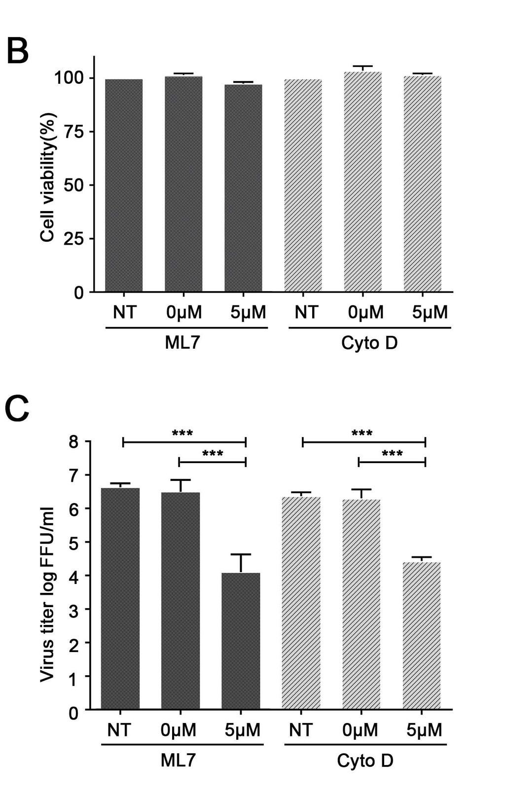
18 Since F-actin and myosin are within nanotube structures, and myosin IIA was identified to interact with certain viral proteins, we further determined whether disrupting the structure of F-actin and myosin II-A could block the intercellular nanotube pathway for cell-cell spreading of the viruses. PRRSV infected cells were treated with ML7, cytochalasin D, or solvent control dimethyl sulfoxide (DMSO; 0 um). ML7 is a specific inhibitor of myosin light chain kinase, regulating myosin IIA function. Cytochalasin D depolymerizes actin by binding to F-actin, which causes breakage of the actin filaments. Using confocal microscopy, PRRSV-infected cells that were treated with ML7 or cytochalasin D showed significantly fewer (80% less) intercellular nanotube connections in comparison to that treated with DMSO and few viral clusters were observed at 24 dpi (Fig. 8A). In the presence of either inhibitor, cell viabilities were not affected at the tested concentrations (5 µm for ML7 and cytochalasin D; Fig. 8B), but viral titers were reduced by several logs (2.56 log reduction for ML7; 1.93 log reduction for cytochalasin D), in comparison to that of DMSO control (Fig. 8C). DISCUSSION The primary pathway for cell-to-cell spread of the PRRSV virus, an enveloped RNA virus, involves assembly, maturation, and release from virus-producing cells and attachment, entry, and disassembly in virus target cells. These processes require abundant cellular resources and also demand that the virus particles evade extracellular host defensive components. In this study, we found that PRRSV can use intercellular nanotube connections as an alternative pathway for cellto-cell spread. Utilizing this pathway to directly access the cytoplasm of a naive cell presents an efficient spreading route that bypasses many of the otherwise critical assembly, budding, and cell entry steps. 18
19 Inside the host cell cytoplasm, the genomic RNA and replicase-associated RTC constitute the infectious core components that elicit a productive infection. Thus, intercellular transport of viral genomic RNA and replicase proteins is sufficient for rapid spreading of the infection. In PRRSV infected cells, confocal images showed that PRRSV replicase proteins nsp1β, nsp2, nsp4, nsp7 and nsp8 were associated with intercellular nanotubes. The presence of PRRSV nsp2 in an intercellular connection was reported previously (33), but the mechanism by which this protein arrived in the connection, and its relevance to virus spread, was unknown at that time. In our study, the movement of viral proteins from cell-to-cell was documented with live cell movies, in which GFP-tagged nsp2 transported from one cell to another in GFP-PRRSV-infected cells. This process was further demonstrated to take place between unrelated MARC-145 and HEK-293T cells. Since nsp1β, nsp2, nsp4, nsp7 and nsp8 are components of viral RTC, the data indicate that PRRSV transports RTC components through intercellular nanotubes. The PRRSV nsp9-12 proteins are also assumed to be part of the RTC. The nsp9 is generated by -1 PRF at ORF1a/1b junction, resulted the nsp9 product that contains nsp8 in its N-terminus. This means the anti-nsp8 mab could also recognize the nsp9; however, future study using the nsp9 C- terminus specific antibody is needed to confirm the presence of nsp9 in the nanotube connections. Due to lack of specific antibodies to nsp1α, nsp5-6, and nsp10-12, whether these replicase-associated proteins are transported through the nanotube connections needs to be determined in the future. Furthermore, double-membrane vesicles (DMVs) were previously reported to be associated with RTC of arterivirus (34); it is possible that DMVs transport with the viral materials. In mouse CAD cells, vesicles of lysosomal origin carrying prion proteins are actively transferred through intercellular nanotubes (35). In HIV type 1 infections, viral materialcontaining endosome cargoes were transported through nanotubes to neighboring uninfected 19
20 macrophages (8). The involvement of cellular vesicles in nanotube transport of viral materials is currently under active investigation in our laboratories. Notably, the novel PRRSV -2 PRF product, nsp2tf, was also observed to associate with intercellular nanotubes, but was not colocalized with the other nanotube-associated nsps (data not shown). This is consistent with our previous findings, in which nsp2tf and nsp2 are not co-localized in infected cells (12). Therefore, there may be multiple independent mechanisms for nsp association with nanotubular components. In addition, it is worth to note here, some of the GFP-tagged nsp2 that we observed could be GFP-nsp2TF or GFP-nsp2N, since -2/-1 PRF in nsp2 region generated ~20% of nsp2tf and ~7% nsp2n (12). Theoretically, viral membrane proteins typically needed for virus-cell egress and entry might be dispensable for virus transport through intercellular nanotubes. Recent studies suggested that PRRSV minor GPs (GP2a, GP3 and GP4) are the primary determinants of host cell binding and may also involve in membrane fusion and entry (37, 38). Using the PRRSV GP4-specific mab, few nanotubes were detected containing GP4, which support the notion that these membrane proteins may not be required for spreading the infection using the intercellular nanotube pathway. In contrast, significant amounts of GP5 proteins were detected to be associated with intercellular nanotubes. It was reported that GP5/M heterodimer of arterivirus play an important role in viral envelope formation (39-41). Gp5 and M have been proposed to be the driving force for virus budding, which may involve in the formation of the highly curved edges of the membrane (reviewed in 42). Our confocal microscopic images showed that GP5 proteins appeared as cell surface membrane-bound structures around the infected cells as well as on the intercellular nanotubes. The result made us suspect that GP5 could be integrated into the extended cell surface membrane during the formation of intercellular tunnels (nanotubes), in 20
21 which GP5 becomes a component of the nanotube for transporting the other viral infectious core materials. It is unknown whether GP5 is directly involved in the transporting process of viral materials or is a more passive component of nanotubes. The unavailability of the anti-m protein antibody and also antibodies to GP2a, E, and GP3 prevented us from analyzing the association of these proteins with nanotubes. Future studies are needed to determine whether M and other membrane proteins play a role in the intercellular spreading of the virus through nanotube connections. In addition, it will be interesting to know whether one or more viral membrane proteins promote nanotubular extension from a cell and fusion with a neighboring cell. Our data showed that PRRSV receptors are not required for nanotubular virus transport, which suggest that viral fusion proteins may not be needed to establish the nanotubular connections. Future indepth analyses are needed to determine whether PRRSV infection and certain viral protein(s) promote nanotube formation. Using the FISH method, viral RNA and N protein were observed to be largely colocalized within the nanotubes. This result is expected, since viral genomic RNA is required to generate the next cycle of viral replication. As discussed above, viral genomic RNA is packaged with N proteins to form the nucleocapsid. Transporting the N protein-bonded RNA from cell-tocell could be a mechanism for protecting the integrity of viral RNA during the transportation process. It still needs to be elucidated whether the assembled nucleocapsid was transported or only unassembled proteins were transported. Intercellular nanotubes are composed of F-actin, and myosin is known as a motor protein binding to the F-actin (reviewed in 32, 43). Myosin is encoded by a multigene family and expressed as multiple isoforms (44). Previously, myosin Va was reported to facilitate the movement of organelles along intercellular nanotubes (reviewed in 9). Recently, myosin X was 21
22 reported to play an important role in the formation of functional intercellular nanotubes within neuronal CAD cells (45). The findings of our study suggest that myosin IIA could be driving the transportation of PRRSV core infectious materials through the intercellular nanotube connections. In live cell movies, we noticed that the movement of GFP-nsp2 within nanotubes proceeded by leaping rather than by gradual transitions, a pattern which is consistent with motorprotein assisted cargo transport. Our co-ip results showed that myosin IIA can be co-precipitated with PRRSV nsp1β, nsp2, nsp4, GP5 and N, suggesting these viral proteins may directly or indirectly bind to myosin IIA during transport. Confocal microscopy also detected PRRSV nsp2tf, nsp7 and nsp8 in the intercellular nanotubes, and using nsp2tf, nsp7 and nsp8 specific antibodies, myosin IIA could be co-precipitated from infected cells. However, using myosin IIAspecific antibody, these viral proteins were not co-precipitated. This could be caused by lower protein expression levels, the insensitivity of our assay, or other unrevealed mechanism(s). Future studies are needed to determine the mechanisms of interaction between GP5 and other viral proteins with cytoskeleton proteins; and the detailed intercellular nanotube transporting process needs to be elucidated, including the process of how the viral materials are initially loaded, transported through the nanotubes, and unloaded in the target cells. The use of intercellular nanotube connections as alternative pathways for viral cell-to-cell transmission could contribute to the pathogenesis of viral infection. Notably, PRRSVneutralizing antibodies could block initial virus entry into the host cells, but could not interfere significantly with cell-to-cell transmission through intercellular nanotube connections. Previous studies also reported that viral spread through intercellular connections allows many viruses to evade neutralizing antibodies, including Influenza A virus and HIV (7, 46). Neutralizing antibodies that effectively inhibit cell-free HIV from infection are less effective or fail entirely to 22
23 inhibit cell-to-cell transmission of the virus (46). In a previous study (47), passive transfer of PRRSV-neutralizing antibodies to young weaned pigs could block viremia in blood, but could not prevent virus replication in peripheral tissues, and viral loads could reach the level similar to that of control pigs. Those pigs could still excrete infectious viruses to sentinels similar to that of control pigs. We speculate that cell-to-cell transmission of PRRSV through intercellular nanotubes could take place in peripheral tissues, a process that is resistant to neutralizing antibodies. This may contribute to viral persistence in peripheral tissues. Future studies are needed to elucidate the extent by which nanotubular intercellular transmission contributes to PRRSV spread in vivo, and whether this process is involved in viral pathogenesis. If this mechanism is truly central to the pathogenesis of PRRSV infection and persistence, the development of inhibitors that directly block this process would be critical to prevent the spread of viral infections. ACKNOWLEDGEMENT We thank Dr. Philine Wangemann and Joel Sanneman at the Confocal Microscopy and Microfluorometry Core facility for assistance on confocal images. Monoclonal antibodies for PRRSV GP4 (NI37) and N protein (SDOW17) were kindly provided by Dr. Eric Nelson at South Dakota State University. This project was supported by Agriculture and Food Research Initiative Competitive Grant no from the USDA National Institute of Food and Agriculture, and Kansas State University research startup fund. The Confocal Core facility is supported by College of Veterinary Medicine at Kansas State University. 23
24 Table 1. List of antibodies used in this study. Virus or cellular proteins PRRSV (SD95-21) Cellular Proteins Antibody name* Protein target Predicted molecular mass (kda) References mab nsp1β 23.0 (22) pab nsp2tf pab nsp2 nsp2tf (epitope CFLKVGVKSAGDLV) nsp2 (C-terminus) (epitope NGLKIRQISKPSGG) (12, 13) This study mab nsp2 (PLP2) (13) mab nsp This study mab nsp This study mab nsp8 4.9 This study mab NI37 GP (21) mab GP This study mab SDOW17 N 13.6 (20) mab N 13.6 This study mab ab8226 F-actin (beta actin) 42 Abcam mab ab55456 non-muscle myosin IIA 215 (heavy chain) Abcam pab HPA non-muscle myosin IIA 215 (heavy chain) Sigma pab sc SV40 large T-antigen of HEK-293T cell * mabs produced in mouse; pabs produced in rabbit. 94 Santa Cruz 24
25 FIGURE LEGEND Figure 1. Detection of intercellular nanotubes containing PRRSV GP5 and N protein. Panels A- D: MARC-145 cells were infected by PRRSV strain SD95-21 at 0.1 moi and fixed at 12 hpi. Panels E-F: Porcine alveolar macrophages were infected with 1 MOI of PRRSV and fixed at 12 hpi. The fixed cells were immunostained for GP5 (green; A, B and E) or N protein (green; C, D and F) together with cytoskeleton protein of F-actin (red; A and C) or myosin-iia (red; B, D, E and F). Images were taken by a confocal microscope (LSM 880, Zeiss). Scale bar for the merge pictures (middle column), 10µm. Scale bar for the zoomed pictures (right column), 5 µm. Figure 2. Intercellular nanotubes contain viral proteins in PRRSV-infected cells. MARC-145 cells were infected by PRRSV strain SD95-21 at 0.1 moi and fixed at 12 hpi. Panels A-N: The fixed cells were immunostained for nsp1β (A, H), nsp2tf (B, I), nsp2 (C, J), nsp4 (D, K), nsp7 (E, L), nsp8 (F, M) or GP4 (G, N) together with F-actin (A-G) or Myosin-IIA (H-N). Viral proteins were labeled with green fluorescence and cytoskeleton proteins were labeled with red fluorescence. Panels O-Q: Double staining of nsp2 with G4 (O and Q) or GP5 (P). The PRRSV nsp2 was labeled with green fluorescence; and GP4 or GP5 was labeled with red fluorescence. Pictures were taken by a confocal microscope (LSM 880, Zeiss). Scale bar for the merge pictures (middle column), 10 µm. Scale bar for the zoomed pictures (right column of each panel), 5 µm. Figure 3. Detection of PRRSV RNA in the intercellular nanotube connections. MARC-145 cells were infected by PRRSV SD95-21 strain at 0.1 moi and fixed at 12 hpi. Viral RNA was detected by fluorescence in-situ hybridization of RNA using CAL594-labeled PRRSV N gene RNA FISH probe (A, red). PRRSV N protein was immunostained using anti-n mab SDOW17 (B, Green). Cell nucleus is stained with DAPI (C, blue). The co-localized foci are readily visible in the 25
26 nanotubes (D, yellow). Images were taken by a confocal microscope (LSM 880, Zeiss). Scale bar 20 µm Figure 4. Intercellular spreading of PRRSV infection in cells with the presence of virusneutralizing antibody. (A) Standard viral neutralizing assay. Immune serum from PRRSVinfected pigs was serial diluted and incubated with the virus for 1 h; and the virus-antibody complex was then added on the MARC-145 cells. At 12 hpi, cells were stained with anti-n mab and virus foci were counted under the fluorescence microscope. The percentage of viral growth reduction was calculated in comparison to the result from cells treated with negative control serum. (B) Cell-to-cell spreading of the viruses under the existence of viral neutralizing antibody. MARC-145 cells were infected with PRRSV SD At 3 hpi, the 1:4 diluted immune serum or negative control serum was added onto the infected cells. At 6, 12, 24 or 36 hpi, cell culture supernatant was harvested and cells were fixed and stained with anti-n mab. Images were taken by a confocal microscope (LSM 880, Zeiss). Scale bar, 50 µm. (C) Virus titers in the harvested cell culture supernatant from experiment described in panel B. Virus titer was determined by counting the virus foci under the fluorescence microscope, and the result was interpreted as fluorescent-focus units (FFU) per ml. NS: negative control serum from a noneinfected pig; PS: immune serum from a PRRSV-infected pig. Figure 5. Live cell images demonstrating intercellular transport of PRRSV GFP-tagged nsp2 through nanotubes. (A) GFP-PRRSV infected cells maintained in cultural medium containing PRRSV neutralizing antibody. (B) GFP-PRRSV infected cells maintained in regular cultural medium (no PRRSV neutralizing antibody). (C) HEK-293T cells were transfected with PRRSV 26
27 full-length cdna infectious clone, pcmv-sd95-21-gfp. At 24 h post infection (A and B) or transfection (C), cells were analyzed using live cell image system of a confocal microscope (LSM 880, Zeiss). In both infected MARC-145 cells and transfected HEK-293T cells, the dotlike GFP-nsp2 proteins were visualized to move through an intercellular nanotube connection into the cytoplasm of a neighboring cell. Insets showed a zoomed interesting area that contains the GFP-nsp2 proteins. Specific nanotube shown between MARC-145 cells is about 20 μm in length, while specific nanotube shown between HEK-293T cells is about 12 μm in length. Scale bar, 10 μm. Figure 6. Intercellular transport of the GFP-nsp2 protein in co-cultured MARC-145 and HEK- 293T cells. (A) GFP-PRRSV infected MARC145 cells were fixed and stained with anti-nsp2 mab and anti-sv40-t pab sc (B) GFP-PRRSV infected HEK-293T cells were fixed and stained with anti-nsp2 mab and anti-sv40-t pab sc (C) GFP-PRRSV infected MARC-145 cells were trypsinized at 12 hpi and mixed with naïve HEK-293T cells. After 36 h post cultivation, cells were fixed and stained with anti-nsp2 mab and anti- SV40-T pab sc The nsp2 was labeled with green fluorescence and SV40 large T was labeled with red fluorescence. Pictures were taken by a confocal microscope (LSM 880, Zeiss). Scale bar, 10 μm. Figure 7. Co-precipitation of PRRSV proteins with cytoskeleton F-actin and myosin IIA. (A-C) PRRSV-infected (+) MARC-145 cell lysates or mock-infected (-) cell lysates were used for immunoprecipitation using PRRSV protein specific mabs as indicated on the top of each panel. Anti-mouse IgG (α-migg) was used as a control. The immunoprecipitated proteins were 27
28 separated by 8-16% Tris-Glycine gradient gel (A) and immunoblotted by anti-myosin-iia pab and anti-f-actin mab (B-C). (D-L) PRRSV-infected MARC-145 cell lysates (+) or mock infected (-) cell lysates were used for immunoprecipitation using anti-myosin-iia pab. The immunoprecipitated proteins were separated by 8-16% Tris-Glycine gradient gel and immunoblotted by PRRSV protein specific mabs as indicated on the bottom of each panel. IP: immunoprecipitation. WB: Western blotting. Arrows in panels A-C pointed F-actin (42 kda), non-muscle myosin heavy chain IIA (215 kda). Arrows in Panels D-E, G-H and J pointed the target protein as indicated at the bottom of each panel. Figure 8. Inhibitors of F-actin and myosin IIA affect the intercellular spread of PRRSV. (A) Confluent MARC-145 cells monolayers were pretreated with ML7 or cytochalasin D (Cyto D) compounds at concentration of 5 µm or 0 µm. Non-treated cells (NT) were used as a control. After 30 min incubation at 37 0 C, the cells were infected with 0.1 moi of PRRSV. After 1 h incubation, the infected cells were washed with PBS and the fresh medium containing Cyto D or ML7 compounds was added. At 24 hpi, cells were fixed and immune-stained with PRRSV N protein specific mab. Images in columns 1 and 3 were taken using the 20x objective lens (20X) with scale bar of 50 µm. Inset at the right bottom corner of each image shows a 40x zoomed region of interest (ROI); scale bar for ROI, 10 um. (B) Effect of Cyto D or ML7 on the viability of the MARC-145 cells compared to the viability of DMSO-treated (0 µm) or non-treated control cells (NT). Graphs show the results (mean and SD) of a representative experiment (n = 3). (C) Viral growth inhibition efficiency of myosin-iia inhibitor ML7 or F-actin inhibitor Cyto D. Virus titration was conducted using cell culture supernatant collected from the experiment in (A). Virus titer was determined by counting the virus foci under the fluorescence microscope, 28
29 and the result was interpreted as fluorescent-focus units (FFU) per ml. Statistical significance between the groups was determined by one-way analysis of variance (ANOVA) test using GraphPad InStat Prism (software version 5.0), and p < 0.01 (***) was considered statistically significant. 29
30 REFERENCES 1. Marsh M, Helenius A Virus Entry into Animal-Cells. Advances in Virus Research 36: Zhong P, Agosto LM, Munro JB, Mothes W Cell-to-cell transmission of viruses. Current Opinion in Virology 3: Mothes W, Sherer NM, Jin J, Zhong P Virus Cell-to-Cell Transmission. Journal of Virology 84: Sattentau Q Avoiding the void: cell-to-cell spread of human viruses. Nature Reviews Microbiology 6: Xu WF, Santini PA, Sullivan JS, He B, Shan MM, Ball SC, Dyer WB, Ketas TJ, Chadburn A, Cohen-Gould L, Knowles DM, Chiu A, Sanders RW, Chen K, Cerutti A HIV-1 evades virus-specific IgG2 and IgA responses by targeting systemic and intestinal B cells via long-range intercellular conduits. Nature Immunology 10:1008- U Gerdes HH, Bukoreshtliev NV, Barroso JFV Tunneling nanotubes: A new route for the exchange of components between animal cells (vol 581, pg 2194, 2007). Febs Letters 581: Roberts KL, Manicassamy B, Lamb RA Influenza A Virus Uses Intercellular Connections To Spread to Neighboring Cells. Journal of Virology 89: Kadiu I, Gendelman HE Human Immunodeficiency Virus type 1 Endocytic Trafficking Through Macrophage Bridging Conduits Facilitates Spread of Infection. Journal of Neuroimmune Pharmacology 6: Rustom A, Saffrich R, Markovic I, Walther P, Gerdes HH Nanotubular highways for intercellular organelle transport. Science 303: Ahmed KA, Xiang J Mechanisms of cellular communication through intercellular protein transfer. Journal of Cellular and Molecular Medicine 15: Fang Y, Snijder EJ The PRRSV replicase: Exploring the multifunctionality of an intriguing set of nonstructural proteins. Virus Research 154: Fang Y, Treffers EE, Li YH, Tas A, Sun Z, van der Meer Y, de Ru AH, van Veelen PA, Atkins JF, Snijder EJ, Firth AE Efficient-2 frameshifting by mammalian ribosomes to synthesize an additional arterivirus protein. Proceedings of the National Academy of Sciences of the United States of America 109:E2920-E Li YH, Treffers EE, Napthine S, Tas A, Zhu LC, Sun Z, Bell S, Mark BL, van Veelen PA, van Hemert MJ, Firth AE, Brierley I, Snijder EJ, Fang Y Transactivation of programmed ribosomal frameshifting by a viral protein. Proceedings of the National Academy of Sciences of the United States of America 111:E2172-E Snijder EJ, Kikkert M, Fang Y Arterivirus molecular biology and pathogenesis. Journal of General Virology 94: Kim HS, Kwang J, Yoon IJ, Joo HS, Frey ML Enhanced Replication of Porcine Reproductive and Respiratory Syndrome (Prrs) Virus in a Homogeneous Subpopulation of Ma-104 Cell-Line. Archives of Virology 133: Welch SKW, Calvert JG A brief review of CD163 and its role in PRRSV infection. Virus Research 154: Nauwynck HJ, Duan X, Favoreel HW, Van Oostveldt P, Pensaert MB Entry of porcine reproductive and respiratory syndrome virus into porcine alveolar 30
31 macrophages via receptor-mediated endocytosis. Journal of General Virology 80: Kreutz LC, Ackermann MR Porcine reproductive and respiratory syndrome virus enters cells through a low ph-dependent endocytic pathway. Virus Research 42: Zeman, D., R. Neiger, M. Yaeger, E. Nelson, D. Benfield, P. Leslie-Steen, J. Thomson, D. Miskimins, R. Daly, and M. Minehart Laboratory investigation of PRRS virus infection in three swine herds. J. Vet. Diagn. Invest. 5: Nelson EA, Christopherhennings J, Drew T, Wensvoort G, Collins JE, Benfield DA Differentiation of United-States and European Isolates of Porcine Reproductive and Respiratory Syndrome Virus by Monoclonal-Antibodies. Journal of Clinical Microbiology 31: Ropp SL, Wees CEM, Fang Y, Nelson EA, Rossow KD, Bien M, Arndt B, Preszler S, Steen P, Christopher-Hennings J, Collins JE, Benfield DA, Faaberg KS Characterization of emerging European-like porcine reproductive and respiratory syndrome virus isolates in the United States. Journal of Virology 78: Chen Z, Lawson S, Sun Z, Zhou X, Guan X, Christopher-Hennings J, Nelson EA, Fang Y Identification of two auto-cleavage products of nonstructural protein 1 (nsp1) in porcine reproductive and respiratory syndrome virus infected cells: nsp1 function as interferon antagonist. Virology 398: Li YH, Tas A, Snijder EJ, Fang Y Identification of porcine reproductive and respiratory syndrome virus ORF1a-encoded non-structural proteins in virus-infected cells. Journal of General Virology 93: Rowland RRR, Chauhan V, Fang Y, Pekosz A, Kerrigan M, Burton MD Intracellular localization of the severe acute respiratory syndrome coronavirus nucleocapsid protein: Absence of nucleolar accumulation during infection and after expression as a recombinant protein in Vero cells. Journal of Virology 79: Li YH, Zhu LC, Lawson SR, Fang Y Targeted mutations in a highly conserved motif of the nsp1 beta protein impair the interferon antagonizing activity of porcine reproductive and respiratory syndrome virus. Journal of General Virology 94: Fang Y, Rowland RRR, Roof M, Lunney JK, Christopher-Hennings J, Nelson EA A full-length cdna infectious clone of North American type 1 porcine reproductive and respiratory syndrome virus: Expression of green fluorescent protein in the Nsp2 region. Journal of Virology 80: Yoon IJ, Joo HS, Goyal SM, Molitor TW A Modified Serum Neutralization Test for the Detection of Antibody to Porcine Reproductive and Respiratory Syndrome Virus in Swine Sera. Journal of Veterinary Diagnostic Investigation 6: Sun Z, Li Y, Ransburgh R, Snijder EJ, Fang Y Nonstructural protein 2 of porcine reproductive and respiratory syndrome virus inhibits the antiviral function of interferon-stimulated gene 15. J Virol 86: Sowinski S, Jolly C, Berninghausen O, Purbhoo MA, Chauveau A, Kohler K, Oddos S, Eissmann P, Brodsky FM, Hopkins C, Onfelt B, Sattentau Q, Davis DM Membrane nanotubes physically connect T cells over long distances presenting a novel route for HIV-1 transmission. Nature Cell Biology 10:
32 Jing Jin NMS, Gisela Heidecker, David Derse, Walther Mothes Assembly of the Murine Leukemia Virus Is Directed towards Sites of Cell- Cell Contact. PLoS Biol 7(7): e doi: /journal.pbio Sun YY, Qi YH, Liu CX, Gao WQ, Chen P, Fu LR, Peng B, Wang HM, Jing ZY, Zhong GC, Li WH Nonmuscle Myosin Heavy Chain IIA Is a Critical Factor Contributing to the Efficiency of Early Infection of Severe Fever with Thrombocytopenia Syndrome Virus. Journal of Virology 88: Taylor MP, Koyuncu OO, Enquist LW Subversion of the actin cytoskeleton during viral infection. Nature Reviews Microbiology 9: Kappes MA, Miller CL, Faaberg KS Highly Divergent Strains of Porcine Reproductive and Respiratory Syndrome Virus Incorporate Multiple Isoforms of Nonstructural Protein 2 into Virions. Journal of Virology 87: Oostra M, Hagemeijer MC, van Gent M, Bekker CPJ, Lintelo EGT, Rottier PJM, de Haan CAM Topology and Membrane Anchoring of the Coronavirus Replication Complex: Not All Hydrophobic Domains of nsp3 and nsp6 Are Membrane Spanning. Journal of Virology 82: Gousset K, Schiff E, Langevin C, Marijanovic Z, Caputo A, Browman DT, Chenouard N, de Chaumont F, Martino A, Enninga J, Olivo-Marin JC, Mannel D, Zurzolo C Prions hijack tunnelling nanotubes for intercellular spread. Nature Cell Biology 11:328-U Snijder EJ, van Tol H, Roos N, Pedersen KW Non-structural proteins 2 and 3 interact to modify host cell membranes during the formation of the arterivirus replication complex. Journal of General Virology 82: Tian DB, Wei ZZ, Zevenhoven-Dobbe JC, Liu RX, Tong GZ, Snijder EJ, Yuan SS Arterivirus Minor Envelope Proteins Are a Major Determinant of Viral Tropism in Cell Culture. Journal of Virology 86: Das PB, Dinh PX, Ansari IH, de Lima M, Osorio FA, Pattnaik AK The Minor Envelope Glycoproteins GP2a and GP4 of Porcine Reproductive and Respiratory Syndrome Virus Interact with the Receptor CD163. Journal of Virology 84: Wieringa R, de Vries AAF, van der Meulen J, Godeke GJ, Onderwater JJM, van Tol H, Koerten HK, Mommaas AM, Snijder EJ, Rottier PJM Structural protein requirements in equine arteritis virus assembly. Journal of Virology 78: Wissink EHJ, Kroese MV, van Wijk HAR, Rijsewijk FAM, Meulenberg JJM, Rottier PJM Envelope protein requirements for the assembly of infectious virions of porcine reproductive and respiratory syndrome virus. Journal of Virology 79: Nam HM, Chae KS, Song YJ, Lee NH, Lee JB, Park SY, Song CS, Seo KH, Kang SM, Kim MC, Choi IS Immune responses in mice vaccinated with virus-like particles composed of the GP5 and M proteins of porcine reproductive and respiratory syndrome virus. Archives of Virology 158: Veit M, Matczuk AK, Sinhadri BC, Krause E, Thaa B Membrane proteins of arterivirus particles: Structure, topology, processing and function. Virus Research 194: Lorenz M, Holmes KC The actin-myosin interface. Proceedings of the National Academy of Sciences of the United States of America 107:
33 Weiss A, Leinwand LA The mammalian myosin heavy chain gene family. Annual Review of Cell and Developmental Biology 12: Gousset K, Marzo L, Commere PH, Zurzolo C Myo10 is a key regulator of TNT formation in neuronal cells. Journal of Cell Science 126: Schiffner T, Sattentau QJ, Duncan CJA Cell-to-cell spread of HIV-1 and evasion of neutralizing antibodies. Vaccine 31: Lopez OJ, Oliveira MF, Garcia EA, Kwon BJ, Doster A, Osorio FA Protection against porcine reproductive and respiratory syndrome virus (PRRSV) infection through passive transfer of PRRSV-neutralizing antibodies is dose dependent. Clinical and Vaccine Immunology 14: Downloaded from on July 24, 2018 by guest 33

34

35

36

37

38

39

40

41

42

43
44
Influenza virus exploits tunneling nanotubes for cell-to-cell spread
 Supplementary Information Influenza virus exploits tunneling nanotubes for cell-to-cell spread Amrita Kumar 1, Jin Hyang Kim 1, Priya Ranjan 1, Maureen G. Metcalfe 2, Weiping Cao 1, Margarita Mishina 1,
Nature Medicine: doi: /nm.4322
 1 2 3 4 5 6 7 8 9 10 11 Supplementary Figure 1. Predicted RNA structure of 3 UTR and sequence alignment of deleted nucleotides. (a) Predicted RNA secondary structure of ZIKV 3 UTR. The stem-loop structure
TSH Receptor Monoclonal Antibody (49) Catalog Number MA3-218 Product data sheet
 Catalog Number MA3-218 Product data sheet") Website: thermofisher.com Customer Service (US): 1 800 955 6288 ext. 1 Technical Support (US): 1 800 955 6288 ext. 441 TSH Receptor Monoclonal Antibody (49) Catalog Number MA3-218 Product data sheet Details
Website: thermofisher.com Customer Service (US): 1 800 955 6288 ext. 1 Technical Support (US): 1 800 955 6288 ext. 441 TSH Receptor Monoclonal Antibody (49) Catalog Number MA3-218 Product data sheet Details
Proteomic profiling of small-molecule inhibitors reveals dispensability of MTH1 for cancer cell survival
 Supplementary Information for Proteomic profiling of small-molecule inhibitors reveals dispensability of MTH1 for cancer cell survival Tatsuro Kawamura 1, Makoto Kawatani 1, Makoto Muroi, Yasumitsu Kondoh,
Supplementary Information POLO-LIKE KINASE 1 FACILITATES LOSS OF PTEN-INDUCED PROSTATE CANCER FORMATION
 Supplementary Information POLO-LIKE KINASE 1 FACILITATES LOSS OF PTEN-INDUCED PROSTATE CANCER FORMATION X. Shawn Liu 1, 3, Bing Song 2, 3, Bennett D. Elzey 3, 4, Timothy L. Ratliff 3, 4, Stephen F. Konieczny
Supplementary data Supplementary Figure 1 Supplementary Figure 2
 Supplementary data Supplementary Figure 1 SPHK1 sirna increases RANKL-induced osteoclastogenesis in RAW264.7 cell culture. (A) RAW264.7 cells were transfected with oligocassettes containing SPHK1 sirna
(a) Significant biological processes (upper panel) and disease biomarkers (lower panel)
 Significant biological processes (upper panel) and disease biomarkers (lower panel)") Supplementary Figure 1. Functional enrichment analyses of secretomic proteins. (a) Significant biological processes (upper panel) and disease biomarkers (lower panel) 2 involved by hrab37-mediated secretory
Supplementary Figure 1. Functional enrichment analyses of secretomic proteins. (a) Significant biological processes (upper panel) and disease biomarkers (lower panel) 2 involved by hrab37-mediated secretory
Recombinant Protein Expression Retroviral system
 Recombinant Protein Expression Retroviral system Viruses Contains genome DNA or RNA Genome encased in a protein coat or capsid. Some viruses have membrane covering protein coat enveloped virus Ø Essential
hexahistidine tagged GRP78 devoid of the KDEL motif (GRP78-His) on SDS-PAGE. This
 on SDS-PAGE. This") SUPPLEMENTAL FIGURE LEGEND Fig. S1. Generation and characterization of. (A) Coomassie staining of soluble hexahistidine tagged GRP78 devoid of the KDEL motif (GRP78-His) on SDS-PAGE. This protein was expressed
SUPPLEMENTAL FIGURE LEGEND Fig. S1. Generation and characterization of. (A) Coomassie staining of soluble hexahistidine tagged GRP78 devoid of the KDEL motif (GRP78-His) on SDS-PAGE. This protein was expressed
Supplementary Information
 Supplementary Information Supplementary Figure 1. CD4 + T cell activation and lack of apoptosis after crosslinking with anti-cd3 + anti-cd28 + anti-cd160. (a) Flow cytometry of anti-cd160 (5D.10A11) binding
SUPPLEMENTARY INFORMATION
 Supplementary Figures Supplementary Figure S1. Binding of full-length OGT and deletion mutants to PIP strips (Echelon Biosciences). Supplementary Figure S2. Binding of the OGT (919-1036) fragments with
Supplemental Information. Autophagy in Oncogenic K-Ras. Promotes Basal Extrusion. of Epithelial Cells by Degrading S1P. Current Biology, Volume 24
 Current Biology, Volume 24 Supplemental Information Autophagy in Oncogenic K-Ras Promotes Basal Extrusion of Epithelial Cells by Degrading S1P Gloria Slattum, Yapeng Gu, Roger Sabbadini, and Jody Rosenblatt
University of Groningen
 University of Groningen Mechanisms of Hemagglutinin Targeted Influenza Virus Neutralization Brandenburg, Boerries; Koudstaal, Wouter; Goudsmit, Jaap; Klaren, Vincent; Tang, Chan; Bujny, Miriam V.; Korse,
Supplementary Material
 Supplementary Material Nuclear import of purified HIV-1 Integrase. Integrase remains associated to the RTC throughout the infection process until provirus integration occurs and is therefore one likely
TFEB-mediated increase in peripheral lysosomes regulates. Store Operated Calcium Entry
 TFEB-mediated increase in peripheral lysosomes regulates Store Operated Calcium Entry Luigi Sbano, Massimo Bonora, Saverio Marchi, Federica Baldassari, Diego L. Medina, Andrea Ballabio, Carlotta Giorgi
7.012 Quiz 3 Answers
 MIT Biology Department 7.012: Introductory Biology - Fall 2004 Instructors: Professor Eric Lander, Professor Robert A. Weinberg, Dr. Claudette Gardel Friday 11/12/04 7.012 Quiz 3 Answers A > 85 B 72-84
Tunneling nanotubes (TNT) as a novel route of cell-to-cell spread of herpesviruses
 as a novel route of cell-to-cell spread of herpesviruses") JVI Accepted Manuscript Posted Online 28 February 2018 J. Virol. doi:10.1128/jvi.00090-18 Copyright 2018 American Society for Microbiology. All Rights Reserved. 1 Tunneling nanotubes (TNT) as a novel route
JVI Accepted Manuscript Posted Online 28 February 2018 J. Virol. doi:10.1128/jvi.00090-18 Copyright 2018 American Society for Microbiology. All Rights Reserved. 1 Tunneling nanotubes (TNT) as a novel route
Supplementary Figure 1: si-craf but not si-braf sensitizes tumor cells to radiation.
 Supplementary Figure 1: si-craf but not si-braf sensitizes tumor cells to radiation. (a) Embryonic fibroblasts isolated from wildtype (WT), BRAF -/-, or CRAF -/- mice were irradiated (6 Gy) and DNA damage
HIV-1 p24 ELISA Pair Set Cat#: orb54951 (ELISA Manual)
") HIV-1 p24 ELISA Pair Set Cat#: orb54951 (ELISA Manual) BACKGROUND Human Immunodeficiency Virus ( HIV ) can be divided into two major types, HIV type 1 (HIV-1) and HIV type 2 (HIV-2). HIV-1 is related to
HIV-1 p24 ELISA Pair Set Cat#: orb54951 (ELISA Manual) BACKGROUND Human Immunodeficiency Virus ( HIV ) can be divided into two major types, HIV type 1 (HIV-1) and HIV type 2 (HIV-2). HIV-1 is related to
Viral structure م.م رنا مشعل
 Viral structure م.م رنا مشعل Viruses must reproduce (replicate) within cells, because they cannot generate energy or synthesize proteins. Because they can reproduce only within cells, viruses are obligate
Protocol for Gene Transfection & Western Blotting
 The schedule and the manual of basic techniques for cell culture Advanced Protocol for Gene Transfection & Western Blotting Schedule Day 1 26/07/2008 Transfection Day 3 28/07/2008 Cell lysis Immunoprecipitation
Islet viability assay and Glucose Stimulated Insulin Secretion assay RT-PCR and Western Blot
 Islet viability assay and Glucose Stimulated Insulin Secretion assay Islet cell viability was determined by colorimetric (3-(4,5-dimethylthiazol-2-yl)-2,5- diphenyltetrazolium bromide assay using CellTiter
SUPPLEMENT. Materials and methods
 SUPPLEMENT Materials and methods Cell culture and reagents Cell media and reagents were from Invitrogen unless otherwise indicated. Antibiotics and Tet-certified serum were from Clontech. In experiments
Supplementary Figure 1 Expression of Crb3 in mouse sciatic nerve: biochemical analysis (a) Schematic of Crb3 isoforms, ERLI and CLPI, indicating the
 Schematic of Crb3 isoforms, ERLI and CLPI, indicating the") Supplementary Figure 1 Expression of Crb3 in mouse sciatic nerve: biochemical analysis (a) Schematic of Crb3 isoforms, ERLI and CLPI, indicating the location of the transmembrane (TM), FRM binding (FB)
Supplementary Figure 1 Expression of Crb3 in mouse sciatic nerve: biochemical analysis (a) Schematic of Crb3 isoforms, ERLI and CLPI, indicating the location of the transmembrane (TM), FRM binding (FB)
Live cell imaging of trafficking of the chaperone complex vaccine to the ER. BMDCs were incubated with ER-Tracker Red (1 M) in staining solution for
 in staining solution for") Live cell imaging of trafficking of the chaperone complex vaccine to the ER. BMDCs were incubated with ER-Tracker Red (1 M) in staining solution for 15 min at 37 C and replaced with fresh complete medium.
Live cell imaging of trafficking of the chaperone complex vaccine to the ER. BMDCs were incubated with ER-Tracker Red (1 M) in staining solution for 15 min at 37 C and replaced with fresh complete medium.
B16-F10 (Mus musculus skin melanoma), NCI-H460 (human non-small cell lung cancer
, NCI-H460 (human non-small cell lung cancer") Electronic Supplementary Material (ESI) for ChemComm. This journal is The Royal Society of Chemistry 2017 Experimental Methods Cell culture B16-F10 (Mus musculus skin melanoma), NCI-H460 (human non-small
Electronic Supplementary Material (ESI) for ChemComm. This journal is The Royal Society of Chemistry 2017 Experimental Methods Cell culture B16-F10 (Mus musculus skin melanoma), NCI-H460 (human non-small
Identification of Microbes Lecture: 12
 Diagnostic Microbiology Identification of Microbes Lecture: 12 Electron Microscopy 106 virus particles per ml required for visualization, 50,000-60,000 magnification normally used. Viruses may be detected
SUPPLEMENTARY INFORMATION
 SUPPLEMENTARY INFORMATION doi:1.138/nature9814 a A SHARPIN FL B SHARPIN ΔNZF C SHARPIN T38L, F39V b His-SHARPIN FL -1xUb -2xUb -4xUb α-his c Linear 4xUb -SHARPIN FL -SHARPIN TF_LV -SHARPINΔNZF -SHARPIN
Supplementary Figures
 Inhibition of Pulmonary Anti Bacterial Defense by IFN γ During Recovery from Influenza Infection By Keer Sun and Dennis W. Metzger Supplementary Figures d a Ly6G Percentage survival f 1 75 5 1 25 1 5 1
Evidence for a biphasic mode of respiratory syncytial virus transmission in permissive HEp2 cell monolayers
 Huong et al. Virology Journal (2016) 13:12 DOI 10.1186/s12985-016-0467-9 RESEARCH Open Access Evidence for a biphasic mode of respiratory syncytial virus transmission in permissive HEp2 cell monolayers
SUPPLEMENTARY INFORMATION. Supplementary Figures S1-S9. Supplementary Methods
 SUPPLEMENTARY INFORMATION SUMO1 modification of PTEN regulates tumorigenesis by controlling its association with the plasma membrane Jian Huang 1,2#, Jie Yan 1,2#, Jian Zhang 3#, Shiguo Zhu 1, Yanli Wang
Supplementary Figure 1. Prevalence of U539C and G540A nucleotide and E172K amino acid substitutions among H9N2 viruses. Full-length H9N2 NS
 Supplementary Figure 1. Prevalence of U539C and G540A nucleotide and E172K amino acid substitutions among H9N2 viruses. Full-length H9N2 NS nucleotide sequences (a, b) or amino acid sequences (c) from
Essential Medium, containing 10% fetal bovine serum, 100 U/ml penicillin and 100 µg/ml streptomycin. Huvec were cultured in
 Supplemental data Methods Cell culture media formulations A-431 and U-87 MG cells were maintained in Dulbecco s Modified Eagle s Medium. FaDu cells were cultured in Eagle's Minimum Essential Medium, containing
Coronaviruses. Virion. Genome. Genes and proteins. Viruses and hosts. Diseases. Distinctive characteristics
 Coronaviruses Virion Genome Genes and proteins Viruses and hosts Diseases Distinctive characteristics Virion Spherical enveloped particles studded with clubbed spikes Diameter 120-160 nm Coiled helical
LESSON 4.4 WORKBOOK. How viruses make us sick: Viral Replication
 DEFINITIONS OF TERMS Eukaryotic: Non-bacterial cell type (bacteria are prokaryotes).. LESSON 4.4 WORKBOOK How viruses make us sick: Viral Replication This lesson extends the principles we learned in Unit
<20 <20 <20 < <20 <20 <20 <20. Mock
 Cross-Lineage Neutralization PRNT 80 Titers Asian Asian West African Indian Ocean Group NHP Strain 181/25 Strain 99659 Strain 37997 Strain LR 142590 80 80 20 40 EILV/CHIKV 150844 640 640 160 320 Mock 150849
Reoviruses. Virion. Genome. Genes and proteins. Viruses and hosts. Diseases. Distinctive characteristics
 Reoviruses Virion Genome Genes and proteins Viruses and hosts Diseases Distinctive characteristics Virion Naked icosahedral capsid (T=13), diameter 60-85 nm Capsid consists of two or three concentric protein
~Lentivirus production~
 ~Lentivirus production~ May 30, 2008 RNAi core R&D group member Lentivirus Production Session Lentivirus!!! Is it health threatening to lab technician? What s so good about this RNAi library? How to produce
SUPPLEMENTAL INFORMATION
 SUPPLEMENTAL INFORMATION EXPERIMENTAL PROCEDURES Tryptic digestion protection experiments - PCSK9 with Ab-3D5 (1:1 molar ratio) in 50 mm Tris, ph 8.0, 150 mm NaCl was incubated overnight at 4 o C. The
Supplementary Figure 1. SC35M polymerase activity in the presence of Bat or SC35M NP encoded from the phw2000 rescue plasmid.
 1 2 3 4 5 6 7 8 9 10 11 12 13 14 15 16 17 18 19 20 21 22 23 24 25 26 27 Supplementary Figure 1. SC35M polymerase activity in the presence of Bat or SC35M NP encoded from the phw2000 rescue plasmid. HEK293T
T H E J O U R N A L O F C E L L B I O L O G Y
 Supplemental material Edens and Levy, http://www.jcb.org/cgi/content/full/jcb.201406004/dc1 T H E J O U R N A L O F C E L L B I O L O G Y Figure S1. Nuclear shrinking does not depend on the cytoskeleton
Supplementary Materials for
 www.sciencesignaling.org/cgi/content/full/8/366/ra25/dc1 Supplementary Materials for Viral entry route determines how human plasmacytoid dendritic cells produce type I interferons Daniela Bruni, Maxime
Superior Fluorescent Labeling Dyes Spanning the Full Visible Spectrum...1. Trademarks: HiLyte Fluor (AnaSpec, Inc.)
") Table of Contents Fluor TM Labeling Dyes Superior Fluorescent Labeling Dyes Spanning the Full Visible Spectrum....1 Fluor TM 405 Dye, an Excellent Alternative to Alexa Fluor 405 & DyLight 405....2 Fluor
Table of Contents Fluor TM Labeling Dyes Superior Fluorescent Labeling Dyes Spanning the Full Visible Spectrum....1 Fluor TM 405 Dye, an Excellent Alternative to Alexa Fluor 405 & DyLight 405....2 Fluor
Figure S1. Western blot analysis of clathrin RNA interference in human DCs Human immature DCs were transfected with 100 nm Clathrin SMARTpool or
 Figure S1. Western blot analysis of clathrin RNA interference in human DCs Human immature DCs were transfected with 100 nm Clathrin SMARTpool or control nontargeting sirnas. At 90 hr after transfection,
Supplementary Methods
 1 2 3 4 5 6 7 8 9 1 11 12 13 14 15 16 17 18 19 2 21 22 23 24 25 26 27 28 29 3 31 32 Supplementary Methods Gentamicin protection assay THP-1 monocytes were seeded onto a 12-well plate and differentiated
Human Immunodeficiency Virus type 1 (HIV-1) p24 / Capsid Protein p24 ELISA Pair Set
 p24 / Capsid Protein p24 ELISA Pair Set") Human Immunodeficiency Virus type 1 (HIV-1) p24 / Capsid Protein p24 ELISA Pair Set Catalog Number : SEK11695 To achieve the best assay results, this manual must be read carefully before using this product
Human Immunodeficiency Virus type 1 (HIV-1) p24 / Capsid Protein p24 ELISA Pair Set Catalog Number : SEK11695 To achieve the best assay results, this manual must be read carefully before using this product
HIV-1 Virus-like Particle Budding Assay Nathan H Vande Burgt, Luis J Cocka * and Paul Bates
 HIV-1 Virus-like Particle Budding Assay Nathan H Vande Burgt, Luis J Cocka * and Paul Bates Department of Microbiology, Perelman School of Medicine at the University of Pennsylvania, Philadelphia, USA
Figure S1. Schematic presentation of genomic replication of idsiv after transfection and infection. After transfection of idsiv plasmid DNA into 293T
 Figure S1. Schematic presentation of genomic replication of idsiv after transfection and infection. After transfection of idsiv plasmid DNA into 293T cells, the RNA genomes with all modifications are generated
Trim29 gene-targeting strategy. (a) Genotyping of wildtype mice (+/+), Trim29 heterozygous mice (+/ ) and homozygous mice ( / ).
 Genotyping of wildtype mice (+/+), Trim29 heterozygous mice (+/ ) and homozygous mice ( / ).") Supplementary Figure 1 Trim29 gene-targeting strategy. (a) Genotyping of wildtype mice (+/+), Trim29 heterozygous mice (+/ ) and homozygous mice ( / ). (b) Immunoblot analysis of TRIM29 in lung primary
Supplementary Figure 1 Trim29 gene-targeting strategy. (a) Genotyping of wildtype mice (+/+), Trim29 heterozygous mice (+/ ) and homozygous mice ( / ). (b) Immunoblot analysis of TRIM29 in lung primary
Virion Genome Genes and proteins Viruses and hosts Diseases Distinctive characteristics
 Hepadnaviruses Virion Genome Genes and proteins Viruses and hosts Diseases Distinctive characteristics Hepatitis viruses A group of unrelated pathogens termed hepatitis viruses cause the vast majority
Gladstone Institutes, University of California (UCSF), San Francisco, USA
, San Francisco, USA") Fluorescence-linked Antigen Quantification (FLAQ) Assay for Fast Quantification of HIV-1 p24 Gag Marianne Gesner, Mekhala Maiti, Robert Grant and Marielle Cavrois * Gladstone Institutes, University of
Fluorescence-linked Antigen Quantification (FLAQ) Assay for Fast Quantification of HIV-1 p24 Gag Marianne Gesner, Mekhala Maiti, Robert Grant and Marielle Cavrois * Gladstone Institutes, University of
Structural vs. nonstructural proteins
 Why would you want to study proteins associated with viruses or virus infection? Receptors Mechanism of uncoating How is gene expression carried out, exclusively by viral enzymes? Gene expression phases?
The Schedule and the Manual of Basic Techniques for Cell Culture
 The Schedule and the Manual of Basic Techniques for Cell Culture 1 Materials Calcium Phosphate Transfection Kit: Invitrogen Cat.No.K2780-01 Falcon tube (Cat No.35-2054:12 x 75 mm, 5 ml tube) Cell: 293
04_polarity. The formation of synaptic vesicles
 Brefeldin prevents assembly of the coats required for budding Nocodazole disrupts microtubules Constitutive: coatomer-coated Selected: clathrin-coated The formation of synaptic vesicles Nerve cells (and
MTC-TT and TPC-1 cell lines were cultured in RPMI medium (Gibco, Breda, The Netherlands)
") Supplemental data Materials and Methods Cell culture MTC-TT and TPC-1 cell lines were cultured in RPMI medium (Gibco, Breda, The Netherlands) supplemented with 15% or 10% (for TPC-1) fetal bovine serum
Supplemental data Materials and Methods Cell culture MTC-TT and TPC-1 cell lines were cultured in RPMI medium (Gibco, Breda, The Netherlands) supplemented with 15% or 10% (for TPC-1) fetal bovine serum
Supplementary Figure 1 Role of Raf-1 in TLR2-Dectin-1-mediated cytokine expression
 Supplementary Figure 1 Supplementary Figure 1 Role of Raf-1 in TLR2-Dectin-1-mediated cytokine expression. Quantitative real-time PCR of indicated mrnas in DCs stimulated with TLR2-Dectin-1 agonist zymosan
Mammalian Membrane Protein Extraction Kit
 Mammalian Membrane Protein Extraction Kit Catalog number: AR0155 Boster s Mammalian Membrane Protein Extraction Kit is a simple, rapid and reproducible method to prepare cellular protein fractions highly
Chapter 6- An Introduction to Viruses*
 Chapter 6- An Introduction to Viruses* *Lecture notes are to be used as a study guide only and do not represent the comprehensive information you will need to know for the exams. 6.1 Overview of Viruses
Identification of Mutation(s) in. Associated with Neutralization Resistance. Miah Blomquist
 in. Associated with Neutralization Resistance. Miah Blomquist") Identification of Mutation(s) in the HIV 1 gp41 Subunit Associated with Neutralization Resistance Miah Blomquist What is HIV 1? HIV-1 is an epidemic that affects over 34 million people worldwide. HIV-1
Identification of Mutation(s) in the HIV 1 gp41 Subunit Associated with Neutralization Resistance Miah Blomquist What is HIV 1? HIV-1 is an epidemic that affects over 34 million people worldwide. HIV-1
Supplementary information. MARCH8 inhibits HIV-1 infection by reducing virion incorporation of envelope glycoproteins
 Supplementary information inhibits HIV-1 infection by reducing virion incorporation of envelope glycoproteins Takuya Tada, Yanzhao Zhang, Takayoshi Koyama, Minoru Tobiume, Yasuko Tsunetsugu-Yokota, Shoji
Supplementary Figure 1
 Supplementary Figure 1 AAV-GFP injection in the MEC of the mouse brain C57Bl/6 mice at 4 months of age were injected with AAV-GFP into the MEC and sacrificed at 7 days post injection (dpi). (a) Brains
Erzsebet Kokovay, Susan Goderie, Yue Wang, Steve Lotz, Gang Lin, Yu Sun, Badrinath Roysam, Qin Shen,
 Cell Stem Cell, Volume 7 Supplemental Information Adult SVZ Lineage Cells Home to and Leave the Vascular Niche via Differential Responses to SDF1/CXCR4 Signaling Erzsebet Kokovay, Susan Goderie, Yue Wang,
PRODUCT INFORMATION & MANUAL
 PRODUCT INFORMATION & MANUAL 0.4 micron for Overall Exosome Isolation (Cell Media) NBP2-49826 For research use only. Not for diagnostic or therapeutic procedures. www.novusbio.com - P: 303.730.1950 - P:
October 26, Lecture Readings. Vesicular Trafficking, Secretory Pathway, HIV Assembly and Exit from Cell
 October 26, 2006 Vesicular Trafficking, Secretory Pathway, HIV Assembly and Exit from Cell 1. Secretory pathway a. Formation of coated vesicles b. SNAREs and vesicle targeting 2. Membrane fusion a. SNAREs
Human SH-SY5Y neuroblastoma cells (A.T.C.C., Manassas, VA) were cultured in DMEM, F-12
 were cultured in DMEM, F-12") SUPPLEMENTARY METHODS Cell cultures Human SH-SY5Y neuroblastoma cells (A.T.C.C., Manassas, VA) were cultured in DMEM, F-12 Ham with 25 mm HEPES and NaHCO 3 (1:1) and supplemented with 10% (v/v) FBS, 1.0
SUPPLEMENTARY METHODS Cell cultures Human SH-SY5Y neuroblastoma cells (A.T.C.C., Manassas, VA) were cultured in DMEM, F-12 Ham with 25 mm HEPES and NaHCO 3 (1:1) and supplemented with 10% (v/v) FBS, 1.0
Chapter 13 Viruses, Viroids, and Prions. Biology 1009 Microbiology Johnson-Summer 2003
 Chapter 13 Viruses, Viroids, and Prions Biology 1009 Microbiology Johnson-Summer 2003 Viruses Virology-study of viruses Characteristics: acellular obligate intracellular parasites no ribosomes or means
MedChem 401~ Retroviridae. Retroviridae
 MedChem 401~ Retroviridae Retroviruses plus-sense RNA genome (!8-10 kb) protein capsid lipid envelop envelope glycoproteins reverse transcriptase enzyme integrase enzyme protease enzyme Retroviridae The
Laboratory diagnosis of congenital infections
 Laboratory diagnosis of congenital infections Laboratory diagnosis of HSV Direct staining Tzanck test Immunostaining HSV isolation Serology PCR Tzanck test Cell scrape from base of the lesion smear on
Overview: Chapter 19 Viruses: A Borrowed Life
 Overview: Chapter 19 Viruses: A Borrowed Life Viruses called bacteriophages can infect and set in motion a genetic takeover of bacteria, such as Escherichia coli Viruses lead a kind of borrowed life between
HCC1937 is the HCC1937-pcDNA3 cell line, which was derived from a breast cancer with a mutation
 SUPPLEMENTARY INFORMATION Materials and Methods Human cell lines and culture conditions HCC1937 is the HCC1937-pcDNA3 cell line, which was derived from a breast cancer with a mutation in exon 20 of BRCA1
Nature Immunology: doi: /ni.3866
 Nature Immunology: doi:10.1038/ni.3866 Supplementary Figure 1 The effect of TIPE2 on chemotaxis. a, The expression of TIPE2 in dhl-60c, dhl-60t, TIPE2-expressing and 15/16Q-expressing dhl-60t neutrophils
Supplementary Figure S1. Venn diagram analysis of mrna microarray data and mirna target analysis. (a) Western blot analysis of T lymphoblasts (CLS)
 Western blot analysis of T lymphoblasts (CLS)") Supplementary Figure S1. Venn diagram analysis of mrna microarray data and mirna target analysis. (a) Western blot analysis of T lymphoblasts (CLS) and their exosomes (EXO) in resting (REST) and activated
Supplementary Figure S1. Venn diagram analysis of mrna microarray data and mirna target analysis. (a) Western blot analysis of T lymphoblasts (CLS) and their exosomes (EXO) in resting (REST) and activated
Supporting Information
 Supporting Information Valkenburg et al. 10.1073/pnas.1403684111 SI Materials and Methods ELISA and Microneutralization. Sera were treated with Receptor Destroying Enzyme II (RDE II, Accurate) before ELISA
MicroRNA sponges: competitive inhibitors of small RNAs in mammalian cells
 MicroRNA sponges: competitive inhibitors of small RNAs in mammalian cells Margaret S Ebert, Joel R Neilson & Phillip A Sharp Supplementary figures and text: Supplementary Figure 1. Effect of sponges on
ab CytoPainter Golgi/ER Staining Kit
 ab139485 CytoPainter Golgi/ER Staining Kit Instructions for Use Designed to detect Golgi bodies and endoplasmic reticulum by microscopy This product is for research use only and is not intended for diagnostic
SUPPLEMENTARY INFORMATION
 DOI:.38/ncb3399 a b c d FSP DAPI 5mm mm 5mm 5mm e Correspond to melanoma in-situ Figure a DCT FSP- f MITF mm mm MlanaA melanoma in-situ DCT 5mm FSP- mm mm mm mm mm g melanoma in-situ MITF MlanaA mm mm
Supplementary Information. Supplementary Figure 1
 Supplementary Information Supplementary Figure 1 1 Supplementary Figure 1. Functional assay of the hcas9-2a-mcherry construct (a) Gene correction of a mutant EGFP reporter cell line mediated by hcas9 or
Influenza A H1N1 HA ELISA Pair Set
 Influenza A H1N1 HA ELISA Pair Set for H1N1 ( A/Puerto Rico/8/1934 ) HA Catalog Number : SEK11684 To achieve the best assay results, this manual must be read carefully before using this product and the
Supplemental information contains 7 movies and 4 supplemental Figures
 1 2 3 4 5 6 7 8 9 10 11 12 13 14 15 16 17 18 19 20 21 22 23 24 25 26 27 Supplemental information contains 7 movies and 4 supplemental Figures Movies: Movie 1. Single virus tracking of A4-mCherry-WR MV
Graveley Lab shrna knockdown followed by RNA-seq Biosample Preparation and Characterization Document
 Graveley Lab shrna knockdown followed by RNA-seq Biosample Preparation and Characterization Document Wet Lab: Sara Olson and Lijun Zhan Computational Lab: Xintao Wei and Michael Duff PI: Brenton Graveley
Fayth K. Yoshimura, Ph.D. September 7, of 7 HIV - BASIC PROPERTIES
 1 of 7 I. Viral Origin. A. Retrovirus - animal lentiviruses. HIV - BASIC PROPERTIES 1. HIV is a member of the Retrovirus family and more specifically it is a member of the Lentivirus genus of this family.
Ali Alabbadi. Bann. Bann. Dr. Belal
 31 Ali Alabbadi Bann Bann Dr. Belal Topics to be discussed in this sheet: Particles-to-PFU Single-step and multi-step growth cycles Multiplicity of infection (MOI) Physical measurements of virus particles
Supplemental Materials and Methods Plasmids and viruses Quantitative Reverse Transcription PCR Generation of molecular standard for quantitative PCR
 Supplemental Materials and Methods Plasmids and viruses To generate pseudotyped viruses, the previously described recombinant plasmids pnl4-3-δnef-gfp or pnl4-3-δ6-drgfp and a vector expressing HIV-1 X4
Materials and Methods , The two-hybrid principle.
 The enzymatic activity of an unknown protein which cleaves the phosphodiester bond between the tyrosine residue of a viral protein and the 5 terminus of the picornavirus RNA Introduction Every day there
STUDIES ON MUSTARD-STIMULATED PROTEASES AND INHIBITORS IN HUMAN EPIDERMAL KERATINOCYTES (HEK): DEVELOPMENT OF ANTIVESICANT DRUGS
: DEVELOPMENT OF ANTIVESICANT DRUGS") STUDIES ON MUSTARD-STIMULATED PROTEASES AND INHIBITORS IN HUMAN EPIDERMAL KERATINOCYTES (HEK): DEVELOPMENT OF ANTIVESICANT DRUGS Xiannu Jin 1, Radharaman Ray 2, Guang Xu 1 and Prabhati Ray 1 1 Department
STUDIES ON MUSTARD-STIMULATED PROTEASES AND INHIBITORS IN HUMAN EPIDERMAL KERATINOCYTES (HEK): DEVELOPMENT OF ANTIVESICANT DRUGS Xiannu Jin 1, Radharaman Ray 2, Guang Xu 1 and Prabhati Ray 1 1 Department
Supporting Information
 Supporting Information Sasportas et al. 10.1073/pnas.0806647106 SI Methods Lentiviral Transduction. MSC and glioma cells were transduced with LVs at varying multiplicity of infection (moi) by incubating
Repulsion of Superinfecting Virions: A Mechanism for Rapid Virus Spread
 www.sciencemag.org/cgi/content/full/science.1183173/dc1 Supporting Online Material for Repulsion of Superinfecting Virions: A Mechanism for Rapid Virus Spread Virginie Doceul, Michael Hollinshead, Lonneke
Fluorescence Microscopy
 Fluorescence Microscopy Imaging Organelles Mitochondria Lysosomes Nuclei Endoplasmic Reticulum Plasma Membrane F-Actin AAT Bioquest Introduction: Organelle-Selective Stains Organelles are tiny, specialized
number Done by Corrected by Doctor Ashraf Khasawneh
 number 3 Done by Mahdi Sharawi Corrected by Doctor Ashraf Khasawneh *Note: Please go back to the slides to view the information that the doctor didn t mention. Prions Definition: Prions are rather ill-defined
19 Viruses BIOLOGY. Outline. Structural Features and Characteristics. The Good the Bad and the Ugly. Structural Features and Characteristics
 9 Viruses CAMPBELL BIOLOGY TENTH EDITION Reece Urry Cain Wasserman Minorsky Jackson Outline I. Viruses A. Structure of viruses B. Common Characteristics of Viruses C. Viral replication D. HIV Lecture Presentation
Elucidation of Viral Replication Mechanisms in an Animal Model for Multiple Sclerosis
 Elucidation of Viral Replication Mechanisms in an Animal Model for Multiple Sclerosis Introduction It is often difficult to research human diseases due to obvious ethical implications. However, when an
SUPPLEMENTARY INFORMATION
 doi:10.1038/nature12652 Supplementary Figure 1. PRDM16 interacts with endogenous EHMT1 in brown adipocytes. Immunoprecipitation of PRDM16 complex by flag antibody (M2) followed by Western blot analysis
Mike Reichelt,* Jennifer Brady, and Ann M. Arvin
 JOURNAL OF VIROLOGY, Apr. 2009, p. 3904 3918 Vol. 83, No. 8 0022-538X/09/$08.00 0 doi:10.1128/jvi.02137-08 Copyright 2009, American Society for Microbiology. All Rights Reserved. The Replication Cycle
Supplementary Materials for
 immunology.sciencemag.org/cgi/content/full/2/16/eaan6049/dc1 Supplementary Materials for Enzymatic synthesis of core 2 O-glycans governs the tissue-trafficking potential of memory CD8 + T cells Jossef
Chapter 13B: Animal Viruses
 Chapter 13B: Animal Viruses 1. Overview of Animal Viruses 2. DNA Viruses 3. RNA Viruses 4. Prions 1. Overview of Animal Viruses Life Cycle of Animal Viruses The basic life cycle stages of animal viruses
Expression of acid base transporters in the kidney collecting duct in Slc2a7 -/-
 Supplemental Material Results. Expression of acid base transporters in the kidney collecting duct in Slc2a7 -/- and Slc2a7 -/- mice. The expression of AE1 in the kidney was examined in Slc26a7 KO mice.
Cell-Cell Contact-Mediated Hepatitis C Virus (HCV) Transfer, Productive. Infection and Replication and Its Requirement for HCV Receptors
 Transfer, Productive. Infection and Replication and Its Requirement for HCV Receptors") JVI Accepts, published online ahead of print on 29 May 2013 J. Virol. doi:10.1128/jvi.01062-13 Copyright 2013, American Society for Microbiology. All Rights Reserved. 1 2 Cell-Cell Contact-Mediated Hepatitis
JVI Accepts, published online ahead of print on 29 May 2013 J. Virol. doi:10.1128/jvi.01062-13 Copyright 2013, American Society for Microbiology. All Rights Reserved. 1 2 Cell-Cell Contact-Mediated Hepatitis
Chromatin IP (Isw2) Fix soln: 11% formaldehyde, 0.1 M NaCl, 1 mm EDTA, 50 mm Hepes-KOH ph 7.6. Freshly prepared. Do not store in glass bottles.
 Fix soln: 11% formaldehyde, 0.1 M NaCl, 1 mm EDTA, 50 mm Hepes-KOH ph 7.6. Freshly prepared. Do not store in glass bottles.") Chromatin IP (Isw2) 7/01 Toshi last update: 06/15 Reagents Fix soln: 11% formaldehyde, 0.1 M NaCl, 1 mm EDTA, 50 mm Hepes-KOH ph 7.6. Freshly prepared. Do not store in glass bottles. 2.5 M glycine. TBS:
Chromatin IP (Isw2) 7/01 Toshi last update: 06/15 Reagents Fix soln: 11% formaldehyde, 0.1 M NaCl, 1 mm EDTA, 50 mm Hepes-KOH ph 7.6. Freshly prepared. Do not store in glass bottles. 2.5 M glycine. TBS:
Supplementary Information
 Supplementary Information Supplementary Figure 1. EBV-gB 23-431 mainly exists as trimer in HEK 293FT cells. (a) Western blotting analysis for DSS crosslinked FLAG-gB 23-431. HEK 293FT cells transfected