DC were seeded into tissue culture dishes in IMDM 2% FCS, and added with PMN. (1:1; PMN: DC) for 16h also in the presence of DNAse (100 U/ml); DC were
|
|
|
- Gloria Evans
- 5 years ago
- Views:
Transcription
1 Supplementary methods Flow cytometric analysis of DCs. DC were seeded into tissue culture dishes in IMDM 2% FCS, and added with PMN (1:1; PMN: DC) for 16h also in the presence of DNAse (100 U/ml); DC were evaluated by flow cytometry for the expression of CD40 and CD86 markers. FITC anti-cd40 (3/23), FITC anti-cd86 (gl1), and PE anti-cd11c (N418), were purchased from ebioscience. Surface staining reactions were performed in PBS supplemented with 2% FCS on ice for 30 min.
 and leukocytes (CD11b staining), mainly PMN")
2 Supplementary Figures Supplementary. Fig. 1 Features of inflammation and PMN recruitment in s.c. implanted agar plugs. A. IHC of the skin surrounding implanted agar blocks showing an inflammatory condition characterized by accumulation of new blood vessels (CD31 staining) and leukocytes (CD11b staining), mainly PMN (GR-1 staining). PMN could be recovered and enriched up to the 95% by washing agar plugs and seeding harvested cells 30 min at 37 C on plastic. Immunocytochemistry (B) for GR-1 and (C) flow cytometry analysis of CD11b and GR-I co-expression in PMN after adhesion on plastic. D. Cumulative
3 data of one representative experiment out of five performed with 3 mice that have had the agar plug removed at the indicated time points.
4 Supplementary Fig. 2
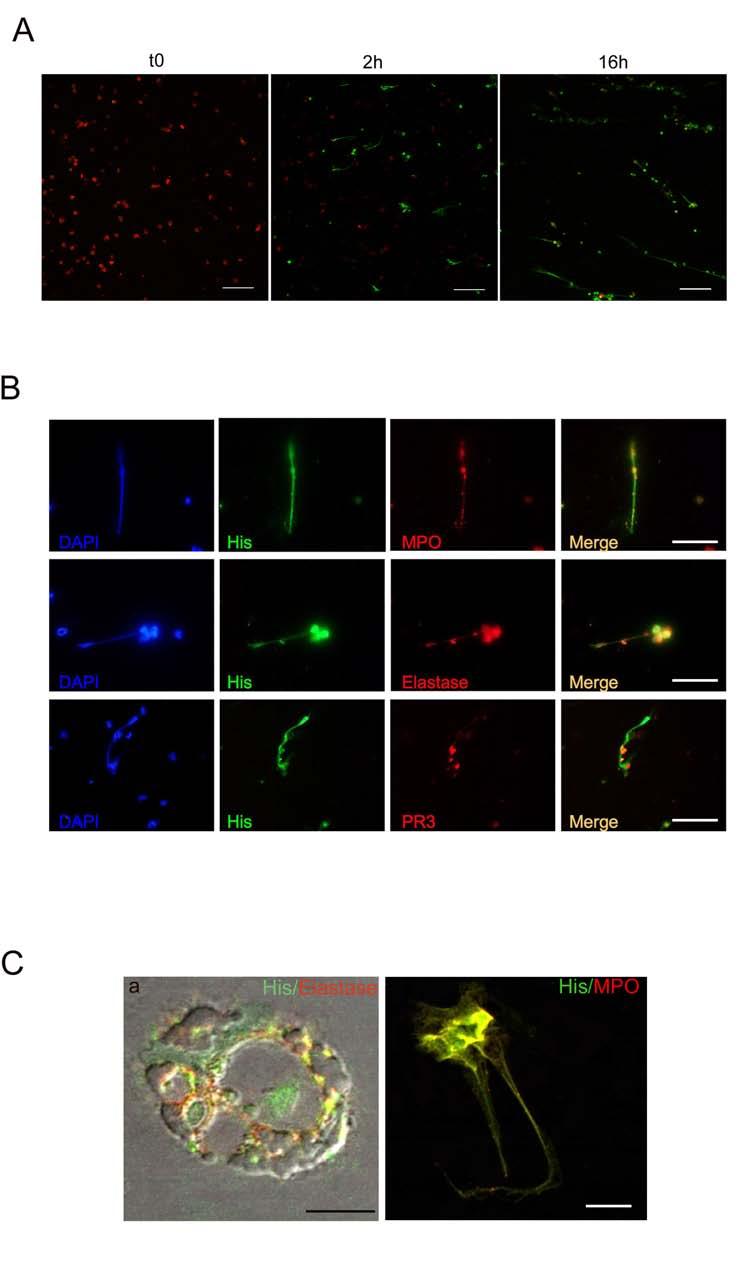
5 Features of NETosis in agar-elicited PMN. A. Confocal microscopy analysis showing the time course of NET formation by agar PMN. PMN undergoing NETosis incorporate the dsdna dye SYTOX green. Red staining: Elastase Ab. Scale bars: 20 µm. B. Characterization of NET produced by agar PMN with Ab to MPO, Elastase and PR3 showing the localization of these proteins in the dsdna backbone (pan Histone Ab). Scale bars: 10 µm. C. Confocal microscopy analysis showing that NETosis by agar PMN initiates with a nuclear permeabilization allowing the colocalization of nuclear material, identified by a staining for histones, with cytoplasmic proteins like elastase (red staining). Scale bar: 5µm. b. The end point of NETosis is visualized as release of DNA-based fibers, decorated with cytoplasmic proteins, here shown with mab to His (green) and MPO (red). Scale bars: 10 µm.

6 Supplementary Fig.3 Quantification of NETosis by confocal microscopy. The method used to quantify NET and to distinguish NETosis from apoptosis is based on measuring the size of nuclear material that is stained with the DNA dye SYTOX green. SYTOX green is a cellimpermeable dye that in live cell imaging experiments, when added before PFA fixation, stains only dsdna released from NET. On the other hand, if added to cells after fixation, this dye stains also apoptotic cells as in the case showed in this picture. Particularly, to evaluate the relative amount of NETosis and apoptosis in our PMN preparation, cells were seeded onto poly-lysine coated glasses, fixed and stained with the SYTOX green, and then imaged through confocal microscopy or immunofluorescence. Using a software-assisted technique the size of nuclear material is measured. As shown in the picture, apoptotic cells (left) have smaller area, whereas NETotic cells (right) are characterized by a broad range of nuclear areas. By
7 plotting SYTOX + nuclear area of a significative number of cells ( ) against the fraction of cells showing a given nuclear area interval, the result obtained is a distribution forming a narrow pick in case of prevalent apoptosis. Apoptotic cells have nuclear areas within the µm 2 interval, whereas NETotic PMN spread their areas from the up to the µm 2 interval.

8 Supplementary Fig. 4 INFγ and TNF are needed for NETosis in agar-pmn. A. Quantification of NET formation by agar PMN obtained from INFγ-KO, TNF-KO and IL6-KO mice. Agar trap collected PMN from INFγ -KO and TNF-KO mice did not form NET. In such deficient mice many cells were still alive and excluded the SYTOX green dye. B. Representative IF analysis of the cells, quantified in A. Reduced NET formation in agar-elicited PMN from INFγ-KO and TNF-KO but not from IL6-KO mice. Number of nuclei analyzed/experiment= 200.
/side scatter (SSC) and annexin V/7-AAD representative plots for CD11b+GR-1+ spleen isolated PMN that have been treated with freeze-thaw or anti-fas mab methods in inducing")
9 Supplementary Fig. 5 DC contacting netting neutrophils are activated and migrate to regional LN. A. Forward scatter (FSC)/side scatter (SSC) and annexin V/7-AAD representative plots for CD11b+GR-1+ spleen isolated PMN that have been treated with freeze-thaw or anti-fas mab methods in inducing necrosis or apoptosis, respectively. B. Flow cytometry analysis of of CD40 expression by mdc co-cultured with NETotic (in
10 presence or absence of DNAse), apoptotic or necrotic neutrophils. Cumulative data from 8 independent experiments are shown. C. Control mdc (unstimulated) or mdc stimulated with CpG or obtained from different mdc-pmn-co-cultures were labeled with fluorescent microbeads and injected i.p. into BALB/c naïve mice. After 24h mesenteric LN were analyzed for the presence of CD11c + /beads + cells. We found that DC treated with CpG or co-cultured with NETotic, apoptotic or necrotic PMN were all able to migrate to mesenteric LN. Representative plot obtained by analyzing a total of 2x10 6 /LN are shown. D. Activation of NADPH oxidase of agar PMN seeded onto poly-d-lysine coated glasses, in presence or absence of DNAse. Activation of NADPH oxidase was not affected by DNAse treatment as evaluated as the nitroblue tetrazolium (NBT) reduction test.
11 Supplementary Fig. 6
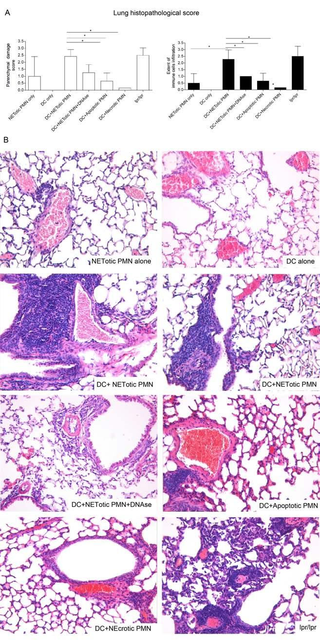
12 NET-loaded mdc immunization induces lung vasculities. A. Histopathological score of parenchymal damage (left) and extent of immune cells infiltration (right) in mice immunized with mdc co-cultured with NETotic, apoptotic or necrotic PMN, and in control mice immunized with DC or PMN alone (n=9/group; p<0.05 and **p<0.01, one-way Anova with post test Dunn s correction. Data are shown as mean ± SD). Autoimmune lpr/lpr mice have been included in the analysis for comparison. B. Representative histological H&E pictures showing the perivascular neutrophilic infiltration characterizing the lungs of mice immunized with DC co-cultured with NEtotic PMN, but not the other groups. This phenotype was prevented in mice immunized with mdc co-cultured with NET in the presence of DNAse. Autoimmune lpr/lpr mice have been included in the analysis for comparison. Original Magnification: x20.
 prominent Gr1+ granulocyte infiltrates are detected throughout the renal parenchyma where they show vascular aggression (arrows).")
13 Supplementary. Fig.7 Histopathological features of the renal small vessel vasculitis induced by immunization with NET-loaded mdc. A) prominent Gr1+ granulocyte infiltrates are detected throughout the renal parenchyma where they show vascular aggression (arrows). B) Foci of granulocyte infiltration are associated with small vessels (arrows) in the renal parenchyma of mice immunized with NET-loaded mdcs. C) The vascular and peri-vascular inflammatory infiltrates show a pleomorphous composition and is populated by polymorphonuclear leukocytes (black arrows). D-E) Small necrotic foci are detected in the glomerular tufts and extraglomerular vessels. Inset: Some glomeruli show signs of extracapillary proliferation and crescent formation (red arrow). The pictures are representative of the 9 mice analyzed for SVV features among those immunized with DC+NETotic PMN.
.")
14 Supplementary Fig. 8 Immunoglobulin deposition in kidney from immunized mice. Renal frozen sections from mice immunized with DC+ NETotic, apoptotic or necrotic PMN as well as from lpr/lpr mice and mice have been stained with Alexa 488 coniugated mab to mouse IgG (H+L). IF analysis shows glomerural IgG deposition in mice immunized with mdc co-cultured with NETotic, apoptotic or necrotic PMN but not in mice immunized with PMN or DC alone or with DC+ NETotic PMN in the presence of
15 DNAse. Overall glomerular IgG deposition in immunized mice is lower than in autoimmune lpr/lpr mice.

16 Supplementary Fig. 9 Correlation between perivascular neutrophils infiltration and development of ANCA in mice immunized with mdc co-cultured with NETotic PMN. A. Immunohistochemical analysis for Gr-1 expression in frozen renal sections from 6 individual mice. B. Overall histological damage and extent of immune cells infiltration in kidneys from the same mice. C. The six mice have been also analyzed for serum MPO-ANCA. The level of circulating ANCA correlated with that of granulocyte/immune cells infiltration in kidneys.
, such to induce or prevent NET formation.")
17 Supplementary Fig. 10 The serum of mice immunized with DC+NETotic PMN recognizes PMA-induce NET. Spleen purified PMN were seeded onto poly-d-lysine coated glasses and treated with PMA, in the presence or absence of DNAse (100U/ml), such to induce or prevent NET formation. PMN were than stained with sera from mice immunized with DC+NETotic PMN or from lpr-lpr mice. Sera from DC+NETotic PMN recognize PMA-treated PMN only in the absence of DNAse, whereas sera from lpr/lpr mice still recognize them even in the presence of DNAse, a result consistent with their high ds- DNA and ss-dna ANA titers. A. Representative IF (Scale bars: 25 µm) and B. Software assisted micrograph quantification of the above staining performed on a total of 200 cells that were imaged by confocal microscopy. One representative experiment out of three performed is shown. D. The serum from mice immunized with DC+NEtotic PMN, similarly to that of lpr/lpr mice is poly-reactive and capable to recognize human neutrophils in peripheral blood smears. Peripheral blood smears from healthy donors have been fixed with PFA 4%, treated with 0.1% TRITON and stained with serum from mice immunized with DC+NETotic PMN or lpr/lpr mice
18 (1/20 dilution). The staining with serum from the lpr/lpr mouse (mouse number 54 of the previous experiments) is characterized by a nuclear and peri-nuclear pattern, whereas that from immune mice shows a more diffuse cytoplasmic staining. The serum from lpr/lpr, but not that from immunized mice, stains also nuclear material in circulating monocytes.
19 Supplementary Table 1. Histophatological and serological parameters of immune mice. PMN alone DC alone DC+Netotic PMN DC+Netotic PMN+DNAse DC+Apoptotic PMN DC+ Necrotic PMN lpr/lpr b) MPO-ANCA 5,57±4,54* 2,89±3,89*** 23,83±14 4,49±4,2** 8,89±6,9* 2,91±1,9** 9,5±10,75* PR3-ANCA 3,2± 3,4* ND 23,40 ±23,44 4,3 ± 3,0* 9,39 ± 15,6 3,7 ± 4,9* 42,5 ±26,12 anti-ss DNA Ab a) 265,8± ,3± ,9±105,3 631,8± ± ,5± ±142 anti-ds DNA Ab a) 103,1±74 152,2±68 503,46± ,9± ,9± ,05± ±293 anti-ana DNA Ab a) 1111± ± ± ,0± ± ± ±1282 Nephritic lesions score Interstitial lesions score 1,00±0 0,33±0,511* 4±1,48 1±0 0,33±0,52* 0,833±1,33* 2,125±1,64 1,33±0,577* ND 8±2,045 2,25±0,957 1,333±1,36* 2,33±1,86 9,25±3,24 Albuminuria 22,27±16,73 24,27±7,65 30,27±20 27,87±10,3 37,7±27 29,15±4,3 32,77±22 Skin lesions(%) ND ND Pulmonary lesions score 22,2±4 ND 8,33±0,0 1,5±2,12 ND 4,71±1,03 2,66±0,577 1,33±1,15* 0,33±0* 5±1,19 For statistical analysis DC-NETotic PMN group is compared to the other type of immunization. Dunn's Multiple Comparison Test. Mean ± SD are shown. * p< 0,05; ** p< 0,01; *** p< 0,001 a) U/ml 1/400 serum dilution; b) Months of age : 9,14±1,41. Mean ± SD ND ND
20 Supplementary table2. Characteristics of subjects with microscopic poliangiitis, SLE, and psoriasis, whose skin lesion biopsies were analyzed for NET/DC spatial interaction. Patient ID Gender Age Clinical diagnosis ANCA pattern (IIF) ANCA type (ELISA) Skin lesions NET/DC interaction #1 F 31 MPA p-anca MPO+ P,E + #2 M 68 MPA n.a. MPO+ E - #3 M 53 MPA p-anca MPO+ P,E + #4 F 76 MPA p-anca MPO+ P,E,U + #5 M 34 MPA p-anca MPO+ P,E - #6 F 42 MPA n.a. MPO+ P + #7 M 47 SLE n.a. n.a. V/B - #8 M 40 SLE n.a. n.a. E,P,M + #9 F 59 SLE n.a. n.a. E,M - #10 F 39 SLE p-anca MPO+ E,P,M + #11 F 42 SLE n.a. n.a. E,PL + #12 F 53 Psoriasis n.a. n.a. E,M - #13 M 26 Psoriasis n.a. n.a. E,M - #14 M 42 Psoriasis n.a. n.a. E - #15 F 55 Psoriasis n.a. n.a. E,PL - Abbreviations: ANCA, anti-neutrophil cytoplasmatic antibodies; MPA, microscopic poliangiitis; SLE, systemic lupus erithematosus; p-anca, peri-nuclear ANCA staining on immunofluorescence; MPO, myeloperoxidase; P, purpura; E, erythema; U, ulcers; V/B, vesciculae/bullae; M, maculae; PL, plaques; NET, neutrophil extracellular trap; DC, dendritic cell.
Supplementary Figure Legends
 Supplementary Figure Legends Supplementary Figure 1. Comparison of RNP IC-mediated NET formation. Quantification of DNA release induced by ICs consisting of SmRNP combined with SLE IgG 961 (n = 10), 1032
Supporting Information
 Supporting Information Desnues et al. 10.1073/pnas.1314121111 SI Materials and Methods Mice. Toll-like receptor (TLR)8 / and TLR9 / mice were generated as described previously (1, 2). TLR9 / mice were
Nature Immunology: doi: /ni eee Supplementary Figure 1
 eee Supplementary Figure 1 Hyphae induce NET release, but yeast do not. (a) NET release by human peripheral neutrophils stimulated with a hgc1 yeast-locked C. albicans mutant (yeast) or pre-formed WT C.
4. KIDNEYS AND AUTOIMMUNE DISEASE
 How to Cite this article: Kidneys and Autoimmune Disease - ejifcc 20/01 2009 http://www.ifcc.org 4. KIDNEYS AND AUTOIMMUNE DISEASE Maksimiljan Gorenjak 4.1 Autoimmune diseases The human immune system limits
Supplementary Figure 1. Characterization of basophils after reconstitution of SCID mice
 Supplementary figure legends Supplementary Figure 1. Characterization of after reconstitution of SCID mice with CD4 + CD62L + T cells. (A-C) SCID mice (n = 6 / group) were reconstituted with 2 x 1 6 CD4
SUPPLEMENTARY INFORMATION
 doi:1.138/nature1554 a TNF-α + in CD4 + cells [%] 1 GF SPF 6 b IL-1 + in CD4 + cells [%] 5 4 3 2 1 Supplementary Figure 1. Effect of microbiota on cytokine profiles of T cells in GALT. Frequencies of TNF-α
LPS CD40 + IL-4. Vorinostat (24 Hours) Vorinostat (24 Hours) Panobinostat (24 Hours) Panobinostat (24 Hours) Romidepsin (48 Hours)
 Vorinostat (24 Hours) Panobinostat (24 Hours) Panobinostat (24 Hours) Romidepsin (48 Hours)") A) CD + IL- B) LPS ( Hours) ( Hours) Cell number (x1-3 ) 1 1 3.7 M 1. M. M.1 M Cell number (x1 - ) 1 1 3. M 1. M.7 M.38 M Cell number (x1-3 ) Cell number (x1-3 ) 3 1 1 1 ( Hours) 7.nM.nM 1.7nM.nM Romidepsin
A) CD + IL- B) LPS ( Hours) ( Hours) Cell number (x1-3 ) 1 1 3.7 M 1. M. M.1 M Cell number (x1 - ) 1 1 3. M 1. M.7 M.38 M Cell number (x1-3 ) Cell number (x1-3 ) 3 1 1 1 ( Hours) 7.nM.nM 1.7nM.nM Romidepsin
Histopathology: Glomerulonephritis and other renal pathology
 Histopathology: Glomerulonephritis and other renal pathology These presentations are to help you identify basic histopathological features. They do not contain the additional factual information that you
and follicular helper T cells is Egr2-dependent. (a) Diagrammatic representation of the
 Diagrammatic representation of the") Supplementary Figure 1. LAG3 + Treg-mediated regulation of germinal center B cells and follicular helper T cells is Egr2-dependent. (a) Diagrammatic representation of the experimental protocol for the
Supplementary Figure 1. LAG3 + Treg-mediated regulation of germinal center B cells and follicular helper T cells is Egr2-dependent. (a) Diagrammatic representation of the experimental protocol for the
Simultaneous comprehensive multiplex autoantibody analysis by CytoBead technology for Rapidly Progressive Glomerulonephritis.
 Simultaneous comprehensive multiplex autoantibody analysis by CytoBead technology for Rapidly Progressive Glomerulonephritis l Assays Indirect Immunofluorescence Goldstandard for Diagnosis of Autoimmune
Aggregated neutrophil extracellular traps limit inflammation by degrading cytokines and chemokines
 CORRECTION NOTICE Nat. Med. doi:10.1038/nm.3547; corrected online 25 August 2014 Aggregated neutrophil extracellular traps limit inflammation by degrading cytokines and chemokines Christine Schauer, Christina
Supplemental Table 1. Primer sequences for transcript analysis
 Supplemental Table 1. Primer sequences for transcript analysis Primer Sequence (5 3 ) Primer Sequence (5 3 ) Mmp2 Forward CCCGTGTGGCCCTC Mmp15 Forward CGGGGCTGGCT Reverse GCTCTCCCGGTTTC Reverse CCTGGTGTGCCTGCTC
Supplementary Figure 1: TSLP receptor skin expression in dcssc. A: Healthy control (HC) skin with TSLP receptor expression in brown (10x
 skin with TSLP receptor expression in brown (10x") Supplementary Figure 1: TSLP receptor skin expression in dcssc. A: Healthy control (HC) skin with TSLP receptor expression in brown (10x magnification). B: Second HC skin stained for TSLP receptor in brown
Supplementary Figure 1: TSLP receptor skin expression in dcssc. A: Healthy control (HC) skin with TSLP receptor expression in brown (10x magnification). B: Second HC skin stained for TSLP receptor in brown
<20 <20 <20 < <20 <20 <20 <20. Mock
 Cross-Lineage Neutralization PRNT 80 Titers Asian Asian West African Indian Ocean Group NHP Strain 181/25 Strain 99659 Strain 37997 Strain LR 142590 80 80 20 40 EILV/CHIKV 150844 640 640 160 320 Mock 150849
Supplementary Figures
 Inhibition of Pulmonary Anti Bacterial Defense by IFN γ During Recovery from Influenza Infection By Keer Sun and Dennis W. Metzger Supplementary Figures d a Ly6G Percentage survival f 1 75 5 1 25 1 5 1
Page 39 of 44. 8h LTA & AT h PepG & AT h LTA
 Page 39 of 44 Fig. S1 A: B: C: D: 8h LTA 8h LTA & AT7519 E: F: 8h PepG G: 8h PepG & AT7519 Fig. S1. AT7519 overrides the survival effects of lipoteichoic acid (LTA) and peptidoglycan (PepG). (A) Human
SUPPLEMENTARY INFORMATION
 doi:10.1038/nature10134 Supplementary Figure 1. Anti-inflammatory activity of sfc. a, Autoantibody immune complexes crosslink activating Fc receptors, promoting activation of macrophages, and WWW.NATURE.COM/NATURE
Endogenous TNFα orchestrates the trafficking of neutrophils into and within lymphatic vessels during acute inflammation
 SUPPLEMENTARY INFORMATION Endogenous TNFα orchestrates the trafficking of neutrophils into and within lymphatic vessels during acute inflammation Samantha Arokiasamy 1,2, Christian Zakian 1, Jessica Dilliway
Evaluation of directed and random motility in microslides Assessment of leukocyte adhesion in flow chambers
 Evaluation of directed and random motility in microslides Motility experiments in IBIDI microslides, image acquisition and processing were performed as described. PMN, which ended up in an angle < 180
Glomerular pathology in systemic disease
 Glomerular pathology in systemic disease Lecture outline Lupus nephritis Diabetic nephropathy Glomerulonephritis Associated with Bacterial Endocarditis and Other Systemic Infections Henoch-Schonlein Purpura
Renal Pathology 1: Glomerulus. With many thanks to Elizabeth Angus PhD for EM photographs
 Renal Pathology 1: Glomerulus With many thanks to Elizabeth Angus PhD for EM photographs Anatomy of the Kidney http://www.yalemedicalgroup.org/stw/page.asp?pageid=stw028980 The Nephron http://www.beltina.org/health-dictionary/nephron-function-kidney-definition.html
Nature Protocols: doi: /nprot Supplementary Figure 1
 Supplementary Figure 1 Traditional electronic gating strategy for analysing cell death based on A5-FITC and 7-AAD. a, Flow cytometry analysis showing the traditional two-stage electronic gating strategy
IKK-dependent activation of NF-κB contributes to myeloid and lymphoid leukemogenesis by BCR-ABL1
 Supplemental Figures BLOOD/2014/547943 IKK-dependent activation of NF-κB contributes to myeloid and lymphoid leukemogenesis by BCR-ABL1 Hsieh M-Y and Van Etten RA Supplemental Figure S1. Titers of retroviral
NK cell flow cytometric assay In vivo DC viability and migration assay
 NK cell flow cytometric assay 6 NK cells were purified, by negative selection with the NK Cell Isolation Kit (Miltenyi iotec), from spleen and lymph nodes of 6 RAG1KO mice, injected the day before with
Assays. New. New. Combinations. Possibilities. Patents: EP , AU
 Assays Patents: EP 2362222, AU 2011217190 New Combinations New Possibilities Technology Classical Handling of Autoimmune Diagnostics 2-Step Diagnostics 1 st Screening 2 nd Confirmation Cell based IFA ELISA
n BASIC-TRANSLATIONAL REVIEW
 n BASIC-TRANSLATIONAL REVIEW At the Bedside: Neutrophil extracellular traps (NETs) as targets for biomarkers and therapies in autoimmune diseases April Barnado,*,1 Leslie J. Crofford,* and Jim C. Oates
Crescentic Glomerulonephritis (RPGN)
") Crescentic Glomerulonephritis (RPGN) Background Rapidly progressive glomerulonephritis (RPGN) is defined as any glomerular disease characterized by extensive crescents (usually >50%) as the principal histologic
Crescentic Glomerulonephritis (RPGN) Background Rapidly progressive glomerulonephritis (RPGN) is defined as any glomerular disease characterized by extensive crescents (usually >50%) as the principal histologic
Supplementary Figure 1. Double-staining immunofluorescence analysis of invasive colon and breast cancers. Specimens from invasive ductal breast
 Supplementary Figure 1. Double-staining immunofluorescence analysis of invasive colon and breast cancers. Specimens from invasive ductal breast carcinoma (a) and colon adenocarcinoma (b) were staining
Supplementary Information. Tissue-wide immunity against Leishmania. through collective production of nitric oxide
 Supplementary Information Tissue-wide immunity against Leishmania through collective production of nitric oxide Romain Olekhnovitch, Bernhard Ryffel, Andreas J. Müller and Philippe Bousso Supplementary
Supplementary Materials for
 www.sciencemag.org/content/348/6241/aaa825/suppl/dc1 Supplementary Materials for A mucosal vaccine against Chlamydia trachomatis generates two waves of protective memory T cells Georg Stary,* Andrew Olive,
SUPPORTING INFORMATIONS
 SUPPORTING INFORMATIONS Mice MT/ret RetCD3ε KO α-cd25 treated MT/ret Age 1 month 3 mnths 6 months 1 month 3 months 6 months 1 month 3 months 6 months 2/87 Survival 87/87 incidence of 17/87 1 ary tumor
Serafino et al. Thymosin α1 activates complement receptor-mediated phagocytosis in human monocyte-derived macrophages. SUPPLEMENTARY FIGURES
 Supplementary Fig. S1. Evaluation of the purity and maturation of macrophage cultures tested by flow cytometry. The lymphocytic/monocytic cellular fraction was isolated from buffy coats of healthy donors
Supplemental Materials for. Effects of sphingosine-1-phosphate receptor 1 phosphorylation in response to. FTY720 during neuroinflammation
 Supplemental Materials for Effects of sphingosine-1-phosphate receptor 1 phosphorylation in response to FTY7 during neuroinflammation This file includes: Supplemental Table 1. EAE clinical parameters of
Suppl Video: Tumor cells (green) and monocytes (white) are seeded on a confluent endothelial
 and monocytes (white) are seeded on a confluent endothelial") Supplementary Information Häuselmann et al. Monocyte induction of E-selectin-mediated endothelial activation releases VE-cadherin junctions to promote tumor cell extravasation in the metastasis cascade
Supplementary Information Häuselmann et al. Monocyte induction of E-selectin-mediated endothelial activation releases VE-cadherin junctions to promote tumor cell extravasation in the metastasis cascade
Eosinophils are required. for the maintenance of plasma cells in the bone marrow
 Eosinophils are required for the maintenance of plasma cells in the bone marrow Van Trung Chu, Anja Fröhlich, Gudrun Steinhauser, Tobias Scheel, Toralf Roch, Simon Fillatreau, James J. Lee, Max Löhning
(a) Significant biological processes (upper panel) and disease biomarkers (lower panel)
 Significant biological processes (upper panel) and disease biomarkers (lower panel)") Supplementary Figure 1. Functional enrichment analyses of secretomic proteins. (a) Significant biological processes (upper panel) and disease biomarkers (lower panel) 2 involved by hrab37-mediated secretory
Supplementary Figure 1. Functional enrichment analyses of secretomic proteins. (a) Significant biological processes (upper panel) and disease biomarkers (lower panel) 2 involved by hrab37-mediated secretory
Elevated Expression of Pentraxin 3 in Anti-neutrophil Cytoplasmic Antibody-associated Glomerulonephritis with Normal Serum C-reactive Protein
 CASE REPORT Elevated Expression of Pentraxin 3 in Anti-neutrophil Cytoplasmic Antibody-associated Glomerulonephritis with Normal Serum C-reactive Protein Risa Ishida 1,KentaroNakai 1, Hideki Fujii 1, Shunsuke
B6/COLODR/SPL/11C/83/LAP/#2.006 B6/COLODR/SPL/11C/86/LAP/#2.016 CD11C B6/COLODR/SPL/11C/80/LAP/#2.011 CD11C
 CD3-specific antibody-induced immune tolerance and suppression of autoimmune encephalomyelitis involves TGF-β production through phagocytes digesting apoptotic T cells Sylvain Perruche 1,3, Pin Zhang 1,
SUPPLEMENTARY METHODS
 SUPPLEMENTARY METHODS Histological analysis. Colonic tissues were collected from 5 parts of the middle colon on day 7 after the start of DSS treatment, and then were cut into segments, fixed with 4% paraformaldehyde,
Supplementary Figure 1
 Supplementary Figure 1 Identification of IFN-γ-producing CD8 + and CD4 + T cells with naive phenotype by alternative gating and sample-processing strategies. a. Contour 5% probability plots show definition
COPD lungs show an attached stratified mucus layer that separate. bacteria from the epithelial cells resembling the protective colonic
 COPD lungs show an attached stratified mucus layer that separate bacteria from the epithelial cells resembling the protective colonic mucus SUPPLEMENTARY TABLES AND FIGURES Tables S1 S8, page 1 and separate
VASCULITIS PRODUCT HIGHLIGHTS
 VASCULITIS PRODUCT HIGHLIGHTS AESKU.DIAGNOSTICS offers a comprehensive and complete diagnostic portfolio in the field of vasculitis diagnostics. Not only are screening and profiling s available but also
Nature Medicine: doi: /nm.2109
 HIV 1 Infects Multipotent Progenitor Cells Causing Cell Death and Establishing Latent Cellular Reservoirs Christoph C. Carter, Adewunmi Onafuwa Nuga, Lucy A. M c Namara, James Riddell IV, Dale Bixby, Michael
As outlined under External contributions (see appendix 7.1), the group of Prof. Gröne at the
, the group of Prof. Gröne at the") 3 RESULTS As outlined under External contributions (see appendix 7.1), the group of Prof. Gröne at the DKFZ in Heidelberg (Dept. of Cellular and Molecular pathology) contributed to this work by performing
3 RESULTS As outlined under External contributions (see appendix 7.1), the group of Prof. Gröne at the DKFZ in Heidelberg (Dept. of Cellular and Molecular pathology) contributed to this work by performing
well for 2 h at rt. Each dot represents an individual mouse and bar is the mean ±
 Supplementary data: Control DC Blimp-1 ko DC 8 6 4 2-2 IL-1β p=.5 medium 8 6 4 2 IL-2 Medium p=.16 8 6 4 2 IL-6 medium p=.3 5 4 3 2 1-1 medium IL-1 n.s. 25 2 15 1 5 IL-12(p7) p=.15 5 IFNγ p=.65 4 3 2 1
Year 2004 Paper one: Questions supplied by Megan
 QUESTION 53 Endothelial cell pathology on renal biopsy is most characteristic of which one of the following diagnoses? A. Pre-eclampsia B. Haemolytic uraemic syndrome C. Lupus nephritis D. Immunoglobulin
GOODPASTURE'S SYNDROME WITH CONCOMITANT IMMUNE COMPLEX MIXED MEMBRANOUS AND PROLIFERATIVE GLOMERULONEFRITIS
 GOODPASTURE'S SYNDROME WITH CONCOMITANT IMMUNE COMPLEX MIXED MEMBRANOUS AND PROLIFERATIVE GLOMERULONEFRITIS VESNA JURČIĆ 1, ANDREJA ALEŠ RIGLER 2, INSTITUTE OF PATHOLOGY, FACULTY OF MEDICINE, UNIVERSITY
Real-time imaging reveals the single steps of brain metastasis fo mation r
 Real-time imaging reveals the single steps of brain metastasis fo mation r Yvonne Kienast, Louisa von Baumgarten, Martin Fuhrmann, Wolfgang E.F. Klinkert, Roland Goldbrunner, Jochen Herms and Frank Winkler
RENAL HISTOPATHOLOGY
 RENAL HISTOPATHOLOGY Peter McCue, M.D. Department of Pathology, Anatomy & Cell Biology Sidney Kimmel Medical College There are no conflicts of interest. 1 Goals and Objectives! Goals Provide introduction
Figure S1. PMVs from THP-1 cells expose phosphatidylserine and carry actin. A) Flow
 Flow") SUPPLEMENTARY DATA Supplementary Figure Legends Figure S1. PMVs from THP-1 cells expose phosphatidylserine and carry actin. A) Flow cytometry analysis of PMVs labelled with annexin-v-pe (Guava technologies)
SUPPLEMENTARY DATA Supplementary Figure Legends Figure S1. PMVs from THP-1 cells expose phosphatidylserine and carry actin. A) Flow cytometry analysis of PMVs labelled with annexin-v-pe (Guava technologies)
- 1 - Cell types Monocytes THP-1 cells Macrophages. LPS Treatment time (Hour) IL-6 level (pg/ml)
 IL-6 level (pg/ml)") Supplementary Table ST1: The dynamic effect of LPS on IL-6 production in monocytes and THP-1 cells after GdA treatment. Monocytes, THP-1 cells and macrophages (5x10 5 ) were incubated with 10 μg/ml of
Supplementary Table ST1: The dynamic effect of LPS on IL-6 production in monocytes and THP-1 cells after GdA treatment. Monocytes, THP-1 cells and macrophages (5x10 5 ) were incubated with 10 μg/ml of
Supplementary Figure 1: Hsp60 / IEC mice are embryonically lethal (A) Light microscopic pictures show mouse embryos at developmental stage E12.
 Light microscopic pictures show mouse embryos at developmental stage E12.") Supplementary Figure 1: Hsp60 / IEC mice are embryonically lethal (A) Light microscopic pictures show mouse embryos at developmental stage E12.5 and E13.5 prepared from uteri of dams and subsequently genotyped.
Supplementary Figure 1: Hsp60 / IEC mice are embryonically lethal (A) Light microscopic pictures show mouse embryos at developmental stage E12.5 and E13.5 prepared from uteri of dams and subsequently genotyped.
Supplementary Figures
 Supplementary Figures Supplementary Fig. 1. Surface thiol groups and reduction of activated T cells. (a) Activated CD8 + T-cells have high expression levels of free thiol groups on cell surface proteins.
Supplemental Figure 1. IL-3 blockade with Fab CSL362 depletes plasmacytoid dendritic cells (pdcs), but not basophils, at higher doses.
, but not basophils, at higher doses.") Supplemental Figure 1. IL-3 blockade with Fab CSL362 depletes plasmacytoid dendritic cells (pdcs), but not basophils, at higher doses. Percentage of viable (A) pdcs (Sytox Blue-, Lin1-, HLADR+, BDCA2++)
Supplemental Figure 1. IL-3 blockade with Fab CSL362 depletes plasmacytoid dendritic cells (pdcs), but not basophils, at higher doses. Percentage of viable (A) pdcs (Sytox Blue-, Lin1-, HLADR+, BDCA2++)
Programmed necrosis, not apoptosis, is a key mediator of cell loss and DAMP-mediated inflammation in dsrna-induced retinal degeneration
 Programmed necrosis, not apoptosis, is a key mediator of cell loss and DAMP-mediated inflammation in dsrna-induced retinal degeneration The Harvard community has made this article openly available. Please
IMO-8400, a novel TLR7, TLR8 and TLR9 antagonist, psoriasis
 IMO-8400, a novel TLR7, TLR8 and TLR9 antagonist, inhibits disease development in mouse models of psoriasis Weiwen e Ja Jiang, Fu-Gang Zhu, Dong Yu, Ekambar R. Kandimalla, a a, Nicola La Monica, and Sudhir
Nature Medicine: doi: /nm.4322
 1 2 3 4 5 6 7 8 9 10 11 Supplementary Figure 1. Predicted RNA structure of 3 UTR and sequence alignment of deleted nucleotides. (a) Predicted RNA secondary structure of ZIKV 3 UTR. The stem-loop structure
Human neutrophils phagocytose and kill Acinetobacter baumannii and A. pittii
 Human neutrophils phagocytose and kill Acinetobacter baumannii and A. pittii María Lázaro-Díez, Itziar Chapartegui-González, Santiago Redondo-Salvo, Chike Leigh, David Merino, David San Segundo, Jesús
How the Innate Immune System Profiles Pathogens
 How the Innate Immune System Profiles Pathogens Receptors on macrophages, neutrophils, dendritic cells for bacteria and viruses Broad specificity - Two main groups of bacteria: gram positive, gram-negative
Blocking antibodies and peptides. Rat anti-mouse PD-1 (29F.1A12, rat IgG2a, k), PD-
, PD-") Supplementary Methods Blocking antibodies and peptides. Rat anti-mouse PD-1 (29F.1A12, rat IgG2a, k), PD- L1 (10F.9G2, rat IgG2b, k), and PD-L2 (3.2, mouse IgG1) have been described (24). Anti-CTLA-4 (clone
Supplementary Methods Blocking antibodies and peptides. Rat anti-mouse PD-1 (29F.1A12, rat IgG2a, k), PD- L1 (10F.9G2, rat IgG2b, k), and PD-L2 (3.2, mouse IgG1) have been described (24). Anti-CTLA-4 (clone
Disclosures. Rheumatological Approaches to Differential Diagnosis, Physical Examination, and Interpretation of Studies. None
 Rheumatological Approaches to Differential Diagnosis, Physical Examination, and Interpretation of Studies Sarah Goglin MD Assistant Professor of Medicine Division of Rheumatology Disclosures None 1 [footer
Interferon γ regulates idiopathic pneumonia syndrome, a. Th17 + CD4 + T-cell-mediated GvH disease
 Interferon γ regulates idiopathic pneumonia syndrome, a Th17 + CD4 + T-cell-mediated GvH disease Nora Mauermann, Julia Burian, Christophe von Garnier, Stefan Dirnhofer, Davide Germano, Christine Schuett,
Pearson r = P (one-tailed) = n = 9
 = n = 9") 8F4-Specific Lysis, % 1 UPN1 UPN3 8 UPN7 6 Pearson r =.69 UPN2 UPN5 P (one-tailed) =.192 4 UPN8 n = 9 2 UPN9 UPN4 UPN6 5 1 15 2 25 8 8F4, % Max MFI Supplementary Figure S1. AML samples UPN1-UPN9 show variable
8F4-Specific Lysis, % 1 UPN1 UPN3 8 UPN7 6 Pearson r =.69 UPN2 UPN5 P (one-tailed) =.192 4 UPN8 n = 9 2 UPN9 UPN4 UPN6 5 1 15 2 25 8 8F4, % Max MFI Supplementary Figure S1. AML samples UPN1-UPN9 show variable
Irf1 fold changes (D) 24h 48h. p-p65. t-p65. p-irf3. t-irf3. β-actin SKO TKO 100% 80% 60% 40% 20%
 24h 48h. p-p65. t-p65. p-irf3. t-irf3. β-actin SKO TKO 100% 80% 60% 40% 20%") Irf7 Fold changes 3 1 Irf1 fold changes 3 1 8h h 8h 8h h 8h p-p6 p-p6 t-p6 p-irf3 β-actin p-irf3 t-irf3 β-actin TKO TKO STKO (E) (F) TKO TKO % of p6 nuclear translocation % % 1% 1% % % p6 TKO % of IRF3
Irf7 Fold changes 3 1 Irf1 fold changes 3 1 8h h 8h 8h h 8h p-p6 p-p6 t-p6 p-irf3 β-actin p-irf3 t-irf3 β-actin TKO TKO STKO (E) (F) TKO TKO % of p6 nuclear translocation % % 1% 1% % % p6 TKO % of IRF3
Renal cell expressed TNF receptor 2, not receptor 1, is essential for the development of glomerulonephritis
 Research article Renal cell expressed TNF receptor 2, not receptor 1, is essential for the development of glomerulonephritis Volker Vielhauer, George Stavrakis, and Tanya N. Mayadas Center of Excellence
Supplementary Materials for
 www.sciencetranslationalmedicine.org/cgi/content/full/8/352/352ra110/dc1 Supplementary Materials for Spatially selective depletion of tumor-associated regulatory T cells with near-infrared photoimmunotherapy
Pathologic Stage. Lymph node Stage
 ASC ASC a c Patient ID BMI Age Gleason score Non-obese PBMC 1 22.1 81 6 (3+3) PBMC 2 21.9 6 6 (3+3) PBMC 3 22 84 8 (4+4) PBMC 4 24.6 68 7 (3+4) PBMC 24. 6 (3+3) PBMC 6 24.7 73 7 (3+4) PBMC 7 23. 67 7 (3+4)
A clinical syndrome, composed mainly of:
 Nephritic syndrome We will discuss: 1)Nephritic syndrome: -Acute postinfectious (poststreptococcal) GN -IgA nephropathy -Hereditary nephritis 2)Rapidly progressive GN (RPGN) A clinical syndrome, composed
Supplementary Figure 1. Efficient DC depletion in CD11c.DOG transgenic mice
 Supplementary Figure 1. Efficient DC depletion in CD11c.DOG transgenic mice (a) CD11c.DOG transgenic mice (tg) were treated with 8 ng/g body weight (b.w.) diphtheria toxin (DT) i.p. on day -1 and every
Procaspase-3. Cleaved caspase-3. actin. Cytochrome C (10 M) Z-VAD-fmk. Procaspase-3. Cleaved caspase-3. actin. Z-VAD-fmk
 Z-VAD-fmk. Procaspase-3. Cleaved caspase-3. actin. Z-VAD-fmk") A HeLa actin - + + - - + Cytochrome C (1 M) Z-VAD-fmk PMN - + + - - + actin Cytochrome C (1 M) Z-VAD-fmk Figure S1. (A) Pan-caspase inhibitor z-vad-fmk inhibits cytochrome c- mediated procaspase-3 cleavage.
A HeLa actin - + + - - + Cytochrome C (1 M) Z-VAD-fmk PMN - + + - - + actin Cytochrome C (1 M) Z-VAD-fmk Figure S1. (A) Pan-caspase inhibitor z-vad-fmk inhibits cytochrome c- mediated procaspase-3 cleavage.
Supplementary Figures
 Supplementary Figures Supplementary Fig. 1. Galectin-3 is present within tumors. (A) mrna expression levels of Lgals3 (galectin-3) and Lgals8 (galectin-8) in the four classes of cell lines as determined
Title of file for HTML: Supplementary Information Description: Supplementary Figures and Supplementary Table
 Title of file for HTML: Supplementary Information Description: Supplementary Figures and Supplementary Table Title of file for HTML: Peer Review File Description: Innate Scavenger Receptor-A regulates
sequences of a styx mutant reveals a T to A transversion in the donor splice site of intron 5
 sfigure 1 Styx mutant mice recapitulate the phenotype of SHIP -/- mice. (A) Analysis of the genomic sequences of a styx mutant reveals a T to A transversion in the donor splice site of intron 5 (GTAAC
a 10 4 Link et al. Supplementary Figure 1 Nature Immunology: doi: /ni.1842 Cells per mouse ( 10 5 ) TRPV2KO anti-gr1 anti-gr anti-f4/80
 TRPV2KO anti-gr1 anti-gr anti-f4/80") a 10 4 WT 10 4 TRPV2KO 10 3 10 3 anti-gr1 10 2 10 1 anti-gr1 10 2 10 1 10 0 10 0 10 1 10 2 10 3 10 4 anti-f4/80 42.3 45.2 10 0 10 0 10 1 10 2 10 3 10 4 anti-f4/80 10 4 10 4 40 42.5 anti-cd11b 10 3 10 2
a 10 4 WT 10 4 TRPV2KO 10 3 10 3 anti-gr1 10 2 10 1 anti-gr1 10 2 10 1 10 0 10 0 10 1 10 2 10 3 10 4 anti-f4/80 42.3 45.2 10 0 10 0 10 1 10 2 10 3 10 4 anti-f4/80 10 4 10 4 40 42.5 anti-cd11b 10 3 10 2
Manchester 2 nd September High Throughput/High Content Screen to Identify Modulators of Neutrophil Extracellular Trap (NET) Formation
 Formation") Manchester 2 nd September 2014 High Throughput/High Content Screen to Identify Modulators of Neutrophil Extracellular Trap (NET) Formation Agenda Neutrophil biology and pathogenicity caused by neutrophil
Manchester 2 nd September 2014 High Throughput/High Content Screen to Identify Modulators of Neutrophil Extracellular Trap (NET) Formation Agenda Neutrophil biology and pathogenicity caused by neutrophil
hexahistidine tagged GRP78 devoid of the KDEL motif (GRP78-His) on SDS-PAGE. This
 on SDS-PAGE. This") SUPPLEMENTAL FIGURE LEGEND Fig. S1. Generation and characterization of. (A) Coomassie staining of soluble hexahistidine tagged GRP78 devoid of the KDEL motif (GRP78-His) on SDS-PAGE. This protein was expressed
SUPPLEMENTAL FIGURE LEGEND Fig. S1. Generation and characterization of. (A) Coomassie staining of soluble hexahistidine tagged GRP78 devoid of the KDEL motif (GRP78-His) on SDS-PAGE. This protein was expressed
Supplementalgfigureg1gSchematicgdiagramgofgtumor1modellingg
 SChinjectionh F:LuchLCLsh IVhinjectionh T:cellsh Monitorhforhtumorh growthhandhxeno: reactivehgvhd GVLgexperimentg kcbgvsgpbgt1cellse Xeno1reactiveg experimentg kcbgvsgpbgt1cellse IVhinjectionh 5xh,N^6
The encephalitogenicity of TH17 cells is dependent on IL-1- and IL-23- induced production of the cytokine GM-CSF
 CORRECTION NOTICE Nat.Immunol. 12, 568 575 (2011) The encephalitogenicity of TH17 cells is dependent on IL-1- and IL-23- induced production of the cytokine GM-CSF Mohamed El-Behi, Bogoljub Ciric, Hong
Supplementary Figure 1. Deletion of Smad3 prevents B16F10 melanoma invasion and metastasis in a mouse s.c. tumor model.
 A B16F1 s.c. Lung LN Distant lymph nodes Colon B B16F1 s.c. Supplementary Figure 1. Deletion of Smad3 prevents B16F1 melanoma invasion and metastasis in a mouse s.c. tumor model. Highly invasive growth
Dr Ian Roberts Oxford
 Dr Ian Roberts Oxford Oxford Pathology Course 2010 for FRCPath Present the basic diagnostic features of the commonest conditions causing renal failure Highlight diagnostic pitfalls. Crescentic GN: renal
Approach to Glomerular Diseases: Clinical Presentation Nephrotic Syndrome Nephritis
 GLOMERULONEPHRITIDES Vivette D Agati Jai Radhakrishnan Approach to Glomerular Diseases: Clinical Presentation Nephrotic Syndrome Nephritis Heavy Proteinuria Renal failure Low serum Albumin Hypertension
Supporting Information. Calculation of the relative contributions of myocyte proliferation, stem cell. Supporting Information Fig 1 (page 9)
") Supporting Information Table of contents Calculation of the relative contributions of myocyte proliferation, stem cell differentiation and cardioprotection (page 2) Supporting Information Fig 1 (page 9)
Supporting Information Table of contents Calculation of the relative contributions of myocyte proliferation, stem cell differentiation and cardioprotection (page 2) Supporting Information Fig 1 (page 9)
a b c Esophageal eosinophilia
 TSLP-elicited basophil responses can mediate the pathogenesis of eosinophilic esophagitis. Mario Noti, Elia D. Tait Wojno, Brian S. Kim, Mark C. Siracusa, Paul R. Giacomin, Meera G. Nair, Alain J. Benitez,
Nature Immunology: doi: /ni Supplementary Figure 1. Production of cytokines and chemokines after vaginal HSV-2 infection.
 Supplementary Figure 1 Production of cytokines and chemokines after vaginal HSV-2 infection. C57BL/6 mice were (a) treated intravaginally with 20 µl of PBS or infected with 6.7x10 4 pfu of HSV-2 in the
Supplementary Table 1
 Supplementary Table 1 Flow Cytometry Antibodies Antibody Fluorochrome Clone Vendor CD45 PE-cyanine 7 30-F11 D ioscience CD3 Pacific lue 17A2 iolegend (San Diego, CA) CD11b APC M1/70 iolegend (San Diego,
Atlas of the Vasculitic Syndromes
 CHAPTER e40 Atlas of the Vasculitic Syndromes Carol A. Langford Anthony S. Fauci Diagnosis of the vasculitic syndromes is usually based upon characteristic histologic or arteriographic findings in a patient
Supporting Information
 Supporting Information Idoyaga et al. 10.1073/pnas.0812247106 SSC a) Single cell suspension 99 Aqua b) Live cells 96 -W c) Singlets 92 -A CD19+ER119 d) CD19 ER119 cells 97 CD3 e) CD3 cells 27 f) DX5 cells
Nature Immunology: doi: /ni Supplementary Figure 1. Cellularity of leukocytes and their progenitors in naive wild-type and Spp1 / mice.
 Supplementary Figure 1 Cellularity of leukocytes and their progenitors in naive wild-type and Spp1 / mice. (a, b) Gating strategies for differentiated cells including PMN (CD11b + Ly6G hi and CD11b + Ly6G
Glomerular diseases mostly presenting with Nephritic syndrome
 Glomerular diseases mostly presenting with Nephritic syndrome 1 The Nephritic Syndrome Pathogenesis: proliferation of the cells in glomeruli & leukocytic infiltrate Injured capillary walls escape of RBCs
Type of file: PDF Title of file for HTML: Supplementary Information Description: Supplementary Figures
 Type of file: PDF Title of file for HTML: Supplementary Information Description: Supplementary Figures Type of file: MOV Title of file for HTML: Supplementary Movie 1 Description: NLRP3 is moving along
Supplementary Figure 1: Fn14 is upregulated in the epidermis and dermis of mice
 Supplementary Figure 1: Fn14 is upregulated in the epidermis and dermis of mice undergoing AD- and psoriasis-like disease. Immunofluorescence staining for Fn14 (green) and DAPI (blue) in skin of naïve
Clinical Laboratory. [None
 Clinical Laboratory Procedure Result Units Ref Interval Accession Collected Received Double-Stranded DNA (dsdna) Ab IgG ELISA Detected * [None 18-289-900151 Detected] Double-Stranded DNA (dsdna) Ab IgG
Acute lung injury in children : from viral infection and mechanical ventilation to inflammation and apoptosis Bern, R.A.
 UvA-DARE (Digital Academic Repository) Acute lung injury in children : from viral infection and mechanical ventilation to inflammation and apoptosis Bern, R.A. Link to publication Citation for published
 antigens co-expressed on granulocytes as well as other leukocyte populations are most often anti-hla class I antibodies. Antigen system Polymorphism Old terminology Location Antigen frequency in Europeans
Supplementary Information
 Supplementary Information Supplementary Figure 1. CD4 + T cell activation and lack of apoptosis after crosslinking with anti-cd3 + anti-cd28 + anti-cd160. (a) Flow cytometry of anti-cd160 (5D.10A11) binding
Supplementary Information
 Nature Immunology doi:1.138/ni.2477 Supplementary Information Capillary and arteriolar pericytes attract innate leukocytes exiting through venules and instruct them with pattern recognition and motility
The toll-like receptor 4 ligands Mrp8 and Mrp14 play a critical role in the development of autoreactive CD8 + T cells
 1 SUPPLEMENTARY INFORMATION The toll-like receptor 4 ligands Mrp8 and Mrp14 play a critical role in the development of autoreactive CD8 + T cells Karin Loser 1,2,6, Thomas Vogl 2,3, Maik Voskort 1, Aloys
Optimizing Intracellular Flow Cytometry:
 Optimizing Intracellular Flow Cytometry: Simultaneous Detection of Cytokines and Transcription Factors An encore presentation by Jurg Rohrer, PhD, BD Biosciences 10.26.10 Outline Introduction Cytokines
Autoimmune Disease. Autoimmunity. Epidemiology. ACR Criteria for Diagnosis. Signs and Symptoms. Autoreactivity: Reactivity to self antigens:
 Autoimmunity Reactivity to self antigens: Autoreactivity: Autoimmune Disease T cells B cells Leading to tissue damage or dysfunction Occurring in the absence of ongoing infection Epidemiology SLE Pathogenesis
Figure S1. Reduction in glomerular mir-146a levels correlate with progression to higher albuminuria in diabetic patients.
 Supplementary Materials Supplementary Figures Figure S1. Reduction in glomerular mir-146a levels correlate with progression to higher albuminuria in diabetic patients. Figure S2. Expression level of podocyte