SUPPLEMENTARY INFORMATION
|
|
|
- Thomasina Turner
- 5 years ago
- Views:
Transcription
1 doi:.38/nature234 Note 1. LINC00961 encodes a lysosomal Type I transmembrane polypeptide In order to determine if the polypeptide encoded by LINC00961 was an integral membrane protein or simply associated with the membrane, cell homogenates were treated with Na 2 CO 3 or NaOH. As shown in Extended Data Fig. 2b, this treatment disrupted the interaction of the membrane associated GM130, while treatment did not affect the localization of the integral membrane protein calnexin (Extended Data Fig. 2b and 2c). Consistent with the transmembrane domain identified by SMART 1, our -tagged LINC00961 encoded polypeptide remained in the membrane fraction upon treatment, indicating that the polypeptide penetrates the membrane via this putative transmembrane domain (Extended Data Fig. 2b and 2c). To further characterize how this polypeptide is integrated with cellular membranes, membrane fractions were treated with proteinase K. This treatment degrades proteins extending into the cytosol, while proteins extending into the luminal space are protected from enzymatic activity. The treatment of the membrane fraction with proteinase K in the absence of Triton X- resulted in the partial digestion of calnexin, while the bands for GM130 and -tagged LINC00961 encoded polypeptide completely disappeared (Extended Data Fig. 2d), indicating that the C terminus of the polypeptide molecule is exposed to the cytosol. Thus, as is the case for Type I transmembrane proteins, the LINC00961 encoded polypeptide harbors a luminal N terminus with a cytoplasmic C terminus. Although the LINC00961 encoded polypeptide localizes to the lysosome it does not appear to have any known lysosomal sorting signals (e.g. di-leucine (DXXLL or [DE]XXXL[LI]) and/or tyrosine motifs (YXXØ) 2, which are normally found at the C terminus of such transmembrane proteins 2. Thus, in order to better characterize the region responsible for lysosomal localization, we generated a series of chimeric proteins using the human CD8 receptor. CD8 is a human T-cell surface marker, a type I transmembrane protein, and is commonly used for chimeric fusions as it is well established as a neutral reporter 3. We generated chimeric fusion proteins with either the transmembrane domain or the C terminus of the LINC00961 encoded polypeptide (Extended Data Fig. 2g). As shown, the transmembrane region or C terminal region of 1
2 RESEARCH SUPPLEMENTARY INFORMATION the CD8 reporter was replaced with the ORF1 transmembrane region or C-terminal region, similar to previous CD8 fusion experiments 3. Overexpression of the of full length CD8 protein (CD8(WT)) in HeLa cells was found to predominantly localize to the plasma membrane (Extended Data Fig. 2h), which was also the case for the CD8 chimeric protein containing the LINC00961 polypeptide transmembrane domain (CD8(N)-ORF1(TM)-CD8(C)) (Extended Data Fig. 2i). The localization of this fusion protein was mutually exclusive with the lysosomal marker LAMP1, demonstrating that this region was not responsible for lysosomal targeting. However, a CD8 chimeric fusion in which the CD8 cytosolic domain was replaced with the C terminal cytoplasmic region of the LINC00961 polypeptide (CD8(N and TM)-ORF1(C)) demonstrated an exquisite localization to intracellular organelles that co-localized with LAMP1 (Extended Data Fig. 2j). Thus, while it is possible that the N terminal region may also contribute to localization of the LINC00961 encoded polypeptide, our data demonstrate that the C terminus is sufficient for lysosomal localization. 2
3 RESEARCH Note 2. The LINC00961 polypeptide does not impact v-atpase localization, assembly or lysosomal functions As HEK 293T do not express LINC00961, we generated cell lines that were stably transduced with empty vector control (Mock), wild type LINC00961 (WT) and LINC00961 with both ATG initiation codons mutated (ΔATG1+2). These cell lines were subsequently utilized to evaluate the affect of the LINC00961 encoded polypeptide on v- ATPase localization and assembly at steady state, and were used to examine lysosomal function upon polypeptide overexpression as measured by lysosomal ph, cathepsin activities (i.e. hydrolase function) and lysosomal morphology. Sucrose gradient fractionation was used to evaluate v-atpase localization and to determine if the LINC00961 polypeptide overexpression could affect this. The sucrose gradient fractionation process enabled us to purify light, medium and heavy membrane fractions, which correspond to a variety of subpopulations of vesicles within the cell. Our data established that the v-atpase complex is enriched in medium membrane fractions (Extended Data Fig. 3a), which corresponds to late endosome/lysosome and endoplasmic reticulum localizations as demonstrated by enrichment of Lamp2 and calnexin in these subcellular fractions (Extended Data Fig. 3a). Importantly, overexpression of the LINC00961 polypeptide had no impact on the localization of any of the v-atpase subunits analyzed. In order for the v-atpase complex to activate its proton pump activity, it is required that the V0 and V1 multi-protein domains associate with one another at the surface of cellular membranes 4. The V0 domain is an integral membrane protein complex that translocates protons, while the V1 domain protein complex transiently associates with the V0 domain and can hydrolyze ATP, and which together are required for proton pump activity 4. In order to determine if at steady state v-atpase assembly was altered upon expression of the LINC00961 polypeptide, we analyzed the levels of the V1 component ATP6V1A in cytoplasmic and membrane fractions, relative to ATP6V0D1, a component or the V0 domain (Extended Data Fig. 3b). In order to measure relative assembly of this complex at steady state, we calculated the ratio of ATP6V1A (V1 marker) to ATP6V0D1 (V0 marker) in membrane pellets, and found to be no difference between Mock, WT or ΔATG1+2 transduced cell lines (Extended Data Fig. 3c). In 3
4 RESEARCH SUPPLEMENTARY INFORMATION contrast, chloroquine is known to promote v-atpase assembly 5, and was used as a positive control for increased assembly in our studies (Extended Data Fig. 3b and 3c). Thus, these data suggest that v-atpase assembly may be unaffected by the LINC00961 polypeptide. As the LINC00961 polypeptide expression appeared to have no affect on v-atpase localization or assembly, we next sought to understand if overexpression could alter lysosomal characteristics and functions associated with v-atpase activity. As illustrated in Extended Data Fig. 3d and 3e, flow cytometry analysis using Lysosensor DND-189 can be utilized to monitor lysosomal acidification. The mean fluorescence intensity (MFI) of Mock or WT overexpressing cells was not altered (Extended Data Fig. 3e), while the v-atpase inhibitor bafilomycin A1 (BafA1) clearly decreased lysosomal acidification (Extended Data Fig. 3d). Similarly, hydrolase function was evaluated by Magic Red TM Cathepsin Assay Kits, which were used to quantify cathepsin L and K activity, and no difference was observed upon LINC00961 overexpression (Extended Data Fig. 3f and 3g). Finally, lysosomal morphology was analyzed by measuring lysosomal diameter upon stable expression of WT LINC While BafA1 clearly showed a significant increase in lysosomal diameter in HEK 293T cells (Extended Data Fig. 3h and 3i), LINC00961 expression had no effect (Extended Data Fig. 3j and 3k). Thus, these data appear to indicate that the LINC00961 polypeptide functions at the lysosome to regulate a process other than v-atpase localization, assembly and lysosomal function at steady state. 4
5 RESEARCH Note 3. Alternative activation of mtor or MAPK is not impacted by LINC00961 While our data established that the LINC00961 encoded polypeptide inhibits mtorc1 activation in response to amino acid signaling, we also examined if this polypeptide could influence activation of mtor by alternative means. Insulin stimulation acts to activate mtorc1 through the canonical PKB/AKT signaling pathway. However, overexpression of LINC00961 (WT) failed to impact mtorc1 activation mediated by insulin stimulation after 24 hours of serum starvation, as evaluated by phosphorylation of, and 4EBP (Extended Data Fig. 5a and 5b). In addition, recent reports reveal Sestrin2 as a cytosolic leucine sensor and CASTOR proteins as a cytosolic arginine sensor, and SLC38A9 as a putative lysosomal arginine sensor for mtorc1 signaling 6-9 (Extended Data Fig. 4a). Therefore, we also examined the effect of leucine-only depletion followed by leucine stimulation, or arginine-only depletion followed by arginine stimulation on mtorc1 activation in the presence of LINC00961 (WT) overexpression. However, we failed to see any affect on mtorc1 signaling in these conditions (Extended Data Fig. 5c-e). Furthermore, overexpression of LINC00961 (WT) showed no significant effects on phosphorylation of AKT at S473, a substrate for mtorc2, in response to insulin stimulation, or phosphorylation of ERK1/2 in response to EGF stimulation (Extended Data Fig. 5f-h). 5
6 RESEARCH SUPPLEMENTARY INFORMATION Note 4. Whole Genome Sequencing Analysis of CRISPR/Cas9 engineered mouse Off-target mutations in Cas9-modified mice have been reported to be low (Lyer et. al., Nature Methods), and for our sgrnas there were few predicted off-target sites (Supplementary Table 2), with the majority of predicted targets having at least 4 mismatched nucleotides (Supplementary Table 2). However, in order to fully address this point, we carried out whole genome sequencing (WGS) on three independent Spar KO mice, which had been backcrossed to the C57BL/6J line for at least 4 generations. No significant alterations were observed in our mice, and none of the alterations observed corresponded with predicted off-targets, indicative of normal and spontaneous variations rather than off-target activity of the Cas9 nickase (Supplementary Table 2). However, there did appear to be an enrichment of SNP nucleotides commonly associated with wild-type genetic background of the LG/J and SM/J strains of mice, rather than the C57BL/6J background of our mice, within the Sfi1 gene (Supplementary Table 2). Although these SNPs are normally found in the wild-type strains, we performed PCR amplification of the region and subsequent Sanger sequencing, and confirmed the presence of two of these observed SNP alleles in the genomes of the three mice subjected to WGS, as well as in five additional wild type and five Spar KO mice (Extended Data Fig. 7f and 7g). Thus, we are confident that the phenotype described in our manuscript is as a direct result of Spar deletion, rather than some other artifact resulting from non-specific CRISPR/Cas9 activity. 6
7 RESEARCH Note 5. The role of rapamycin in muscle regeneration In order to confirm that mtorc1 signaling is required for efficient regeneration of muscle, we treated wild type mice with rapamycin (2 mg/kg/day) to block mtorc1 activation prior to the administration of a single dose of cardiotoxin (CTX) by intramuscular injection in the tibialis anterior (TA) muscle (Extended Data Fig. 8a). Following injury, TA muscles were collected at 4 days post injury to monitor mtorc1 activation by western blot analysis (Extended Data Fig. 8b and 8c), and collected at 14 days post injury to monitor the muscle regeneration by histological analysis (Extended Data Fig. 8d-f). Rapamycin treatment greatly suppressed mtorc1 activation and muscle regeneration in this setting. Importantly, treatment with rapamycin had no impact on total body weight over this time, and therefore does not account for the decrase in TA muscle weight in response to CTX and rapamycin treatment (Extended Data Fig. 8g). Thus, these data establish a clear role for mtorc1 in muscle regeneration. 7
8 RESEARCH SUPPLEMENTARY INFORMATION Note 6. SPAR inactivation promotes muscle regenerative capacity As shown in Fig. 4d, treatment of TA muscle with CTX results in a strong activation of mtorc1 that is enhanced by the absence of Spar. Importantly, the activation of mtorc1 in the TA muscle of both Spar +/+ and Spar -/- mice is completely inhibited by the deprivation of leucine. We carefully evaluated TA muscle weight as a measure of regeneration in this tissue according to the schematic outlined in Extended Data Fig. 8i. The body weight of mice was taken at 7 days post-ctx administration, and while no difference in weight was observed between Spar +/+ or Spar -/- cohorts, leucine deprivation did result in a decrease in total body weight for both Spar +/+ or Spar -/- mice (Extended Data Fig. 8j). As mtor plays a role in growth and metabolism, we evaluated food intake over the 7-day period post-ctx administration, and found no significant difference in food intake relative to body weight for Spar +/+ and Spar -/- mice fed on a control or leucine-free diet (Extended Data Fig. 8k). As might be expected in the case of mice fed a leucine-free diet, both Spar +/+ and Spar -/- mice showed a decrease in control lateral uninjured TA muscle (TA(-)) weight, corresponding to the decrease observed for total body weight (Extended Data Fig. 8l). However, when normalized for total body weight, no difference was observed between control or leucine-fed Spar +/+ or Spar -/- mice (Extended Data Fig. 8m). Importantly, in the CTXinduced injured TA muscle (TA(+)) Spar -/- mice on the control diet showed a significant increase in both absolute weight (Extended Data Fig. 8n) and normalized weight (Extended Data Fig. 8o) at 7 days post-ctx administration relative to Spar +/+ mice, as a result of increased mtorc1 activity. In contrast, both Spar +/+ and Spar -/- mice fed a leucine free diet showed no difference in TA(+) muscle weights, indicative of a complete block in mtorc1 activity, as shown in Fig. 4d. 8
9 RESEARCH References 1 Letunic, I., Doerks, T. & Bork, P. SMART: recent updates, new developments and status in. Nucleic acids research, doi:.93/nar/gku949 (14). 2 Braulke, T. & Bonifacino, J. S. Sorting of lysosomal proteins. Biochim Biophys Acta 1793, , doi:.16/j.bbamcr (09). 3 Ihrke, G., Gray, S. R. & Luzio, J. P. Endolyn is a mucin-like type I membrane protein targeted to lysosomes by its cytoplasmic tail. Biochem J 345 Pt 2, (00). 4 McGuire, C., Cotter, K., Stransky, L. & Forgac, M. Regulation of V-ATPase assembly and function of V-ATPases in tumor cell invasiveness. Biochim Biophys Acta 1857, , doi:.16/j.bbabio (16). 5 Stransky, L. A. & Forgac, M. Amino Acid Availability Modulates Vacuolar H+- ATPase Assembly. J Biol Chem 290, , doi:.74/jbc.m (). 6 Chantranupong, L. et al. The CASTOR Proteins Are Arginine Sensors for the mtorc1 Pathway. Cell 165, 3-164, doi:.16/j.cell (16). 7 Jung, J., Genau, H. M. & Behrends, C. Amino Acid-Dependent mtorc1 Regulation by the Lysosomal Membrane Protein SLC38A9. Mol Cell Biol 35, , doi:.1128/mcb.001- (). 8 Rebsamen, M. et al. SLC38A9 is a component of the lysosomal amino acid sensing machinery that controls mtorc1. Nature 519, , doi:.38/nature147 (). 9 Wolfson, R. L. et al. Sestrin2 is a leucine sensor for the mtorc1 pathway. Science 351, 43-48, doi:.1126/science.aab2674 (16). 9


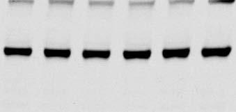


10 RESEARCH SUPPLEMENTARY INFORMATION Supplementary Figure 1. Original immunoblot images Fig. 1b Fig. 1f Fig. 1g ORF1 ORF1 Fig. 1c (Short exp.) 1 LAMP (Long exp.) Fig. 2a Fig. 2b Fig. 2c 2 1 ATP6V0A ATP6V0A2 ORF1 HA 2 1 ATP6V0A1 2 1 ATP6V0A2 RagC


11 RESEARCH Supplementary Figure 1. Original immunoblot images (continued) Fig. 2d Fig. 2f p- p- p-4ebp p-4ebp 4EBP 4EBP Fig. 3c Fig. 3e Fig. 3g Atp6V1A p- p


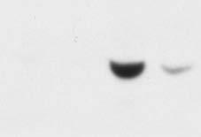


12 RESEARCH SUPPLEMENTARY INFORMATION Supplementary Figure 1. Original immunoblot images (continued) Fig. 4a Fig. 4d Extended Data Fig. 1k Extended Data Fig. 2a Spar Lamp1 Fig. 4c 2 1 (Short exp.) (Long exp.) p- ΔATG2 ΔATG1 Calnexin 2 1 p- Extended Data Fig. 2b Extended Data Fig. 2d Extended Data Fig. 2l GM ATP6V0A1 1 Calnexin 1 Calnexin 2 1 ATP6V0A2 1 GM ORF1 (Antibody No.2) 12


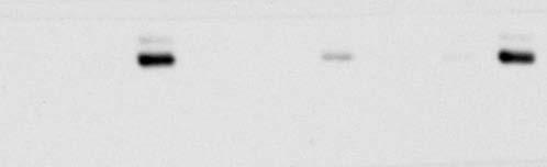
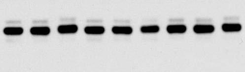


13 RESEARCH Supplementary Figure 1. Original immunoblot images (continued) Extended Data Fig. 3a ATP6V0A1 ATP6V0A2 ATP6V0D1 1 Extended Data Fig. 3b ATP6V1A ATP6V0D1 LAMP2 1 Extended Data Fig. 4c p- Extended Data Fig. 4d ATP6V1A PC3 LAMP2 Calnexin 1 2 HeLa EEA1 1 Extended Data Fig. 4e Extended Data Fig. 4g Extended Data Fig. 4i p- p- p- p-4ebp 4EBP 13


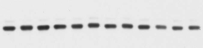


14 RESEARCH SUPPLEMENTARY INFORMATION Supplementary Figure 1. Original immunoblot images (continued) Extended Data Fig. 5a Extended Data Fig. 5c Extended Data Fig. 5f p-akt (S473) Akt p- p- p-erk1/2 ERK1/2 p-4ebp 4EBP Extended Data Fig. 6c Extended Data Fig. 6d Extended Data Fig. 6e p- p- 14


15 RESEARCH Supplementary Figure 1. Original immunoblot images (continued) Extended Data Fig. 6f Extended Data Fig. 6i Extended Data Fig. 7j Extended Data Fig. 7k Spar Lamp1 Gapdh 2 1 Atp6v0a1 Atp6v0a Atp6v0a1 Atp6v0a2 Atp6v1a Atp6v1a Atp6v0d1 Lamp1 1 Spar Atp6v0d1 1 Lamp1 Extended Data Fig. 8b Extended Data Fig. 9g Extended Data Fig. 9o Pax7 Myog p- p- GAPDH
SUPPLEMENTARY INFORMATION
 DOI:.38/ncb2822 a MTC02 FAO cells EEA1 b +/+ MEFs /DAPI -/- MEFs /DAPI -/- MEFs //DAPI c HEK 293 cells WCE N M C P AKT TBC1D7 Lamin A/C EEA1 VDAC d HeLa cells WCE N M C P AKT Lamin A/C EEA1 VDAC Figure
Supplementary Fig. 1. GPRC5A post-transcriptionally down-regulates EGFR expression. (a) Plot of the changes in steady state mrna levels versus
 Plot of the changes in steady state mrna levels versus") Supplementary Fig. 1. GPRC5A post-transcriptionally down-regulates EGFR expression. (a) Plot of the changes in steady state mrna levels versus changes in corresponding proteins between wild type and Gprc5a-/-
Supplementary Fig. 1. GPRC5A post-transcriptionally down-regulates EGFR expression. (a) Plot of the changes in steady state mrna levels versus changes in corresponding proteins between wild type and Gprc5a-/-
SUPPLEMENTARY INFORMATION
 doi:10.1038/nature22314 Supplementary Discussion In mammals, BCAAs are essential amino acids that must be supplied from food. These dietary BCAAs are subsequently used for protein synthesis or are catabolized
SUPPLEMENTARY FIGURES
 SUPPLEMENTARY FIGURES Supplementary Figure 1. (A) Left, western blot analysis of ISGylated proteins in Jurkat T cells treated with 1000U ml -1 IFN for 16h (IFN) or left untreated (CONT); right, western
SUPPLEMENTARY INFORMATION
 doi:10.1038/nature12652 Supplementary Figure 1. PRDM16 interacts with endogenous EHMT1 in brown adipocytes. Immunoprecipitation of PRDM16 complex by flag antibody (M2) followed by Western blot analysis
Supplementary Fig. 1 V-ATPase depletion induces unique and robust phenotype in Drosophila fat body cells.
 Supplementary Fig. 1 V-ATPase depletion induces unique and robust phenotype in Drosophila fat body cells. a. Schematic of the V-ATPase proton pump macro-complex structure. The V1 complex is composed of
Supplementary Figure 1.TRIM33 binds β-catenin in the nucleus. a & b, Co-IP of endogenous TRIM33 with β-catenin in HT-29 cells (a) and HEK 293T cells
 and HEK 293T cells") Supplementary Figure 1.TRIM33 binds β-catenin in the nucleus. a & b, Co-IP of endogenous TRIM33 with β-catenin in HT-29 cells (a) and HEK 293T cells (b). TRIM33 was immunoprecipitated, and the amount of
Supplementary Figure 1.TRIM33 binds β-catenin in the nucleus. a & b, Co-IP of endogenous TRIM33 with β-catenin in HT-29 cells (a) and HEK 293T cells (b). TRIM33 was immunoprecipitated, and the amount of
Receptor mediated Signal Transduction
 Receptor mediated Signal Transduction G-protein-linked receptors adenylyl cyclase camp PKA Organization of receptor protein-tyrosine kinases From G.M. Cooper, The Cell. A molecular approach, 2004, third
Molecular Cell Biology Problem Drill 16: Intracellular Compartment and Protein Sorting
 Molecular Cell Biology Problem Drill 16: Intracellular Compartment and Protein Sorting Question No. 1 of 10 Question 1. Which of the following statements about the nucleus is correct? Question #01 A. The
1. endoplasmic reticulum This is the location where N-linked oligosaccharide is initially synthesized and attached to glycoproteins.
 Biology 4410 Name Spring 2006 Exam 2 A. Multiple Choice, 2 pt each Pick the best choice from the list of choices, and write it in the space provided. Some choices may be used more than once, and other
SUPPLEMENTARY INFORMATION
 SUPPLEMENTARY INFORMATION doi:1.138/nature9814 a A SHARPIN FL B SHARPIN ΔNZF C SHARPIN T38L, F39V b His-SHARPIN FL -1xUb -2xUb -4xUb α-his c Linear 4xUb -SHARPIN FL -SHARPIN TF_LV -SHARPINΔNZF -SHARPIN
Practice Exam 2 MCBII
 1. Which feature is true for signal sequences and for stop transfer transmembrane domains (4 pts)? A. They are both 20 hydrophobic amino acids long. B. They are both found at the N-terminus of the protein.
T H E J O U R N A L O F C E L L B I O L O G Y
 Supplemental material Chairoungdua et al., http://www.jcb.org/cgi/content/full/jcb.201002049/dc1 T H E J O U R N A L O F C E L L B I O L O G Y Figure S1. Expression of CD9 and CD82 inhibits Wnt/ -catenin
MCB130 Midterm. GSI s Name:
 1. Peroxisomes are small, membrane-enclosed organelles that function in the degradation of fatty acids and in the degradation of H 2 O 2. Peroxisomes are not part of the secretory pathway and peroxisomal
Intracellular MHC class II molecules promote TLR-triggered innate. immune responses by maintaining Btk activation
 Intracellular MHC class II molecules promote TLR-triggered innate immune responses by maintaining Btk activation Xingguang Liu, Zhenzhen Zhan, Dong Li, Li Xu, Feng Ma, Peng Zhang, Hangping Yao and Xuetao
Organization of ATPases
 The Primary Active Transporter II: The ATPase Objectives: Organization P type with NPA domains Proton pumps of the rotary V type ATPase 1 Organization of P type, solute transport, found in plasma membranes
DECLARATION OF CONFLICT OF. No disclosure INTEREST
 DECLARATION OF CONFLICT OF No disclosure INTEREST ESC Congress 2011 2011.8.28 Impaired vacuolar H + -ATPase function causes cardiomyocyte death with extensive vacuolation and impaired autophagic degradation
SUPPLEMENTARY INFORMATION
 SUPPLEMENTARY INFORMATION doi:10.10/nature10195 NCBI gene: Tagged Subunit(s: HA-Vpx; FLAG-Cul4 HA-DCAF1 FLAG-Cul4 HA-FLAG-Vpx Mock Vpx (SIVmac 100 (a ; 0.159 (b ; 0.05 DCAF1 DDB1 DDA1 Cul4A 1; 0.024591
Zool 3200: Cell Biology Exam 4 Part I 2/3/15
 Name: Trask Zool 3200: Cell Biology Exam 4 Part I 2/3/15 Answer each of the following questions in the space provided, explaining your answers when asked to do so; circle the correct answer or answers
THE ROLE OF ALTERED CALCIUM AND mtor SIGNALING IN THE PATHOGENESIS OF CYSTINOSIS
 Research Foundation, 18 month progress report THE ROLE OF ALTERED CALCIUM AND mtor SIGNALING IN THE PATHOGENESIS OF CYSTINOSIS Ekaterina Ivanova, doctoral student Elena Levtchenko, MD, PhD, PI Antonella
SUPPLEMENTARY INFORMATION
 doi:10.1038/nature10353 Supplementary Figure 1. Mutations of UBQLN2 in patients with ALS and ALS/dementia. (a) A mutation, c.1489c>t (p.p497s), was identified in F#9975. The pedigree is shown on the left
Supplementary Figure 1. Spatial distribution of LRP5 and β-catenin in intact cardiomyocytes. (a) and (b) Immunofluorescence staining of endogenous
 and (b) Immunofluorescence staining of endogenous") Supplementary Figure 1. Spatial distribution of LRP5 and β-catenin in intact cardiomyocytes. (a) and (b) Immunofluorescence staining of endogenous LRP5 in intact adult mouse ventricular myocytes (AMVMs)
Supplementary Figure 1. Spatial distribution of LRP5 and β-catenin in intact cardiomyocytes. (a) and (b) Immunofluorescence staining of endogenous LRP5 in intact adult mouse ventricular myocytes (AMVMs)
SUPPLEMENTARY INFORMATION
 SUPPLEMENTARY INFORMATION doi:10.1038/nature11429 S1a 6 7 8 9 Nlrc4 allele S1b Nlrc4 +/+ Nlrc4 +/F Nlrc4 F/F 9 Targeting construct 422 bp 273 bp FRT-neo-gb-PGK-FRT 3x.STOP S1c Nlrc4 +/+ Nlrc4 F/F casp1
04_polarity. The formation of synaptic vesicles
 Brefeldin prevents assembly of the coats required for budding Nocodazole disrupts microtubules Constitutive: coatomer-coated Selected: clathrin-coated The formation of synaptic vesicles Nerve cells (and
Zool 3200: Cell Biology Exam 4 Part I 2/3/15
 Name: Key Trask Zool 3200: Cell Biology Exam 4 Part I 2/3/15 Answer each of the following questions in the space provided, explaining your answers when asked to do so; circle the correct answer or answers
The clathrin adaptor Numb regulates intestinal cholesterol. absorption through dynamic interaction with NPC1L1
 The clathrin adaptor Numb regulates intestinal cholesterol absorption through dynamic interaction with NPC1L1 Pei-Shan Li 1, Zhen-Yan Fu 1,2, Ying-Yu Zhang 1, Jin-Hui Zhang 1, Chen-Qi Xu 1, Yi-Tong Ma
BMDCs were generated in vitro from bone marrow cells cultured in 10 % RPMI supplemented
 Supplemental Materials Figure S1. Cultured BMDCs express CD11c BMDCs were generated in vitro from bone marrow cells cultured in 10 % RPMI supplemented with 15 ng/ml GM-CSF. Media was changed and fresh
MISSION: understanding the mechanisms of therapeutic strategies
 Telethon Institute of Genetics and Medicine MISSION: understanding the mechanisms of genetic diseases to develop preventive and therapeutic strategies G E N O T Y P E G E Researcher N 1 3 5 O T Y P E 8
Nature Genetics: doi: /ng Supplementary Figure 1. Clinical timeline for the discovery WES cases.
 Supplementary Figure 1 Clinical timeline for the discovery WES cases. This illustrates the timeline of the disease events during the clinical course of each patient s disease, further indicating the available
Supplementary Figure 1
 Supplementary Figure 1 Supplementary Figure 1 Schematic depiction of the tandem Fc GDF15. Supplementary Figure 2 Supplementary Figure 2 Gfral mrna levels in the brains of both wild-type and knockout Gfral
Supplementary Figure 1
 Supplementary Figure 1 how HFD how HFD Epi WT p p Hypothalamus p p Inguinal WT T Liver Lean mouse adipocytes p p p p p p Obese mouse adipocytes Kidney Muscle Spleen Heart p p p p p p p p Extracellular
Supplementary Materials for
 www.sciencesignaling.org/cgi/content/full/9/430/ra57/dc1 Supplementary Materials for The 4E-BP eif4e axis promotes rapamycinsensitive growth and proliferation in lymphocytes Lomon So, Jongdae Lee, Miguel
Insulin Resistance. Biol 405 Molecular Medicine
 Insulin Resistance Biol 405 Molecular Medicine Insulin resistance: a subnormal biological response to insulin. Defects of either insulin secretion or insulin action can cause diabetes mellitus. Insulin-dependent
A particular set of insults induces apoptosis (part 1), which, if inhibited, can switch to autophagy. At least in some cellular settings, autophagy se
, which, if inhibited, can switch to autophagy. At least in some cellular settings, autophagy se") A particular set of insults induces apoptosis (part 1), which, if inhibited, can switch to autophagy. At least in some cellular settings, autophagy serves as a defence mechanism that prevents or retards
A particular set of insults induces apoptosis (part 1), which, if inhibited, can switch to autophagy. At least in some cellular settings, autophagy serves as a defence mechanism that prevents or retards
Supporting Information Table of Contents
 Supporting Information Table of Contents Supporting Information Figure 1 Page 2 Supporting Information Figure 2 Page 4 Supporting Information Figure 3 Page 5 Supporting Information Figure 4 Page 6 Supporting
Nature Genetics: doi: /ng Supplementary Figure 1. Details of sequencing analysis.
 Supplementary Figure 1 Details of sequencing analysis. (a) Flow chart showing which patients fall into each category and were used for analysis. (b) Graph showing the average and median coverage for all
Supplementary Material
 Supplementary Material The Androgen Receptor is a negative regulator of eif4e Phosphorylation at S209: Implications for the use of mtor inhibitors in advanced prostate cancer Supplementary Figures Supplemental
Supplementary Figure 1. Normal T lymphocyte populations in Dapk -/- mice. (a) Normal thymic development in Dapk -/- mice. Thymocytes from WT and Dapk
 Normal thymic development in Dapk -/- mice. Thymocytes from WT and Dapk") Supplementary Figure 1. Normal T lymphocyte populations in Dapk -/- mice. (a) Normal thymic development in Dapk -/- mice. Thymocytes from WT and Dapk -/- mice were stained for expression of CD4 and CD8.
Supplementary Figure 1. Normal T lymphocyte populations in Dapk -/- mice. (a) Normal thymic development in Dapk -/- mice. Thymocytes from WT and Dapk -/- mice were stained for expression of CD4 and CD8.
Explain that each trna molecule is recognised by a trna-activating enzyme that binds a specific amino acid to the trna, using ATP for energy
 7.4 - Translation 7.4.1 - Explain that each trna molecule is recognised by a trna-activating enzyme that binds a specific amino acid to the trna, using ATP for energy Each amino acid has a specific trna-activating
Effects of Second Messengers
 Effects of Second Messengers Inositol trisphosphate Diacylglycerol Opens Calcium Channels Binding to IP 3 -gated Channel Cooperative binding Activates Protein Kinase C is required Phosphorylation of many
SUPPLEMENTARY INFORMATION
 DOI: 10.1038/ncb2607 Figure S1 Elf5 loss promotes EMT in mammary epithelium while Elf5 overexpression inhibits TGFβ induced EMT. (a, c) Different confocal slices through the Z stack image. (b, d) 3D rendering
SUPPLEMENTARY INFORMATION
 Supplementary Figures Supplementary Figure S1. Binding of full-length OGT and deletion mutants to PIP strips (Echelon Biosciences). Supplementary Figure S2. Binding of the OGT (919-1036) fragments with
Homework Hanson section MCB Course, Fall 2014
 Homework Hanson section MCB Course, Fall 2014 (1) Antitrypsin, which inhibits certain proteases, is normally secreted into the bloodstream by liver cells. Antitrypsin is absent from the bloodstream of
Cells and reagents. Synaptopodin knockdown (1) and dynamin knockdown (2)
 and dynamin knockdown (2)") Supplemental Methods Cells and reagents. Synaptopodin knockdown (1) and dynamin knockdown (2) podocytes were cultured as described previously. Staurosporine, angiotensin II and actinomycin D were all obtained
Supplemental Methods Cells and reagents. Synaptopodin knockdown (1) and dynamin knockdown (2) podocytes were cultured as described previously. Staurosporine, angiotensin II and actinomycin D were all obtained
Lysosomes and endocytic pathways 9/27/2012 Phyllis Hanson
 Lysosomes and endocytic pathways 9/27/2012 Phyllis Hanson General principles Properties of lysosomes Delivery of enzymes to lysosomes Endocytic uptake clathrin, others Endocytic pathways recycling vs.
Supplementary Figure 1 Chemokine and chemokine receptor expression during muscle regeneration (a) Analysis of CR3CR1 mrna expression by real time-pcr
 Analysis of CR3CR1 mrna expression by real time-pcr") Supplementary Figure 1 Chemokine and chemokine receptor expression during muscle regeneration (a) Analysis of CR3CR1 mrna expression by real time-pcr at day 0, 1, 4, 10 and 21 post- muscle injury. (b)
Supplementary Figure 1 Chemokine and chemokine receptor expression during muscle regeneration (a) Analysis of CR3CR1 mrna expression by real time-pcr at day 0, 1, 4, 10 and 21 post- muscle injury. (b)
T H E J O U R N A L O F C E L L B I O L O G Y
 T H E J O U R N A L O F C E L L B I O L O G Y Supplemental material Krenn et al., http://www.jcb.org/cgi/content/full/jcb.201110013/dc1 Figure S1. Levels of expressed proteins and demonstration that C-terminal
RAW264.7 cells stably expressing control shrna (Con) or GSK3b-specific shrna (sh-
 or GSK3b-specific shrna (sh-") 1 a b Supplementary Figure 1. Effects of GSK3b knockdown on poly I:C-induced cytokine production. RAW264.7 cells stably expressing control shrna (Con) or GSK3b-specific shrna (sh- GSK3b) were stimulated
1 a b Supplementary Figure 1. Effects of GSK3b knockdown on poly I:C-induced cytokine production. RAW264.7 cells stably expressing control shrna (Con) or GSK3b-specific shrna (sh- GSK3b) were stimulated
1. This is the location where N-linked oligosaccharide is initially synthesized and attached to glycoproteins.
 Biology 4410 Name Spring 2006 Exam 2 A. Multiple Choice, 2 pt each Pick the best choice from the list of choices, and write it in the space provided. Some choices may be used more than once, and other
SUPPLEMENTAL FIGURE LEGENDS
 SUPPLEMENTAL FIGURE LEGENDS Supplemental Figure S1: Endogenous interaction between RNF2 and H2AX: Whole cell extracts from 293T were subjected to immunoprecipitation with anti-rnf2 or anti-γ-h2ax antibodies
Title: Cytosolic DNA-mediated, STING-dependent pro-inflammatory gene. Fig. S1. STING ligands-mediated signaling response in MEFs. (A) Primary MEFs (1
 Primary MEFs (1") 1 Supporting Information 2 3 4 Title: Cytosolic DNA-mediated, STING-dependent pro-inflammatory gene induction necessitates canonical NF-κB activation through TBK1 5 6 Authors: Abe et al. 7 8 9 Supporting
1 Supporting Information 2 3 4 Title: Cytosolic DNA-mediated, STING-dependent pro-inflammatory gene induction necessitates canonical NF-κB activation through TBK1 5 6 Authors: Abe et al. 7 8 9 Supporting
Name: Multiple choice questions. Pick the BEST answer (2 pts ea)
") Exam 1 202 Oct. 5, 1999 Multiple choice questions. Pick the BEST answer (2 pts ea) 1. The lipids of a red blood cell membrane are all a. phospholipids b. amphipathic c. glycolipids d. unsaturated 2. The
Exam 1 202 Oct. 5, 1999 Multiple choice questions. Pick the BEST answer (2 pts ea) 1. The lipids of a red blood cell membrane are all a. phospholipids b. amphipathic c. glycolipids d. unsaturated 2. The
Tumor suppressor Spred2 interaction with LC3 promotes autophagosome maturation and induces autophagy-dependent cell death
 www.impactjournals.com/oncotarget/ Oncotarget, Supplementary Materials 2016 Tumor suppressor Spred2 interaction with LC3 promotes autophagosome maturation and induces autophagy-dependent cell death Supplementary
Supplemental Figures:
 Supplemental Figures: Figure 1: Intracellular distribution of VWF by electron microscopy in human endothelial cells. a) Immunogold labeling of LC3 demonstrating an LC3-positive autophagosome (white arrow)
AP VP DLP H&E. p-akt DLP
 A B AP VP DLP H&E AP AP VP DLP p-akt wild-type prostate PTEN-null prostate Supplementary Fig. 1. Targeted deletion of PTEN in prostate epithelium resulted in HG-PIN in all three lobes. (A) The anatomy
A. Generation and characterization of Ras-expressing autophagycompetent
 Supplemental Material Supplemental Figure Legends Fig. S1 A. Generation and characterization of Ras-expressing autophagycompetent and -deficient cell lines. HA-tagged H-ras V12 was stably expressed in
SUPPLEMENTARY INFORMATION
 Supplementary Discussion The cell cycle machinery and the DNA damage response network are highly interconnected and co-regulated in assuring faithful duplication and partition of genetic materials into
MOLECULAR CELL BIOLOGY
 1 Lodish Berk Kaiser Krieger scott Bretscher Ploegh Matsudaira MOLECULAR CELL BIOLOGY SEVENTH EDITION CHAPTER 13 Moving Proteins into Membranes and Organelles Copyright 2013 by W. H. Freeman and Company
Summary of Endomembrane-system
 Summary of Endomembrane-system 1. Endomembrane System: The structural and functional relationship organelles including ER,Golgi complex, lysosome, endosomes, secretory vesicles. 2. Membrane-bound structures
Regulation of Lamp2a Levels in the Lysosomal Membrane
 Traffic 2000 1: 570 583 Munksgaard International Publishers Regulation of Lamp2a Levels in the Lysosomal Membrane A.M. Cuervo and J.F. Dice* Department of Physiology, Tufts University School of Medicine,
Stewart et al. CD36 ligands promote sterile inflammation through assembly of a TLR 4 and 6 heterodimer
 NFκB (fold induction) Stewart et al. ligands promote sterile inflammation through assembly of a TLR 4 and 6 heterodimer a. mrna (fold induction) 5 4 3 2 1 LDL oxldl Gro1a MIP-2 RANTES mrna (fold induction)
Bioluminescence Resonance Energy Transfer (BRET)-based studies of receptor dynamics in living cells with Berthold s Mithras
-based studies of receptor dynamics in living cells with Berthold s Mithras") Bioluminescence Resonance Energy Transfer (BRET)-based studies of receptor dynamics in living cells with Berthold s Mithras Tarik Issad, Ralf Jockers and Stefano Marullo 1 Because they play a pivotal role
Bioluminescence Resonance Energy Transfer (BRET)-based studies of receptor dynamics in living cells with Berthold s Mithras Tarik Issad, Ralf Jockers and Stefano Marullo 1 Because they play a pivotal role
293T cells were transfected with indicated expression vectors and the whole-cell extracts were subjected
 SUPPLEMENTARY INFORMATION Supplementary Figure 1. Formation of a complex between Slo1 and CRL4A CRBN E3 ligase. (a) HEK 293T cells were transfected with indicated expression vectors and the whole-cell
(a) Schematic diagram of the FS mutation of UVRAG in exon 8 containing the highly instable
 Schematic diagram of the FS mutation of UVRAG in exon 8 containing the highly instable") Supplementary Figure 1. Frameshift (FS) mutation in UVRAG. (a) Schematic diagram of the FS mutation of UVRAG in exon 8 containing the highly instable A 10 DNA repeat, generating a premature stop codon
Supplementary Figure 1. Frameshift (FS) mutation in UVRAG. (a) Schematic diagram of the FS mutation of UVRAG in exon 8 containing the highly instable A 10 DNA repeat, generating a premature stop codon
Supplementary Figure 1.
 Supplementary Figure 1. Visualization of endoplasmic reticulum-mitochondria interaction by in situ proximity ligation assay. A) Illustration of targeted proteins in mitochondria (M), endoplasmic reticulum
SUPPLEMENTARY INFORMATION
 sirna pool: Control Tetherin -HA-GFP HA-Tetherin -Tubulin Supplementary Figure S1. Knockdown of HA-tagged tetherin expression by tetherin specific sirnas. HeLa cells were cotransfected with plasmids expressing
Supporting Information
 Supporting Information Chen et al. 10.1073/pnas.0807991106 SI Methods Sucrose Gradient Fractionation. Fractionation by sucrose gradient was performed as described by Gasparini et al. [(2001) J Neurosci
Lecture: CHAPTER 13 Signal Transduction Pathways
 Lecture: 10 17 2016 CHAPTER 13 Signal Transduction Pathways Chapter 13 Outline Signal transduction cascades have many components in common: 1. Release of a primary message as a response to a physiological
October 26, Lecture Readings. Vesicular Trafficking, Secretory Pathway, HIV Assembly and Exit from Cell
 October 26, 2006 Vesicular Trafficking, Secretory Pathway, HIV Assembly and Exit from Cell 1. Secretory pathway a. Formation of coated vesicles b. SNAREs and vesicle targeting 2. Membrane fusion a. SNAREs
Leucine Deprivation Reveals a Targetable Liability
 Cancer Cell, 19 Supplemental Information Defective Regulation of Autophagy upon Leucine Deprivation Reveals a Targetable Liability of Human Melanoma Cells In Vitro and In Vivo Joon-Ho Sheen, Roberto Zoncu,
Expanded View Figures
 Expanded View Figures A B C D E F G H I J K L Figure EV1. The dysregulated lipid metabolic phenotype of mouse models of metabolic dysfunction is most pronounced in the fasted state. A L Male 12-weeks-old
Supplementary Fig. 1 eif6 +/- mice show a reduction in white adipose tissue, blood lipids and normal glycogen synthesis. The cohort of the original
 Supplementary Fig. 1 eif6 +/- mice show a reduction in white adipose tissue, blood lipids and normal glycogen synthesis. The cohort of the original phenotypic screening was n=40. For specific tests, the
SUPPLEMENTARY INFORMATION. Supp. Fig. 1. Autoimmunity. Tolerance APC APC. T cell. T cell. doi: /nature06253 ICOS ICOS TCR CD28 TCR CD28
 Supp. Fig. 1 a APC b APC ICOS ICOS TCR CD28 mir P TCR CD28 P T cell Tolerance Roquin WT SG Icos mrna T cell Autoimmunity Roquin M199R SG Icos mrna www.nature.com/nature 1 Supp. Fig. 2 CD4 + CD44 low CD4
Professor Christopher Proud
 South Australian Health and Medical Research Institute Professor Christopher Proud Cell Signalling & Gene Regulation Professor Christopher G. Proud Nutrition and Metabolism Theme Leader South Australian
Life Sciences 1A Midterm Exam 2. November 13, 2006
 Name: TF: Section Time Life Sciences 1A Midterm Exam 2 November 13, 2006 Please write legibly in the space provided below each question. You may not use calculators on this exam. We prefer that you use
Supplementary Information
 Supplementary Information Supplementary Figure 1: cholesterol manipulation alters the positioning of autophagosomes in cells, related to figure 1. (a) HeLa cells were treated for 24h under conditions reducing
Muscular Dystrophy. Biol 405 Molecular Medicine
 Muscular Dystrophy Biol 405 Molecular Medicine Duchenne muscular dystrophy Duchenne muscular dystrophy is a neuromuscular disease that occurs in ~ 1/3,500 male births. The disease causes developmental
UNIVERSITY OF YORK BSc Stage 2 Degree Examinations Department: BIOLOGY. Title of Exam: Cell Biology
 Examination Candidate Number: Desk Number: UNIVERSITY OF YORK BSc Stage 2 Degree Examinations 2017-18 Department: BIOLOGY Title of Exam: Cell Biology Time allowed: 1 hour and 30 minutes Total marks available
Chapter 3. Expression of α5-megfp in Mouse Cortical Neurons. on the β subunit. Signal sequences in the M3-M4 loop of β nachrs bind protein factors to
 22 Chapter 3 Expression of α5-megfp in Mouse Cortical Neurons Subcellular localization of the neuronal nachr subtypes α4β2 and α4β4 depends on the β subunit. Signal sequences in the M3-M4 loop of β nachrs
Cell Quality Control. Peter Takizawa Department of Cell Biology
 Cell Quality Control Peter Takizawa Department of Cell Biology Cellular quality control reduces production of defective proteins. Cells have many quality control systems to ensure that cell does not build
General information. Cell mediated immunity. 455 LSA, Tuesday 11 to noon. Anytime after class.
 General information Cell mediated immunity 455 LSA, Tuesday 11 to noon Anytime after class T-cell precursors Thymus Naive T-cells (CD8 or CD4) email: lcoscoy@berkeley.edu edu Use MCB150 as subject line
Enzyme-coupled Receptors. Cell-surface receptors 1. Ion-channel-coupled receptors 2. G-protein-coupled receptors 3. Enzyme-coupled receptors
 Enzyme-coupled Receptors Cell-surface receptors 1. Ion-channel-coupled receptors 2. G-protein-coupled receptors 3. Enzyme-coupled receptors Cell-surface receptors allow a flow of ions across the plasma
Supplementary Figure 1. PD-L1 is glycosylated in cancer cells. (a) Western blot analysis of PD-L1 in breast cancer cells. (b) Western blot analysis
 Western blot analysis of PD-L1 in breast cancer cells. (b) Western blot analysis") Supplementary Figure 1. PD-L1 is glycosylated in cancer cells. (a) Western blot analysis of PD-L1 in breast cancer cells. (b) Western blot analysis of PD-L1 in ovarian cancer cells. (c) Western blot analysis
Supplementary Figure 1. PD-L1 is glycosylated in cancer cells. (a) Western blot analysis of PD-L1 in breast cancer cells. (b) Western blot analysis of PD-L1 in ovarian cancer cells. (c) Western blot analysis
TITLE: Improved Therapy for Breast Cancer by Inhibiting Autophagy. CONTRACTING ORGANIZATION: Trustees of Dartmouth College Hanover, NH
 AD Award Number: W81XWH-07-1-0684 TITLE: Improved Therapy for Breast Cancer by Inhibiting Autophagy PRINCIPAL INVESTIGATOR: Alan Eastman CONTRACTING ORGANIZATION: Trustees of Dartmouth College Hanover,
Types of cells. Cell size comparison. The Jobs of Cells 10/5/2015. Cells & Cell Organelles. Doing Life s Work
 Types of cells Prokaryote Cells & Cell Organelles bacteria cells Doing Life s Work Eukaryotes 2009-2010 animal cells plant cells Cell size comparison Animal cell Bacterial cell most bacteria (prokaryotic)
SUPPLEMENTARY INFORMATION
 doi:10.1038/nature11095 Supplementary Table 1. Summary of the binding between Angptls and various Igdomain containing receptors as determined by flow cytometry analysis. The results were summarized from
Lipids and Membranes
 Lipids and Membranes Presented by Dr. Mohammad Saadeh The requirements for the Pharmaceutical Biochemistry I Philadelphia University Faculty of pharmacy Membrane transport D. Endocytosis and Exocytosis
Molecular Cell Biology 5068 In Class Exam 1 October 3, 2013
 Molecular Cell Biology 5068 In Class Exam 1 October 3, 2013 Exam Number: Please print your name: Instructions: Please write only on these pages, in the spaces allotted and not on the back. Write your number
Supplementary Information. Supplementary Figure 1
 Supplementary Information Supplementary Figure 1 1 Supplementary Figure 1. Functional assay of the hcas9-2a-mcherry construct (a) Gene correction of a mutant EGFP reporter cell line mediated by hcas9 or
Supplementary Figure 1. Generation of knockin mice expressing L-selectinN138G. (a) Schematics of the Sellg allele (top), the targeting vector, the
 Schematics of the Sellg allele (top), the targeting vector, the") Supplementary Figure 1. Generation of knockin mice expressing L-selectinN138G. (a) Schematics of the Sellg allele (top), the targeting vector, the targeted allele in ES cells, and the mutant allele in
Supplementary Figure 1. Generation of knockin mice expressing L-selectinN138G. (a) Schematics of the Sellg allele (top), the targeting vector, the targeted allele in ES cells, and the mutant allele in
Supplementary Figure 1. SC35M polymerase activity in the presence of Bat or SC35M NP encoded from the phw2000 rescue plasmid.
 1 2 3 4 5 6 7 8 9 10 11 12 13 14 15 16 17 18 19 20 21 22 23 24 25 26 27 Supplementary Figure 1. SC35M polymerase activity in the presence of Bat or SC35M NP encoded from the phw2000 rescue plasmid. HEK293T
SUPPLEMENTARY INFORMATION
 DOI: 10.1038/ncb3311 A B TSC2 -/- MEFs C Rapa Hours WCL 0 6 12 24 36 pakt.s473 AKT ps6k S6K CM IGF-1 Recipient WCL - + - + - + pigf-1r IGF-1R pakt ps6 AKT D 1 st SILAC 2 nd SILAC E GAPDH FGF21 ALKPGVIQILGVK
REGULATION OF ENZYME ACTIVITY. Medical Biochemistry, Lecture 25
 REGULATION OF ENZYME ACTIVITY Medical Biochemistry, Lecture 25 Lecture 25, Outline General properties of enzyme regulation Regulation of enzyme concentrations Allosteric enzymes and feedback inhibition
Chapt. 10 Cell Biology and Biochemistry. The cell: Student Learning Outcomes: Describe basic features of typical human cell
 Chapt. 10 Cell Biology and Biochemistry Cell Chapt. 10 Cell Biology and Biochemistry The cell: Lipid bilayer membrane Student Learning Outcomes: Describe basic features of typical human cell Integral transport
Supplementary Materials for
 www.sciencesignaling.org/cgi/content/full/6/283/ra57/dc1 Supplementary Materials for JNK3 Couples the Neuronal Stress Response to Inhibition of Secretory Trafficking Guang Yang,* Xun Zhou, Jingyan Zhu,
doi: /nature10632
 SUPPLEMENTARY INFORMATION doi:10.1038/nature10632 Supplementary Figure 1 Lyn mediates neutrophil wound responses as a redox sensor. a, A schematic model. Wounded epithelial cells release H 2 O 2 by an
Lysosomal metabolomics reveals V-ATPase- and mtordependent regulation of amino acid efflux from lysosomes
 Lysosomal metabolomics reveals V-ATPase- and mtordependent regulation of amino acid efflux from lysosomes The MIT Faculty has made this article openly available. Please share how this access benefits you.
Type of file: PDF Title of file for HTML: Supplementary Information Description: Supplementary Figures
 Type of file: PDF Title of file for HTML: Supplementary Information Description: Supplementary Figures Type of file: MOV Title of file for HTML: Supplementary Movie 1 Description: NLRP3 is moving along
TFEB-mediated increase in peripheral lysosomes regulates. Store Operated Calcium Entry
 TFEB-mediated increase in peripheral lysosomes regulates Store Operated Calcium Entry Luigi Sbano, Massimo Bonora, Saverio Marchi, Federica Baldassari, Diego L. Medina, Andrea Ballabio, Carlotta Giorgi
Supplemental Data. Prolonged Rapamycin Treatment Inhibits mtorc2 Assembly and Akt/PKB. Supplemental Experimental Procedures
 Supplemental Data Prolonged Rapamycin Treatment Inhibits mtorc2 Assembly and Akt/PKB Dos D. Sarbassov, Siraj M. Ali, Shomit Sengupta, Joon-Ho Sheen, Peggy P. Hsu, Alex F. Bagley, Andrew L. Markhard, and