DORSAL MUSCLES OF THE HINDLIMB (ca)
|
|
|
- Aldous McKenzie
- 5 years ago
- Views:
Transcription
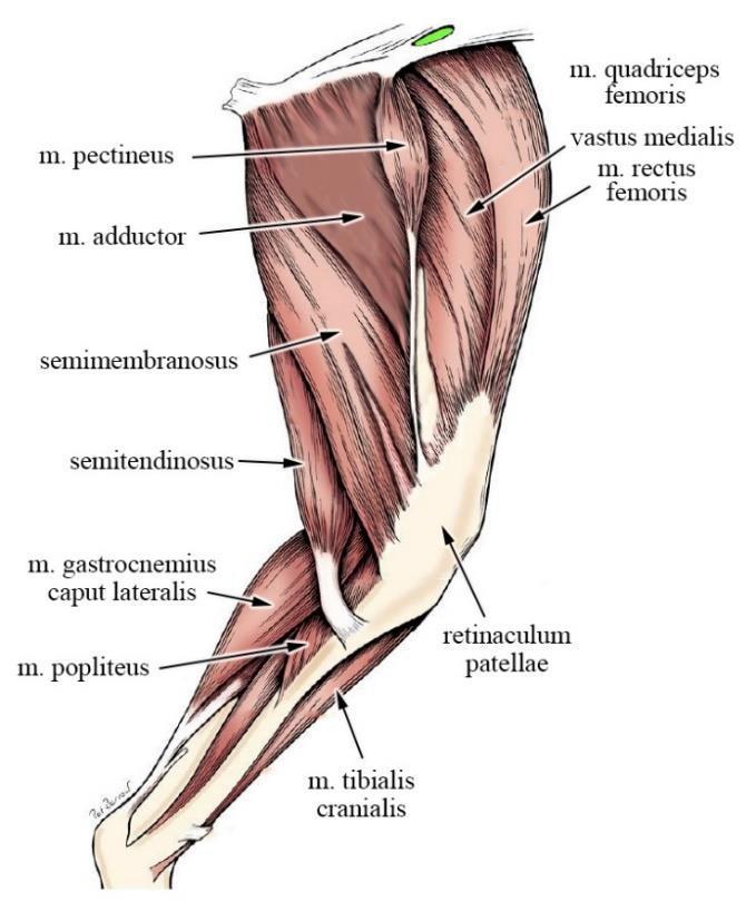
1 Fascia thoracolumbalis Fascia glutea Fascia lata Lamina superficialis Lamina profunda Fascia cruris DORSAL MUSCLES OF THE HINDLIMB (ca) M. gluteus superficialis o Origin: sacrum and first caudal vertebrae, partly from sacrotuberous ligament; (and by means of deep gluteal fascia also from cranial dorsal iliac spine) o Insertion: on tuberositas glutea (below greater trochanter) o Action: extension of hip M. gluteus medius o Origin: crista iliaca and gluteal surface of iliac bone o Insertion: greater trochanter of femur o Action: strongest extensor of hip joint M. piriformis o Origin: last sacral and first caudal vertebrae o Insertion: greater trochanter of femur o Action: extension of hip joint M. gluteus profundus o Origin: gluteal surface and body of iliac bone o Insertion: greater trochanter of femur o Action: extension of hip joint Interspecies differences M. gluteus superficialis in bo, su: fused with m. biceps femoris and they form m. gluteobiceps, eq: inserts on trochanter tertius M. piriformis in eq, bo, su: fused with m. gluteus medius 1


2 2
3 DEEP MUSCLES OF THE HINDLIMB (ca) M. obturatorius externus o Origin: outer surface of pelvis, around foramen obturatum o Insertion: trochanteric fossa of femur o Action: lateral rotation (supination) of hindlimb M. quadratus femoris o Origin: ventral surface of tabula ossis ischii (medial to tuber ischiadicum) o Insertion: trochanteric fossa of femur o Action: extension of hip joint and lateral rotation of hindlimb M. obturatorius internus o Origin: inner surface of pelvis around for. obturatum (from regions of ramus cranialis et caudalis ossis pubis, ramus ossis ischii and tabula ossis ischii) o Insertion: after crossing lesser sciatic notch it will attach in trochanteric fossa of femur; its tendon runs over the muscle belly of m. gemelli o Action: lateral rotation (supination) of hindlimb M. gemelli o Origin: lateral side on corpus ossis ischii (ventral to lesser sciatic notch incisura ischiadica minor) o Insertion: trochanteric fossa of femur o Action: lateral rotation (supination) of hindlimb 3

4 4
5 CAUDAL MUSCLES OF THE HINDLIMB (ca) M. biceps femoris o Origin: its cranial part on sacrotuberous ligament, caudal part on tuber ischiadicum o Insertion: by means of fascia lata and crural fascia to patella, patellar ligament, and tibial tuberosity and tibial crest, tuber calcanei by an accessory tendon o Action: extends hip, and hock; its cranial part extends, caudal part flexes stifle M. abductor cruris caudalis o Origin: distal part of sacrotuberous ligament o Insertion: disappears in crural fascia on lateral side (it runs first medial, then lateral to m. biceps femoris) o Action: abducts hindlimb, flexes stifle M. semitendinosus o Origin: tuber ischiadicum o Insertion: tibial crest (medially), and tuber calcanei by means of an accessory tendon o Action: extends hip, flexes stifle and extends hock M. semimembranosus o Origin: ventral aspect of tuber ischiadicum o Insertion: cranial belly distally on medial lip and medial condyle of femur, caudal muscle belly on medial condyle of tibia (behind medial collateral ligament) o Action: extends hip and flexes stifle joint Interspecies differences M. abductor cruris caudalis - not present in eq, bo 5
6 6
7 CRANIAL MUSCLES OF THE HINDLIMB (ca) M. quadriceps femoris It has four muscle bellies. o Origin: m. rectus femoris from body of ilium (area m. recti femoris medialis et lat.), the three vastus proximally on femur: vastus lateralis from its craniolateral, vastus medialis from its craniomedial and vastus intermedius from its cranial surface o Insertion: patella, and by means of patellar ligament on tibial tuberosity o Action: strongest extensor of stifle joint; m. rectus femoris also flexes hip M. tensor fasciae latae o Origin: tuber coxae (cranial ventral iliac spine and spina alaris) o Insertion: radiates into fascia lata and fascia cruris. Its superficial fascial layer covers thigh, deep fascial layer surrounds m. quadriceps femoris and attaches to femur o Action: tenses fascia lata, flexes hip and extends stifle 7
8 MEDIAL MUSCLES OF THE HINDLIMB (ca) M. sartorius In dogs it has a cranial and caudal part. o Origin: iliac crest, cranial ventral iliac spine, thoracolumbar fascia o Insertion: radiating into fascia lata and crural fascia, cranial part inserts above stifle joint, caudal part inserts below stifle joint (the latter medially reaches tibial crest) o Action: flexes hip, extends or flexes stifle (depending on which part contracts more) M. adductor o Origin: m. adductor longus from ramus cranialis ossis pubis, m. adductor magnus et brevis are arising from symphysis pelvis (tendo symphysialis), ramus caudalis ossis pubis and ramus ossis ischi o Insertion: facies aspera and labium laterale of femur o Action: adducts hindlimb and extends hip M. pectineus o Origin: pecten ossis pubis, prepubic tendon o Insertion: medial lip of rough surface on femoral shaft (labium mediale ossis femoris, next to facies aspera) o Action: adduction of thigh M. gracilis o Origin: symphysis pelvis (by means of tendo symphysialis) o Insertion: medially on tibial crest, fascia cruris, tuber calcanei by an accessory tendon to common calcaneal tendon o Action: adducts hindlimb, extends hip, flexes stifle, extends tarsus 8

9 9
 Foramen ischiadicum majus (Un) Foramen ischiadicum minus (Un) Articulatio sacroiliaca type: amphiarthrosis Ligg. sacroiliaca ventralia Ligg. sacroiliaca interossea Ligg.")
10 ARTICULATIONES MEMBRI PELVINI all species Membrana obturatoria (eq) Lig. sacrotuberale (ca) Lig. sacrotuberale latum (Un) Foramen ischiadicum majus (Un) Foramen ischiadicum minus (Un) Articulatio sacroiliaca type: amphiarthrosis Ligg. sacroiliaca ventralia Ligg. sacroiliaca interossea Ligg. sacroiliaca dorsalia Symphysis pelvina Symphysis pubica Symphysis ischiadica Articulatio coxae type: diarthrosis, articulatio simplex, articulatio spheroidea, multiaxial Capsula articularis Labrum acetabulare Lig. capitis ossis femoris Lig. accessorium ossis femoris (eq) 10
11 Articulatio genus (ca) Articulatio femoropatellaris type: diarthrosis, articulatio simplex, articulatio delabens, uniaxial Articulatio femorotibialis type: diarthrosis, articulatio duplex, articulatio condylaris, biaxial Extracapsular ligaments (Ligg. extracapsularia) of stifle joint: o Retinaculum patellae (1): from fascia lata and aponeurosis of muscles, covers stifle o Lig. patellae (2): Continuation of m. quadriceps femoris s tendon, the patella is a sesamoid bone in the tendon before patellar ligament inserts on tibial tuberosity o Lig. collaterale laterale (3): from lateral epicondyle of femur to head of fibula, a lesser part to lateral condyle of tibia; underneath there is the tendon of m. popliteus o Lig. collaterale mediale (4): from medial epicondyle of femur to distal part of tibia s medial condyle; it also attaches strongly to medial meniscus Capsular ligaments (Ligg. capsularia) of stifle joint: o Lig. femoropatellare laterale (5): from lateral Vesalius (sesamoid) bone to patella, also attaches to lateral femoral epicondyle; reinforcement of joint s capsule o Lig. femoropatellare mediale (6): from medial Vesalius (sesamoid) bone to patella, also attaches to medial femoral epicondyle; reinforcement of joint s capsule o Lig. popliteum obliquum (23): fibrous reinforcement in caudal wall of joint capsule Intracapsular ligaments (Ligg. intracapsularia) of stifle joint: o Lig. transversum genus (15): connects cranial angles of the two meniscus o Ligg. meniscotibiales: from cranial and caudal ends of both meniscus to tibia lig. craniale menisci laterale (16): area intercondylaris cranialis lateralis lig. craniale menisci mediale (17): area intercondylaris cranialis medialis lig. caudale menisci laterale (18): incisura poplitea lig. caudale menisci mediale (19): area intercondylaris caudalis o Lig. cruciatum craniale (20): from lateral femoral condyle s caudal axial surface to area intercondylaris cranialis of tibia (Nickel, Nomenclatura, does not specify it by species! Only explains horse!) o Lig. cruciatum caudale (21): from medial femoral condyle s cranial axial surface to area intercondylaris caudalis and incisura poplitea of tibia o Lig. meniscofemorale (22): from medial femoral condyle s caudal axial surface to caudal end of lateral meniscus Synovial, protecting and fibrocartilaginous structures of stifle joint: o Corpus adiposum infrapatellare (7): fatty tissue under lig. patellae o Bursa infrapatellaris (8): under lig. patellae at tibial tuberosity o Recessus suprapatellaris (9): behind and above patella o Recessus subextensorius (10): around tendon of m. ext. dig. longus o Recessus subpopliteus (11): around tendon of m. popliteus o Meniscus medialis (12): fibrocartilage between femur and tibia o Meniscus lateralis (13): fibrocartilage between femur and tibia o Fibrocartilago parapatellaris (14): around patella, on both sides 11

 Lig.")
12 Interspecies differences Lig. transversum genus- not present in eq, bo Lig. patellae mediale (eq, bo) Lig. patellae intermedium (eq, bo) Lig. patellae laterale (eq, bo) Trochlea ossis femoris 12
13 Tuberculum trochleae ossis femoris (eq) Cavities of the joint capsule (eq): femorotibial joint has separated medial, lateral cavities (which forms proximal, distal sacs), medial femorotibial joint cavity communicates with femoropatellar joint cavity Lig. cruciatum craniale (eq): from lateral femoral condyle s caudal axial surface to area intercondylaris centralis of tibia STIFLE EXTENSORS (ca) M. quadriceps femoris o Action: strongest extensor of stifle joint; m. rectus femoris also flexes hip M. tensor fasciae latae o Action: tenses fascia lata, flexes hip and extends stifle M. sartorius cranial part o Action: flexes hip, extends stifle M. biceps femoris o Action: extends hip, and hock; its cranial part extends stifle STIFLE FLEXORS (ca) M. biceps femoris o Action: extends hip, and hock; caudal part flexes stifle M. abductor cruris caudalis o Action: abducts hindlimb, flexes stifle M. semitendinosus o Action: extends hip, flexes stifle and extends hock M. semimembranosus o Action: extends hip and flexes stifle joint M. sartorius pars caudalis o Action: flexes hip, flexes stifle M. gracilis o Action: adducts hindlimb, extends hip, flexes stifle, extends tarsus M. popliteus o Origin: fossa m. poplitei on lateral femoral condyle o Insertion: it twists caudally under lig. collaterale laterale, then inserts caudally on proximal part of tibia, on linea m. poplitei o Action: flexes stifle, rotates leg medially M. flexor digitorum superficialis o Origin: fossa supracondylaris of femur, partly fused with m. gastrocnemius caput lateralis o Insertion: its plantar tendon (tendo plantaris) turns from medial to lateral direction around common calcaneal tendon, on the surface of tuber calcanei it 13
14 makes a cap (galea calcanea), then finally attaches to middle phalanx of digits II-V. on plantar surface o Action: flexes stifle, extends hock, flexes joints of digits II-V DIGITAL EXTENSORS (ca) M. extensor digitorum longus o Origin: fossa extensoria on lateral femoral condyle o Insertion: extensor processes of distal phalanx of digits II-V. o Action: extends joints of digits and flexes tarsus M. extensor digitorum lateralis o Origin: proximal third of fibula o Insertion: unites with long digital extensor s tendon to reach digit V. distal phalanx o Action: extension and abduction of digit V. (and flexes tarsus) M. extensor digiti I. [hallucis] longus o Origin: from middle third of fibula o Insertion: radiates into fascia over metacarpophalangeal joint of digit II. o Action: extends digit II. (and digit I. if it s present) (and flexes tarsus) TARSAL FLEXORS (ca) M. tibialis cranialis o Origin: cranial margin of lateral condyle and crest of tibia o Insertion: medial side on os tarsi I. and Mt I-II. o Action: flexes tarsus M. peroneus longus also called: m. fibularis longus o Origin: cranial margin of tibia s lateral condyle, head of fibula, lateral collateral ligament o Insertion: first attaches to os tarsi 4+5 (on its plantar surface), then extends to all metatarsal bones plantar base o Action: flexes tarsus M. peroneus brevis also called: m. fibularis brevis o Origin: laterally on distal third of fibula (and partly on tibia) o Insertion: base of Mt. V. o Action: flexes tarsus 14
 turns from medial to lateral direction around common calcaneal tendon, on the surface of tuber calcanei it makes a cap")
15 DIGITAL FLEXORS (ca) M. flexor digitorum superficialis o Origin: fossa supracondylaris of femur, partly fused with m. gastrocnemius caput lateralis o Insertion: its plantar tendon (tendo plantaris) turns from medial to lateral direction around common calcaneal tendon, on the surface of tuber calcanei it makes a cap (galea calcanea), then finally attaches to middle phalanx of digits II-V. on plantar surface o Action: flexes stifle, extends hock, flexes joints of digits II-V M. flexor digitorum profundus o Origin: m. flexor digitorum lateralis (former: m. flexor hallucis longus) from proximal caudal half of fibula, proximal caudolateral border of tibia and from interosseous membrane; m. flexor digitorum medialis (former: m. flexor digitorum longus) starts caudo-proximally from tibia. Below tarsus the two parts unite. 15
16 o Insertion: plantar surface of distal phalanx of digits II-V. (on tuberculum flexorium) o Action: extends tarsus, flexes joint if digits II-V. TARSAL EXTENSORS (ca) M. gastrocnemius o Origin: medial and lateral supracondylar tuberosities of femur (inside their heads caput mediale and laterale there are sesamoid /Vesalius/ bones, former: fabellae) o Insertion: on tuber calcanei (as a part of common calcaneal tendon) o Action: flexes stifle, extends tarsus M. tibialis caudalis o Origin: caudally on head of fibula o Insertion: medially at tarsus it radiates into tarsal fascia o Action: extends tarsus M. flexor digitorum superficialis M. flexor digitorum profundus Tendo accessorius (m. biceps femoris, m. semitendinosus, m. gracilis) 16


17 17
18 TENDO CALCANEUS COMMUNIS (ca) o It comprise three main tendons: - tendo m. gastrocnemii [Achilli] (ex: m. gastrocnemius) - tendo plantaris (ex: m. flexor dig. superficialis) - tendo accessorius, from the following parts: - lateral: m. biceps femoris - medial: m. gracilis and m. semitendinosus o Between skin and tendo plantaris there is a protecting bursa subcutanea calcanea, between tendo plantaris and tendo m. gastrocnemii bursa subtendinea calcanea, and between tendo accessorius and tendo m. gastrocnemii bursa tendinis calcanea. o As tendo plantaris widens, it forms galea calcanea o Action: strongest extensor of tarsus Interspecies differences eq - m. triceps surae: m. gastrocnemius+ m. soleus - m. peroneus longus, m. peroneus brevis- not present - m. peroneus tertius- purely tendinous, o Origin: together with m. extensor digitorum longus from fossa extensoria o Insertion: on distal tarsal bones, proximal end of Mt o Action: flex the tarsus, important part of passive stay apparatus - m. extensor hallucis longus- not present - m. tibialis caudalis is a part of the m. flexor digitorum profundus 18
lesser trochanter of femur lesser trochanter of femur iliotibial tract (connective tissue) medial surface of proximal tibia
 medial surface of proximal tibia") LOWER LIMB MUSCLES OF THE APPENDICULAR SKELETON The muscles that act on the lower limb fall into three groups: those that move the thigh, those that move the lower leg, and those that move the ankle, foot,
LOWER LIMB MUSCLES OF THE APPENDICULAR SKELETON The muscles that act on the lower limb fall into three groups: those that move the thigh, those that move the lower leg, and those that move the ankle, foot,
Muscles of the lower extremities. Dr. Nabil khouri MD, MSc, Ph.D
 Muscles of the lower extremities Dr. Nabil khouri MD, MSc, Ph.D Posterior leg Popliteal fossa Boundaries Biceps femoris (superior-lateral) Semitendinosis and semimembranosis (superior-medial) Gastrocnemius
Muscles of Lesson Five. Muscular Nomenclature and Kinesiology - Two. Muscles of Lesson Five, cont. Chapter 16
 Chapter 16 Muscular Nomenclature and Kinesiology - Two Lessons 5-6 Muscles of Lesson Five Iliopsoas (psoas major, iliacus) Hip outward rotators (piriformis, gemellus superior, gemellus inferior, obturator
Lower limb summary. Anterior compartment of the thigh. Done By: Laith Qashou. Doctor_2016
 Lower limb summary Done By: Laith Qashou Doctor_2016 Anterior compartment of the thigh Sartorius Anterior superior iliac spine Upper medial surface of shaft of tibia 1. Flexes, abducts, laterally rotates
The Muscular System. Chapter 10 Part D. PowerPoint Lecture Slides prepared by Karen Dunbar Kareiva Ivy Tech Community College
 Chapter 10 Part D The Muscular System Annie Leibovitz/Contact Press Images PowerPoint Lecture Slides prepared by Karen Dunbar Kareiva Ivy Tech Community College Table 10.14: Muscles Crossing the Hip and
Anatomy & Physiology. Muscles of the Lower Limbs.
 Anatomy & Physiology Muscles of the Lower Limbs http://www.ishapeup.com/musclecharts.html Muscles of the Lower Limbs Among the strongest muscles in the body. Because pelvic girdle is composed of heavy,
Muscles of the Hip 1. Tensor Fasciae Latae O: iliac crest I: lateral femoral condyle Action: abducts the thigh Nerve: gluteal nerve
 Muscles of the Hip 1. Tensor Fasciae Latae O: iliac crest I: lateral femoral condyle Action: abducts the thigh Nerve: gluteal nerve 2. Gluteus Maximus O: ilium I: femur Action: abduct the thigh Nerve:
The Hip (Iliofemoral) Joint. Presented by: Rob, Rachel, Alina and Lisa
 Joint. Presented by: Rob, Rachel, Alina and Lisa") The Hip (Iliofemoral) Joint Presented by: Rob, Rachel, Alina and Lisa Surface Anatomy: Posterior Surface Anatomy: Anterior Bones: Os Coxae Consists of 3 Portions: Ilium Ischium Pubis Bones: Pubis Portion
The Hip (Iliofemoral) Joint Presented by: Rob, Rachel, Alina and Lisa Surface Anatomy: Posterior Surface Anatomy: Anterior Bones: Os Coxae Consists of 3 Portions: Ilium Ischium Pubis Bones: Pubis Portion
Human Anatomy Biology 351
 Human Anatomy Biology 351 Lower Limb Please place your name on the back of the last page of this exam. You must answer all questions on this exam. Because statistics demonstrate that, on average, between
Human Anatomy Biology 351
 Human Anatomy Biology 351 Lower Limb Please place your name on the back of the last page of this exam. You must answer all questions on this exam. Because statistics demonstrate that, on average, between
Anatomy of the lower limb
 Anatomy of the lower limb 1. Bones of the lower limb Pelvis Hip bone/coxal bone Acetabulum o Acetabular margin o Acetabular fossa o Acetabular notch o Lunate surface Ischiopubic ramus Obturator foramen
MUSCLES OF THE LOWER LIMBS
 MUSCLES OF THE LOWER LIMBS Naming, location and general function Dr. Nabil khouri ROLES THAT SHOULD NOT BE FORGOTTEN Most anterior compartment muscles of the hip and thigh Flexor of the femur at the hip
Anatomage Table Instructors Guide- Lower Limb
 The Lower Limb Anatomage Table Instructors Guide- Lower Limb Table of Contents Lower Limb 1- The Skeletal System...3 1: Hip Bone...3 2: Hip Joint and Femur...4 3: Patella and Knee Joint...7 4: Tibia, Fibula,
Topic 7: Hip and pelvis. Parts of the hip. Parts of the femur
 Topic 7: Hip and pelvis Parts of the hip Parts of the femur Classifying the hip joint Ball and socket Synovial Multiaxial Movements of the hip: Abduction/adduction Flexion/extension Medial/lateral rotation
16) M. quadriceps femoris, m. tensor fasciae latae, m. sartorius
 M. quadriceps femoris, m. tensor fasciae latae, m. sartorius") Anatomy 1 - Lesson XIII: Myologia Part IV Objective: Students will examine the muscles of a canine in order to identify the musculature of the body.. Practical Tasks 15-18: 15) Medial muscles of the thigh,
Anatomy 1 - Lesson XIII: Myologia Part IV Objective: Students will examine the muscles of a canine in order to identify the musculature of the body.. Practical Tasks 15-18: 15) Medial muscles of the thigh,
DISSECTION SCHEDULE. Session I - Hip (Front) & Thigh (Superficial)
 & Thigh (Superficial)") DISSECTION SCHEDULE Session I - Hip (Front) & Thigh (Superficial) Surface anatomy Inguinal region Gluteal region Thigh Leg Foot bones Hip bone Femur Superficial fascia Great saphenous vein Superficial
DISSECTION SCHEDULE Session I - Hip (Front) & Thigh (Superficial) Surface anatomy Inguinal region Gluteal region Thigh Leg Foot bones Hip bone Femur Superficial fascia Great saphenous vein Superficial
Muscles of the Gluteal Region
 Muscles of the Gluteal Region 1 Some of the most powerful in the body Extend the thigh during forceful extension Stabilize the iliotibial band and thoracolumbar fascia Related to shoulders and arms because
Gluteal region DR. GITANJALI KHORWAL
 Gluteal region DR. GITANJALI KHORWAL Gluteal region The transitional area between the trunk and the lower extremity. The gluteal region includes the rounded, posterior buttocks and the laterally placed
Skeleton of lower extremity Ossa membri inferioris
 Skeleton of lower extremity Ossa membri inferioris Skeleton of lower extremity consists of lower extremity girdle (cingulum membri inferioris) created by pelvis bone (os coxae) and bones of free lower
Skeleton of the lower limb
 Skeleton of the lower limb (ossa membri inferioris) RNDr. Michaela Račanská, Ph.D. Lecture 3 DENTISTRY Autumn 2016 Skeleton of the lower limb support the weight of the body and transfers that support to
Muscles of the Thigh. 6.1 Identify, describe the attachments of and deduce the actions of the muscles of the thigh: Anterior group
 Muscles of the Thigh 6.1 Identify, describe the attachments of and deduce the actions of the muscles of the thigh: Anterior group Sartorius: This is a long strap like muscle with flattened tendons at each
Hip joint and pelvic girdle. Lower Extremity. Pelvic Girdle 6/5/2017
 Hip joint and pelvic girdle Lower Extremity The relationship between the pelvic girdle and hip is similar to that between the shoulder girdle and shoulder joint. The lower limbs are attached to the axial
rotation of the hip Flexion of the knee Iliac fossa of iliac Lesser trochanter Femoral nerve Flexion of the thigh at the hip shaft of tibia
 Anatomy of the lower limb Anterior & medial compartments of the thigh Dr. Hayder The fascia lata encloses the entire thigh like a sleeve/stocking. Three intramuscular fascial septa (lateral, medial, and
Lectures of Human Anatomy
 Lectures of Human Anatomy Lower Limb Gluteal Region and Hip Joint By DR. ABDEL-MONEM AWAD HEGAZY M.B. with honor 1983, Dipl."Gynecology and Obstetrics "1989, Master "Anatomy and Embryology" 1994, M.D.
The thigh. Prof. Oluwadiya KS
 The thigh Prof. Oluwadiya KS www.oluwadiya.com The Thigh: Boundaries The thigh is the region of the lower limb that is approximately between the hip and knee joints Anteriorly, it is separated from the
The Hip Joint. Shenequia Howard David Rivera
 The Hip Joint Shenequia Howard David Rivera Topics Of Discussion Movement Bony Anatomy Ligamentous Anatomy Muscular Anatomy Origin/Insertion/Action/Innervation Common Injuries MOVEMENT Flexion Extension
Human Anatomy Biology 255
 Human Anatomy Biology 255 Exam #4 Please place your name and I.D. number on the back of the last page of this exam. You must answer all questions on this exam. Because statistics demonstrate that, on average,
MUSCLES OF THE LOWER EXTREMITY
 MUSCLES OF THE LOWER EXTREMITY Muscles of the lower extremity are divisible into groups, corresponding with the different regions of the limb. I. Muscles of the Iliac Region II. Muscles of the Thigh III.
Contents of the Posterior Fascial Compartment of the Thigh
 Contents of the Posterior Fascial Compartment of the Thigh 1-Muscles: B i c e p s f e m o r i s S e m i t e n d i n o s u s S e m i m e m b r a n o s u s a small part of the adductor magnus (h a m s t
Due in Lab weeks because of Thanksgiving Prelab #10. Homework #8. Both sides! Both sides!
 Lab 8 MUSCLES Due in Lab 10 2 weeks because of Thanksgiving Prelab #10 Both sides! Homework #8 Both sides! Refer to Muscles 22-23 Naming of muscles Origin Site of muscle attachment that doesn t move during
Acland's DVD Atlas of Human Anatomy. Transcript for Volume Robert D Acland
 Acland's DVD Atlas of Human Anatomy Transcript for Volume 2 2007 Robert D Acland This free downloadable pdf file is to be used for individual study only. It is not to be reproduced in any form without
LOWER LIMB. As we know the bony part of the body is divided into Axial and Appendicular (upper and lower Limbs)
") LOWER LIMB As we know the bony part of the body is divided into Axial and Appendicular (upper and lower Limbs) Bones of the Lower limb: 1-Pelvic Girdle: composed of: 1. Right hip bone : is formed by 3
LOWER LIMB As we know the bony part of the body is divided into Axial and Appendicular (upper and lower Limbs) Bones of the Lower limb: 1-Pelvic Girdle: composed of: 1. Right hip bone : is formed by 3
Identify the muscles associated with the medial compartment of the thigh. Identify the attachment points of the medial thigh muscles.
 L 8 A B O R A T O R Y Thigh MEDIAL THIGH Identify the muscles associated with the medial compartment of the thigh. Identify the attachment points of the medial thigh muscles. Identify the actions of these
Bones of the Lower Limb Bone Structure Description Notes. border of the superior ramus. inferolaterally from the pubic symphysis
 Bones of the Lower Limb Bone Structure Description Notes pubis an angulated bone the forms the anterior part of the pelvis one of three bones that form the os coxae: ilium, ischium, pubis; its forms 1/5
TABLE OF MUSCLES OF LOWER EXTREMITY 2018zillmusom ANTERIOR THIGH
 TABLE OF MUSCLES OF LOWER EXTREMITY 2018zillmusom ANTERIOR THIGH MUSCLE ORIGIN INSERTION ACTION NERVE Iliopsoas Ilium, vertebra Femur Flex hip joint Femoral nerve (T12-L5) Pectineus Pubis Femur Flex hip
The functional and phylogenetic implications of the myology of the lumbar region,
 The functional and phylogenetic implications of the myology of the lumbar region, tail, and hind limbs of the lesser grison (Galictis cuja) Journal of Mammalian Evolution Marcos D. Ercoli, Sebastián Echarri,
Lower Limb Dr. Robin Paudel
 Lower Limb n What is a limb? n Skeleton n Joints n Pelvis or limb girdle n Hip/Hip Muscles n Lumber and sacral plexus getting spinal nerves out onto limb n Muscles anterior and posterior compartments n
TABLES OF MUSCLE ACTIONS, INNERVATIONS, AND ATTACHMENTS
 TABLES OF MUSCLE ACTIONS, INNERVATIONS, AND ATTACHMENTS Table 1-1 ERECTOR SPINAE MUSCLES Intrinsic muscles producing extension and/or lateral of the spine Muscle Joint and Action Innervation Inferior Attachment
Main Menu. Joint and Pelvic Girdle click here. The Power is in Your Hands
 1 Hip Joint and Pelvic Girdle click here Main Menu K.6 http://www.handsonlineeducation.com/classes//k6entry.htm[3/23/18, 2:01:12 PM] Hip Joint (acetabular femoral) Relatively stable due to : Bony architecture
11/15/2018. Temporalis Elevates & retracts mandible. Masseter = Prime mover of jaw closure. Levator scapulae Supraspinatus Clavicle.
 Due in Lab 10 Lab 8 MUSCLES 2 weeks because of Thanksgiving Prelab #10 Both sides! Homework #8 Both sides! Refer to Muscles 22-23 Examples of Origin & Insertion Naming of muscles Origin Site of muscle
The University Of Jordan Faculty Of Medicine THE LOWER LIMB. Dr.Ahmed Salman Assistant Prof. of Anatomy. The University Of Jordan
 The University Of Jordan Faculty Of Medicine THE LOWER LIMB Dr.Ahmed Salman Assistant Prof. of Anatomy. The University Of Jordan Gluteal Region Cutaneous nerve supply of (Gluteal region) 1. Lateral cutaneous
Lesson 24. A & P Hip
 Lesson 24 A & P Hip 1 Aims of the Session This session will allow candidates to have an understanding of the bony prominences and soft tissues of the hip 2 Learning Outcomes By the end of the lesson the
Musculature of Indian Elephant Part II. Musculature of the Hindlimb
 Musculature of Indian Elephant Part II. Musculature of the Hindlimb Tokuichi Shindo (Emeritus Professor) and Masaru Mori Department of Anatomy (Prof. Marasu Mori) Faculty of Medicine, Kyushu University,
this makes sense, however this is lower order thinking and does not solve the lower leg
 Functional Knee Valgus in a Barbell Squat 1 One of the most common lower leg dysfunction we see in athletes, particularly general population is functional knee valgus, or better referred to as the knees
In-Depth Foundations: Anatomy Terms to Know
 Be familiar with / able to identify and define all the following parts. The Spine Cranium Vertebrae Cervical, Thoracic, Lumbar Sacrum Coccyx Bones of Upper Body Cranium Mastoid process; Occipital condyle,
The Hay is in the Barn
 Anatomy 1 Practical 1 Review Made by Forrest Allen (nerd) Edited by TJ Williamson (not nerd) The Hay is in the Barn 2019 Thunderbringers Too much to handle https://www.youtube.com/watch?v=glii-kaza d8
Sports Medicine 15. Unit I: Anatomy. The knee, Thigh, Hip and Groin. Part 4 Anatomies of the Lower Limbs
 Sports Medicine 15 Unit I: Anatomy Part 4 Anatomies of the Lower Limbs The knee, Thigh, Hip and Groin Anatomy of the lower limbs In Part 3 of this section we focused upon 11 of the 12 extrinsic muscles
CHAPTER 8: THE BIOMECHANICS OF THE HUMAN LOWER EXTREMITY
 CHAPTER 8: THE BIOMECHANICS OF THE HUMAN LOWER EXTREMITY _ 1. The hip joint is the articulation between the and the. A. femur, acetabulum B. femur, spine C. femur, tibia _ 2. Which of the following is
The Leg. Prof. Oluwadiya KS
 The Leg Prof. Oluwadiya KS www.oluwadiya.sitesled.com Compartments of the leg 4 Four Compartments: 1. Anterior compartment Deep fibular nerve Dorsiflexes the foot and toes 2. Lateral Compartment Superficial
Lowe w r e r B ody Resistance Training
 Lower Body Resistance Training Tibialis Anterior Extensor Hallucis Longus Extensor Digitorum Longus Proneus Tertius AROM 25-40 degrees Extensor active-insufficiency Flexion & Eversion (Pronation) Flexion
Hind limb myology of the common hippopotamus, Hippopotamus amphibius (Artiodactyla: Hippopotamidae)
") Zoological Journal of the Linnean Society, 2010, 158, 661 682. With 9 figures Hind limb myology of the common hippopotamus, Hippopotamus amphibius (Artiodactyla: Hippopotamidae) REBECCA E. FISHER 1,2 *,
Zoological Journal of the Linnean Society, 2010, 158, 661 682. With 9 figures Hind limb myology of the common hippopotamus, Hippopotamus amphibius (Artiodactyla: Hippopotamidae) REBECCA E. FISHER 1,2 *,
5.1 Identify, describe the attachments of and deduce the actions of the muscles of the thigh:
 5.1 Identify, describe the attachments of and deduce the actions of the muscles of the thigh: Anterior group Proximal attachment Distal attachment Sartorius ASIS» Upper part of shaft tibia (middle surface)»
Muscles to know. Lab 21. Muscles of the Pelvis and Lower Limbs. Muscles that Position the Lower Limbs. Generally. Muscles that Move the Thigh
 Muscles to know Lab 21 Muscles of the Pelvis, Leg and Foot psoas major iliacus gluteus maximus gluteus medius sartorius quadriceps femoris (4) gracilus adductor longus biceps femoris semitendinosis semimembranosus
As for the forelimb, treatment of condition of the hindlimb may be treated by both localised therapy, applying the laser
 MLS Master Class - Veterinary Imaging Presented by CelticSMR Ltd Free Phone (UK): 0800 279 9050 International: +44 (0) 1646 603150 AUTHOR DETAILS Carl Gorman BVSc MRCVS PUBLISHER DETAILS Mike Howe B Vet
Myology of the Knee. PTA 105 Kinesiology
 Myology of the Knee PTA 105 Kinesiology Objectives Describe the planes of motion and axes of rotation of the knee joint Visualize the origins and insertions of the muscles about the knee List the innervations
Muscle Anatomy Review Chart
 Muscle Anatomy Review Chart BACK Superficial (5) Trapezius Transverse cervical a. Latissimus dorsi Thoracodorsal a. Rhomboideus major Dorsal scapular a. Rhomboideus minor Levator scapulae Intermediate
BLUE SKY SCHOOL OF PROFESSIONAL MASSAGE AND THERAPEUTIC BODYWORK Musculoskeletal Anatomy & Kinesiology KNEE & ANKLE MUSCLES
 BLUE SKY SCHOOL OF PROFESSIONAL MASSAGE AND THERAPEUTIC BODYWORK Musculoskeletal Anatomy & Kinesiology KNEE & ANKLE MUSCLES MSAK201-I Session 3 1) REVIEW a) THIGH, LEG, ANKLE & FOOT i) Tibia Medial Malleolus
Bio 113 Anatomy and Physiology The Muscles. Muscles of the Head and Neck. Masseter. Orbicularis occuli. Orbicularis oris. Sternocleidomastoid
 Bio 113 Anatomy and Physiology The Muscles Muscles of the Head and Neck Masseter Orbicularis occuli Orbicularis oris Sternocleidomastoid Temporalis BIO 113 Fall 2011 Muscles Page 1 of 5 Muscles of the
Practical 1 Worksheet
 Practical 1 Worksheet ANATOMICAL TERMS 1. Use the word bank to fill in the missing words. reference side stand body arms palms anatomical forward All anatomical terms have a(n) point which is called the
BONES JOINTS MUSCLES OF THE LOWER LIMB
 BONES JOINTS MUSCLES OF THE LOWER LIMB LOWER LIMB: BONES LOWER LIMB GLUTEAL REGION consists of 6 major segments: FEMORAL REGION (THIGH) KNEE REGION LEG REGION TALOCRURAL REGION (ANKLE) FOOT REGION LOWER
Scapula Spine Lateral edge of clavicle. Medial border Scapula. Medial border of Scapula, between superior angle and root of spine. Scapula.
 Muscle attachments and actions answer sheet Muscle Origins insertions Movements Joints crossed Trapezius Base of skull Spinous process of C7 Thoracic Spine Lateral edge of clavicle Elevation Retraction
Plexus lumbalis et sacralis
 Plexus lumbalis et sacralis Plexus lumbalis L1-L4 Sensory innervation skin of the lumbar area and skin of anterior and medial surface of thigh and crus Motor innervation posterior and partly lateral group
Lab Exercise #5 The Muscular System Student Performance Objectives
 Student Performance Objectives The material that you are required to learn in this exercise can be found in either the lecture text or the supplemental materials provided in lab. Prior to coming to class,
HUMAN BODY COURSE LOWER LIMB NERVES AND VESSELS
 HUMAN BODY COURSE LOWER LIMB NERVES AND VESSELS October 22, 2010 D. LOWER LIMB MUSCLES 2. Lower limb compartments ANTERIOR THIGH COMPARTMENT General lfunction: Hip flexion, knee extension, other motions
Appendix. Useful Anatomical Data of Clinical Significance
 Appendix Useful Anatomical Data of Clinical Significance Appendix Outline Respiratory System 426 Table I. Important Airway Distances (Adult) 426 Table II. Important Data Concerning the Trachea 426 Musculoskeletal
Figure 1 - Hip and Pelvis
 Hip Figure 1 - Hip and Pelvis The terms hip and pelvis are frequently used interchangeably, but strictly speaking, the pelvis is a girdle of bones and the hip is a joint. The pelvis consists of The sacrum
Lecture 08 THIGH MUSCLES ANTERIOR COMPARTMENT. Dr Farooq Khan Aurakzai. Dated:
 Lecture 08 THIGH MUSCLES ANTERIOR COMPARTMENT BY Dr Farooq Khan Aurakzai Dated: 11.02.2017 INTRODUCTION to the thigh Muscles. The musculature of the thigh can be split into three sections by intermuscular
Organization of the Lower Limb Audrone Biknevicius, Ph.D. Dept. Biomedical Sciences, OU HCOM at Dublin Clinical Anatomy Immersion 2014
 Organization of the Lower Limb Audrone Biknevicius, Ph.D. Dept. Biomedical Sciences, OU HCOM at Dublin Clinical Anatomy Immersion 2014 www.thestudio1.co.za LIMB FUNCTION choco-locate.com blog.coolibar.com
ANATYOMY OF The thigh
 ANATYOMY OF The thigh 1- Lateral cutaneous nerve of the thigh Ι) Skin of the thigh Anterior view 2- Femoral branch of the genitofemoral nerve 5- Intermediate cutaneous nerve of the thigh 1, 2 and 3 are
The Lower Limb. Sevda LAFCI FAHRİOĞLU, MD.PhD.
 The Lower Limb Sevda LAFCI FAHRİOĞLU, MD.PhD. The Lower Limb The bones of the lower limb form the inferior part of the appendicular skeleton the organ of locomotion for bearing the weight of body stronger
Head & Neck The muscle names are followed by the chapter number
 Head & Neck The muscle names are followed by the chapter number. Splenius capitis (9) 2. Occipitalis (2) Temporalis () 3. Temporalis () 4. Semispinalis capitis (9) Facial / Scalp (2) 5. Temporalis () Facial
Peripheral Nervous System: Lower Body
 Peripheral Nervous System: Lower Body MSTN121 - Neurophysiology Session 11 Department of Myotherapy Lumbar Plexus Iliohypogastric nerve (T12-L1) Motor: Transverse abdominis and internal obliques Sensory:
5 Testing the Muscles of the Lower Extremity
 C H A P T E R 5 Testing the Muscles of the Lower Extremity Hip Flexion Hip Flexion, Abduction, and External Rotation with Knee Flexion Hip Extension Hip Abduction Hip Abduction from Flexed Position Hip
musculoskeletal system anatomy nerves of the lower limb 2 done by: Dina sawadha & mohammad abukabeer
 musculoskeletal system anatomy nerves of the lower limb 2 done by: Dina sawadha & mohammad abukabeer #Sacral plexus : emerges from the ventral rami of the spinal segments L4 - S4 and provides motor and
Anterior and Medial compartments of the thigh. Dr. Heba Kalbouneh Associate Professor of Anatomy and Histology
 Anterior and Medial compartments of the thigh Dr. Heba Kalbouneh Associate Professor of Anatomy and Histology Terms Related to Movements Movement Flexion Extension Abduction Adduction Medial (internal)
MUSCULOSKELETAL LOWER LIMB
 MUSCULOSKELETAL LOWER LIMB Spinal Cord Lumbar and Sacral Regions Spinal cord Dorsal root ganglion Conus medullaris Cauda equina Dorsal root ganglion of the fifth lumbar nerve End of subarachnoid space
ANATOMY TEAM GLUTEAL REGION & BACK OF THIGH
 ANATOMY TEAM GLUTEAL REGION & BACK OF THIGH OBJECTIVES By the end of this lecture, the student should be able to identify and discuss: Contents of gluteal region: Groups of Glutei muscles and small muscles
ACTIVITIES 5 & 6: APPENDICULAR AND AXIAL MUSCLES
 ACTIVITIES 5 & 6: APPENDICULAR AND AXIAL MUSCLES Objectives: 1) How to get ready: Read Chapter 11 & 12, McKinley et al., Human Anatomy, 4e. All text references are for this textbook. Begin identifying
~, /' ~::'~ EXTENSOR HALLUCIS LONGUS. Leg-anterolateral :.:~ / ~\,
 TIBIALIS ANTERIOR Lateral condyle of tibia, upper half of lateral surface of tibia, interosseous membrane Medial side and plantar surface of medial cuneiform bone, and base of first metatarsal bone Dorsiflexes
In which arm muscle are intramuscular injections most often given? (not in text)
") AP1 Lab 9 - Muscles of the Arms and Legs Locate the following muscles on the models and on yourself. Recall anatomical position. Directional terms such as anterior, posterior, lateral, etc. all assume
AP1 Lab 9 - Muscles of the Arms and Legs Locate the following muscles on the models and on yourself. Recall anatomical position. Directional terms such as anterior, posterior, lateral, etc. all assume
Pelvic cavity. Gross anatomy of the lower limb. Walking. Sándor Katz M.D.,Ph.D.
 Pelvic cavity. Gross anatomy of the lower limb. Walking. Sándor Katz M.D.,Ph.D. Lower limb Pelvic girdle Free lower extremity Hip bone Definitive fusion of the Y- shaped growth plate occurs 16th -18th
The Lower Limb II. Anatomy RHS 241 Lecture 3 Dr. Einas Al-Eisa
 The Lower Limb II Anatomy RHS 241 Lecture 3 Dr. Einas Al-Eisa Tibia The larger & medial bone of the leg Functions: Attachment of muscles Transfer of weight from femur to skeleton of the foot Articulations
The psoas minor is medial to the psoas major. The iliacus is a fan-shaped muscle that when contracted helps bring the swinging leg forward in walking
 1 p.177 2 3 The psoas minor is medial to the psoas major. The iliacus is a fan-shaped muscle that when contracted helps bring the swinging leg forward in walking and running. The iliopsoas and adductor
Bones of Lower Limb. Dr. Heba Kalbouneh Associate Professor of Anatomy and Histology
 Bones of Lower Limb Dr. Heba Kalbouneh Associate Professor of Anatomy and Histology Bones of the lower limb Hip Bone Made up of 3 bones: 1) Ilium (flat), superior in position 2) Ischium (L), postero-inferior
Mohammad Ashraf. Abdulrahman Al-Hanbali. Ahmad Salman. 1 P a g e
 - 7 Mohammad Ashraf Abdulrahman Al-Hanbali Ahmad Salman 1 P a g e Structures under the cover of Gluteus Maximus: 1-Bones: Ileum, Femur (Head, greater trochanter and gluteal tuberosity), Ischium (ischial
Human Anatomy Lab #7: Muscles of the Cadaver
 Human Anatomy Lab #7: Muscles of the Cadaver Table of Contents: Expected Learning Outcomes.... 1 Introduction...... 1 Identifying Muscles on Yourself.... 2 Muscles of the Anterior Trunk and Arm.. 2 Muscles
Applied anatomy of the hip and buttock
 CHAPTER CONTENTS The hip joint e9 Capsule and ligaments e9 s e0 Flexor muscles................... e0 Extensor muscles.................. e Abductor muscles.................. e Adductor muscles..................
Muscles of Gluteal Region
 1 The Gluteal Region In the gluteal region the skin is tough with many layers underneath. Directly under it is the superficial fascia followed by the deep fascia then the muscles and the bones of the thigh.
!"#$%&'(")*' '('+",' -.#/-.0*% #/-%#-#$"1% *'.+"2%
*' '('+,' -.#/-.0*% #/-%#-#$1% *'.+2%") !"#$%&'#(%( ) *%+,&-.) /%.#0#"12# 3-2)4(%( 5)62+ 7. 89-1+$12#!"#$%&'(")*' '('+",' -.#/-.0*% #/-%#-#$"1% *'.+"2% 5+2(+&12-.#1%&(-0#:- *%+,&-., 2012 University of Belgrade School of Medicine Du!ko V. Spasovski
!"#$%&'#(%( ) *%+,&-.) /%.#0#"12# 3-2)4(%( 5)62+ 7. 89-1+$12#!"#$%&'(")*' '('+",' -.#/-.0*% #/-%#-#$"1% *'.+"2% 5+2(+&12-.#1%&(-0#:- *%+,&-., 2012 University of Belgrade School of Medicine Du!ko V. Spasovski
Muscle Testing of Knee Extensors. Yasser Moh. Aneis, PhD, MSc., PT. Lecturer of Physical Therapy Basic Sciences Department
 Muscle Testing of Knee Extensors Yasser Moh. Aneis, PhD, MSc., PT. Lecturer of Physical Therapy Basic Sciences Department Muscle Testing of Knee Extensors othe Primary muscle Quadriceps Femoris -Rectus
SKELETAL MUSCLE ANATOMY
 SKELETAL MUSCLE ANATOMY OUTLINE I. Anatomical Terms of Motion II. Head, Face & Neck Muscles III. Anterior Torso Muscles IV. Posterior Torso Muscles V. Arm & Shoulder Muscles VI. Leg & Hip Muscles 2 ANATOMICAL
1-Muscles: 2-Blood supply: Branches of the profunda femoris artery. 3-Nerve supply: Sciatic nerve
 1-Muscles: B i c e p s f e m o r i s S e m i t e n d i n o s u s S e m i m e m b r a n o s u s a small part of the adductor magnus (h a m s t r i n g p a r t o r i s c h i a l p a r t ) 2-Blood supply:
The Appendicular Skeleton
 8 The Appendicular Skeleton PowerPoint Lecture Presentations prepared by Jason LaPres Lone Star College North Harris 8-1 The Pectoral Girdle The Pectoral Girdle Also called shoulder girdle Connects the
A&P 1 Muscle In-Lab Guide
 A&P 1 Muscle In-Lab Guide This lab guide includes a table with all the muscles you need to ID, along with their origins, insertions and actions Dashed lines means ignore. If several actions are listed,
Bony Anatomy. Femur. Femoral Head Femoral Neck Greater Trochanter Lesser Trochanter Intertrochanteric Crest Intertrochanteric Line Gluteal Tuberosity
 Hip Anatomy Bony Anatomy Femur Femoral Head Femoral Neck Greater Trochanter Lesser Trochanter Intertrochanteric Crest Intertrochanteric Line Gluteal Tuberosity Bony Anatomy Pelvic Girdle Acetabulum 3 bones
Anatomy images for MSS practical exam- 2019
 Anatomy images for MSS practical exam- 2019 Ilium Ischium Pubis Acetabulaum Iliac crest Iliac tubercle ASIS (muscle and ligament attached) AIIS (muscle attached) PSIS PIIS Ischial spine Ischial tuberosity
First practical session. Bones of the gluteal region
 First practical session 2017 Bones of the gluteal region The Hip bone The hip bone is made of: 1 The ilium: superior in position 2 The ischium:postero-inferior in position 3 The pubis: antero-inferior
Lumbar Plexus. Ventral rami L1 L4 Supplies: Major nerves.. Abdominal wall External genitalia Anteromedial thigh
 Lower Limb Nerves Lectures Objectives Describe the structure and relationships of the plexuses of the lower limb. Describe the course, relationships and structures supplied for the major nerves of the
Leg. Dr. Heba Kalbouneh Associate Professor of Anatomy and Histology
 Leg Dr. Heba Kalbouneh Associate Professor of Anatomy and Histology Skin of the Leg Cutaneous Nerves Medially: The saphenous nerve, a branch of the femoral nerve supplies the skin on the medial surface
Baraa Ayed حسام أبو عوض. Ahmad Salman. 1 P a g e
 4 Baraa Ayed حسام أبو عوض Ahmad Salman 1 P a g e Today we are going to cover these concepts: Iliotibial tract Anterior compartment of the thigh and the hip Medial compartment of the thigh Femoral triangle