Anatomy Made Easy MSS
|
|
|
- Susan Rice
- 6 years ago
- Views:
Transcription


1 Anatomy Made Easy MSS part #: 11 هذا البارت يشمل التفريغ 13 ونص 14 Done By :Kenan Al-Rijjal Edited by: Awn Academic team


2 SOME N OTES Note 2.. I rearranged some topics in the sheet order to gather them at one place.
 - Hamstrings muscles (Semitendinosis and semimembranosis) (superior-medial) - Bifurcated")
3 The popliteal region It s a very important region located posterior to the knee joint. Rhomboidal in shape.bordered by : -Biceps femoris (superior-lateral) - Hamstrings muscles (Semitendinosis and semimembranosis) (superior-medial) - Bifurcated Gastrocnemius muscle (inferior)

4 . Contents :Popliteal artery and vein posterior tibial nerve
 After that the tibial nerve.")
5 From medial to lateral,you can see : the popliteal artery (called femoral when in the thigh and it s a continuation of it) which passes through the adductor hiatus. Then you ll see the vein (popliteal vein) After that the tibial nerve. Before it becomes the tibial nerve, it s a nerve that comes from the sciatic nerve in the upper posterior aspect of the thigh and gluteal region. Femoral artery and vein passing through hiatus of adductor magnus muscle to become popliteal artery and vein
 nerve. Fibular = peroneal.")
6 Sciatic nerve Sciatic nerve Popliteal vein and artery Tibial nerve This area containing the artery,vein and nerve is very important in surface anatomy.there is a common peroneal (or fibular) nerve. Fibular = peroneal.

7
8 The figure in the slide above shows the thigh muscles in cross section and the corresponding function or movement that each one it makes. - The thigh is flexed or extended according to anterior angle between thigh and abdomen. -When we say thigh extension, it's the same as the leg flexion and vice versa. - Dorsum of the foot is the superior surface of it, whereas the plantar surface is the inferior surface of the foot. - At the ankle joint, when we do dorsi flex that means also the plantar extension because it increases the angle between the plantar surface and the posterior surface of the leg. New
9 New Muscles of the lower extrimities * The lower muscles are subdivided according to regional origin which indicates the bone of lower limb that is affected by muscle contraction into: 1. Muscles of gluteal region - that move the thigh. 2. Muscles of the thigh - that move thigh & the leg bones (tibia and fibula). 3. Muscles of the leg - that move the foot and toes.

10 * We call the muscle by "extensor" or "flexor" according to its action on leg. *we call the medial muscles as "glutei antagonists" because these muscles adduct thigh whereas the glutei abduct the thigh. New

11 -the extensor muscles on the upper extremity are posterior while in the lower extremity they are Anterior.The flexor muscles are anterior in the upper extremity while posterior in the lower extremity. Notes : -When you want to talk about the action of a muscle you should put in mind where this action occurs and at what joint.*some muscles flex the thigh towards the abdomen but at the same time extend the knee because they pass through more than one joint. So muscles that pass through more than one joint can perform more than one action.*the upper muscles have limited movements;they act in flexion and sometimes lateral rotation. -concentrate on the prime action of each muscle,not the additive action, in the exam you ll be asked about the prime action
")
12 Muscles of hip - the gluteal region The gluteal region is located posterior to the iliac fossa.and we know that the posterior lateral iliac fossa has lines, posterior,middle and inferior lines, these are gluteal lines Ala of ilium ( gluteal surface ) Gluteal lines ; posterior anterior inferior Ala of ilium (Iliac fossa )
13 New **The posterior part of the hip is called the buttocks. There are inferior buttock lines or gluteal folds which are the inferior borders of the gluteus Maximus muscles. **Inferior to the gluteal region anteriorly, there are anterior compartment of the thigh, knee joint, anterior compartment of the leg dorsum of the foot. On the other hand, posteriorly, there are the posterior compartment of the thigh, popliteal fossa, the posterior compartment of the leg and the plantar surface of the foot.

14

15 New
16 * The previous figure is important showing the anterior surface of the thigh under the skin and fascia. here you will find directly what we call the "femoral triangle". * The femoral triangle has 3 boundaries which are 1. The inguinal ligament. 2. Medial surface of Sartorius muscle (the tailor). 3. Lateral surface of the adductor longus muscle. * There are 3 structures that pass through the femoral triangle: (N-A-V) 1. Femoral nerve 2. Femoral artery 3. Femoral vein (The medial). * The femoral artery is easily accessible and palpable within the triangle and it's the site for insertion in catheters. New
17 New * Immediately below the inguinal ligament there is tough lower border of the Aponeurosis of external oblique muscle. * The landmark for the indication of the precise site of femoral artery and vein in the femoral triangle is the anterior superior iliac spine. When you go medially and inferiorly to this spine you will find the femoral artery and vein. * The femoral triangle is covered by the fascia of the tensor fasciae latae muscle which is tough enough to make the femoral artery more palpable.
18 The buttock is formed by three muscles : The gluteus maximus. -the largest and heaviest of the three gluteal muscles -one of the largest muscles in the body -is the chief extensor of the thigh (prime action) -laterally rotates the thigh (additive action) Deep to the gluteus maximus is the gluteus medius. medially rotates the thigh (prime action) a powerful abductor of the thigh (additive action) intramuscular injections are often given here The smallest of the gluteal muscles is the gluteus minimus. lies deep to the gluteus medius works with the gluteus medius to abduct (prime action) and medially rotate (additive action) the thigh

19 *So the 3 muscles rotate the thigh at the hip joint but they have separate actions when they act alone,one is an extensor,one is a medial rotator and one is an abductor (prime actions)
20 Gluteus maximus -The gluteus maximus originates from the iliac crest(this muscle practically extends from the posterior aspect of iliac fossa, goes along the way to the greater trochanter.),the superior more medial aspect to the posterior gluteal line and from the posterior aspect of the sacrum. - It is ligated with the lateral protector of then thigh which is a lateral muscle is called tensor fasciae latae that comes from the anterior superior iliac spine. -Both gluteus Maximus and tensor latae contribute to form the iliotibial fasciae of the thigh after they are ligated inferiorly. New * Gluteus Maximus is a big muscle that tend to be oriented toward the midline and it covers 2 other muscles below it, which are the gluteus minimus and gluteus medius.
21 Iliotibial fascia extends to condyle of femur forming the iliotibial tract. From this band or fasciae, attachments go all over the places and hold all the muscles that exist. *iliotibial tract :The iliotibial tract (or iliotibial band ) is a longitudinal fibrous reinforcement of the fascia lata originates from iliac crest and inserts at the lateral condyle of tibia. New * practically, the gluteal Maximus continues downward as iliotibial tract, and the tensor fasciae latae muscle anteriorly will go all the way and insert into the iliotibial tract.
22 Back to the pelvis girdle anatomy, there three lines called the gluteal line in the lateral surface of the ilium. These lines are the site where the glutei muscles originate. The gluteus Maximus has a broad origin. It originate from the ilium (posterior border of iliac crest where the gluteal lines present), from the sacrum going on the way to the coccyx. The superior border of gluteus muscle is free. insertion : -its upper fibers inserts at iliotibial tract of the facia lata* -Its lower fibers inserts at gluteal tuberosity Action : extends,laterally rotates and abducts thigh at the hip joint. Innervtion: innervated by inferior gluteal nerve. New


23


24
25 Tensor facia lata Origin : iliac crest and anterior inferior iliac spine Insertion : iliotibial tract Action - Flex thigh, abduct thigh, medial rotation of thigh *This muscle has a small belly and it s mainly made of fasciae *Located in the lateral aspect of the thigh *The actions of the muscles in the gluteal region depend on the tensor fasciae muscle. * Part of the tensor fasciae lata provides what we call septa.these septa divide the compartments of the thigh into anterior and posterior compartments mainly.the medial compartment has no differentiation.
26 **the tensor fascia latae originates from anterior aspect of the crest. It will never reach the tip of anterior superior iliac spine because external oblique muscle overlaps and over there the inguinal ligament begins. **continues from the iliotibial tract going down and blends with the lateral aspect of the knee joint that will form a lateral reinforcement for the knee. ** Although the tensor latae extends laterally, it is considered a muscle of anterior compartment of the thigh because it has the same effect of them which is flexion. New

27
28 Gluteus medius Covered by gluteus maximus,it is over the minimus and partially covers it. originates in the outer surface of the ilium between the posterior and middle gluteal lines. It inserts at the lateral surface of greater trochanter. Action: - powerful abductor at hip (prime action) and medially rotate the thigh
29 Gluteus minimus It originates in outer surface of the ilium between the middle and anterior gluteal lines. Inserts at the greater trochanter of femur It is a powerful abductor and a medial Rotator **entirely covered by gluteus maximus (cannot be seen until the gluteus maximus is removed!)


30

31
32 The inferior border of gluteus medius together with Piriformis muscle makes a very important landmark. Above the superior border of the piriformis the superior gluteal artery, vein and nerve pass into the medius and minimus. And in the inferior aspect of the performis,the sciatic nerve goes all the way to the posterior aspect of the gluteal region into the thigh.

33
34 Between the anterior border of the minimus,medius and piriformis an artery which is part of the internal iliac artery passes through the greater sciatic foramen to be divided into superior and inferior gluteal arteries. the nerve that is inferior to the piriformis is the sciatic nerve.while the one that is superior to the piriformis is the superior gluteal nerve.


35

36 Short Lateral Rotators ofthigh -Piriformis (pear-shaped) -obturator internus -superior and inferior gamellus -quadratus femoris
37 Those muscles are deep rotators of the thigh.(deep gluteal muscles) The piriformis will originate from pelvic surface of the sacrum at the 2nd and 3rd sacral vertebra.and insert into the upper border of the greater trochanter Just know it inserts into the greater trochanter. The nerve is the anterior rami of the S1 and S2 (sacral nerves); It s called the inferior gluteal nerve.
38 The gemelli muscles are satellite with the obturator internus. -The obturator internus Originates from the body of the ischium. It inserts into a common tendon that goes through the gluteal region throug the lesser sciatic foramen and it will be attached to the upper border of the greater trochanter of the femur. -As you can see the obturator internus,superior and inferior gemelli all share the same insertion which is the common tendon. - The obturator internus, quadratus femoris, superior and inferior gemelli are all originated from the Ischial tuberosity. So these 4 rotator muscles that are located deeper to the gluteus maximus begin at the ischial tuberosity and end at the greater trochanter of the femur.

39

40 - The quadrate tubercle is part of greater trochanter.

41
42 Piriformis : * passes through the greater sciatic foramen and posterior aspect of the sacrum. * It's a guide for nerves that come from the sacral plexus as sciatic nerve which passes below piriformis New **At the level of the posterior aspect of the thigh and before the sciatic nerve reaches the popliteal fossa it will divide into common peroneal nerve or (fibular nerve) and tibial nerve. **The common peroneal will go lateral and into the lateral aspect of the leg while the tibial will continue down to the posterior aspect of the leg. *** Piriformis divides the external iliac artery into superior and inferior gluteal artery. The superior gluteal artery will pass above the piriformis belly and the inferior gluteal one will pass below the piriformis.
43 Immediately below the inferior border of the gluteus medius and minimus at the superior border of the piriformis muscle you will find the superior gluteal nerve and artery. This (look at the red arrow) is the inferior gluteal artery that gives irrigation (feeds) to more than one structure as the deep rotators or lateral rotators at the deep portion of the gluteal region. The pudendal nerve goes to the genitalia so therefore this nerve might give certain few branches to certain muscles such as obturator internus

44 --Here is the sciatic nerve pathway, note that its branches present in one fasciae. --Also, superior and inferior gluteal arteries are present here.

45 Thigh extensors (posterior)
46 The thigh region is divided into three regions: i) anterior region ii) medial region which is the adductor region iii)posterior region.here we have three muscles that are called Hamstring muscles (*The semitendinosus, semimembranosus and *biceps femoris ) * The gluteus maximus covers the upper one third of the hamstring muscles

47

48
49 How to differnetiate between the semitendinosus and semimembranosus? -The semitendinosus has a very long tendon while the semimembranosus has a wide aponeurosis (which is a wide tendon) so it's called aponeurotic tendo. semimembranosus m uscle is wider and has a bigger insertion and origin If you take out the semitendinosus and the semimembranosus you will reach another region of the posterior medial aspect of the thigh which is called the adductor region. New - Hamstrings are antagonist of the quadriceps femoris because they have opposite actions on the thigh and leg. That thing will reinforce the knee and provide the stability against dislocation anteriorly or posteriorly.

50 * This is the biceps femoris muscle with two heads: - The long head originate from the ischial tuberosity. - The short head originate from the linea aspera of the femur. - Both heads of biceps femoris insert into the fibular head. - They are agonist of the gluteus Maximus because of the lateral rotation they Cause. New
 - It mingle with Sartorius and gracilis in the medial condyle of the tibia - It's called chief flexor of knee plus it has a lateral rotation action in the semi flexed knee")
51 this is the semitendinosus, it has long tendons from both sides of the belly, this muscle originates from the ischial tuberosity and insert into the tibial part (not fibular part as the biceps femoris) - It mingle with Sartorius and gracilis in the medial condyle of the tibia - It's called chief flexor of knee plus it has a lateral rotation action in the semi flexed knee New
 - the action of this muscle is the same with the semitendinosus : knee flexion and lateral")
52 New --the semimembranosus, most of the muscle is flat with more flatter tendon comes from the ischial tuberosity and insert into the medial condyle of posterior surface of tibia (similar to semitendosus muscle) - the action of this muscle is the same with the semitendinosus : knee flexion and lateral rotation
53 Flexors of the hip joint Four muscles : -Two of them act only on the hip joint which are :psoas major and illiacus -Two of them act on hip joint and knee joint which are :rectus femoris (the superficial part of quadriceps*) and sartorious -pectenius muscle and tensor faciae latae also help in hip flexion *quadriceps :we ll talk about it later

54
55 New Anterior compartment muscle at the hip: * these muscle originate from the pelvic region and vertebral column and pass anterior to hip joint to cause the flexion of the thigh at the hip joint level. * this compartment include: iliopsoas muscle, Sartorius, tensor fasciae lata, rectus femoris, and the pectineus.
56 Multiple muscles insert on the anterior thigh and flex the coxal (hip) joint. the psoas major and the iliacus have different origins, but they share the common insertion, they insert into a common tendon that will insert at the lesser trochanter of the femur they merge and insert on the femur as the iliopsoas work synergistically to flex and laterally rotate the thigh the sartorius crosses over the anterior thigh and helps flex the thigh

57
58 The muscles that act on the hip region and flexes the hip joint are psoas major and the iliacus.these two m uscles combined are called iliopsoas. The iliopsoas muscle flexes thigh muscle in an opposite manner to that of the posterior compartment of hip muscles (glutei muscle and the others). New The Psoas muscle comes from the vertebral body and transverse processes of the lower 4 lumbers (till the sacral vertebrae) and goes all the way to the lesser trochanter of the femur. iliacus muscle originates from the anterior medial surface of the ilium. They are flexor muscles and also they laterally rotate the thigh. The lateral rotation of the thigh is done with the help of the anterior fibers of the belly of tensor fascia lata muscle.

59 The anterior muscles located on the anterior compartment of the thigh include Sartorius which is considered to be the longest muscle in the body.sometimes it s called the tailor s muscle because the tailor mainly uses this muscle.

60 adductors of hip joint (medial thigh compartment) include: Adductor magnus which covers most of the medial aspect of the medial surface. Adductor brives. Adductor longus. Gracilis Pectineus * These muscles originate either from pubis or ischium. No muscle originate from the ilium. * The primary action of these muscle is the adduction of the thigh, but each one has a secondary action (either flexion or extension of the thigh ). * All of them (except adductor Magnus) flex the thigh at hip joint. New
61 Adductor magnus has a canal called adductor canal with the presence of a hiatus which makes the opening for femoral artery and vein.once femoral artery and vein pass through this hiatus, they will be called popliteal artery and vein. -The pectineous muscle (medial muscle adductor-) lies superior to all of them, practically belongs to this adductor group but the action of this muscle is minimized by the presence of the adductor longus, adductor magnus,adductor brevis,and the gracilis muscle.the main action of the pectineus is not just adduction, but also medial rotation of the thigh and flexes the thigh.

62 Adduct the thigh and perform additional functions. Adductor longus, adductor brevis, gracilis, and pectineus also flex the thigh. Adductor magnus extends and laterally rotates the thigh.
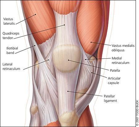
63 Con.Anterior compartment of the thigh Quadriceps femoris is made of four parts: 1.Rectus femoris which is the most superficial part. 2.Vastus lateralis. 3.Vastus Medialis. 4.Vastus Inetmedialis. All these muscles will form a tendon called Patellar tendon. In which the patella is embedded. This tendon continues downward to be inserted into the tibial tuberosity

64

65
66 Quadriceps femoris *-- Rectus femoris muscle is the only muscle of quadriceps femoris that originates from the pelvis region. The others (vasti muscles) originate at the thigh level. Rectus femoris Origin anterior inferior iliac spine,margin of acetabulum Insertion patella and tibial tuberosity via the patellar ligament Action extends knee,flexes thigh (it crosses two joints) Vastus lateralis Vastus medialis Vastus intermedius Origin - femur They share a common tendon that is called patellar ( ligament) which insert into the tibial tuberosity. - the common patellar ligament which is the continuation of the quadriceps tendon. - The patellar ligament reinforces the knee joint anteriorly. Insertion patella and tibial tuberosity via the patellar ligament Action extends knee (they cross one joint) Those are the anterior compartment muscles of the thigh.if you contract them they will extend the knee joint and flex the hip joint towards the abdomen.so these are the main actions of those muscles.

67 * As shown here: the common patellar ligament which is the continuation of the quadriceps tendon. New

68 Spotlights.. Many muscles participate in one function. - Flexion, abduction and medial rotation are caused by the group of muscles located at the lateral aspect which are the gluteus minimus, medius,and the tensor fasciae latae. -Extensors are the gluteus group which is the gluteus maximus and a small part of the gluteus medius.these muscles also assist abduction. -The adductors are the gracilis,adductor magnus,adductor brevis,and the adductor longus -The lateral rotators located in the lateral compartment of the thigh are the psoasiliac muscle group.

69
70 The tensor fascae lata divides the thigh into two compartments.the anterior compartment,holding the vasti muscles and the abductor magnus. The posterior compartment holds the biceps femoris,semitendonous,and semimembranous.these compartments are made by the iliotibial tract, which is the aponeurosis of the tensor fascae latae.

71

72 The knee extensors are the rectus femoris,vastus lateralis,vastus medialis and vastus intermiedius
73 Leg muscles In the leg,there are three compartments: Anterior,posterior and lateral. The posterior divides into two layers, superficial and deep.(the posterior muscles mainly have one insertion.) *Most of the tendon insertions in these compartments are attached in the foot, except for the superficial posterior layer. *These tendons perform dorsiflexion,plantar flexion, inversion, or eversion of the foot.at the ankle joint
74 **planter extension = Dorsiflexion New all the anterior group do extension and all the posterior group do flexion at the level of the foot; planter extension and flexion. The lateral group is important because we have two muscles (fibularis longus and brevis) that go to the planter of the foot and do two unique movements of the foot; eversion (lateral rotation) and inversion (medial rotation), these two muscles participate in forming the arches of the foot.

75 found in the lateral aspect of the leg (anterior-lateral). We have the tibialis anterior muscle which moves the foot and toes, then we have the extensor digitorum longus and medial to it we have the extensor hallicus longus. New
76 ** Tibialis anterior: New - The biggest muscle of the anterior aspect of the leg. - The origin: tuberosity of tibia - The insertion: tarsals (the medial aspect of the foot) -The action: it helps the muscles of the lateral group (fibularis longus and brevis) to do the eversion and the inversion of the foot ** Extensor digitorum longus: - A long muscle which has a long wide fibers. - The origin: the anterior aspect of tibia and fibula - The insertion: phalanges The action: extension of toes. **extensor hallicus longus it is the same but it inserts to the base of the first phalange (the big toe).
77 Just like the forearm muscles, they will pass into a retinaculum, either an extensor retinaculumn or a flexor retinaculum. -The extensor retinaculum is more anterior and Y shaped, located superior to the lateral and medial malleolus, in what is called the communicating line. -The flexor retinaculum is located in the medial aspect, posterior to the medial malleolus. -The lateral compartment has its own retinaculum called the fibular retinaculum or peroneal retinaculum which is located at the level of the lateral malleolus and makes a tunnel for the peroneus(fibularis) longus and peroneus(fibularis) brevis *In the medial aspect,the medial retinaculum contains only flexor muscles. * The lateral retinaculum contains evertor muscles * The anterior retinaculum contains extensor muscles
78 Any muscle that doesn t have capturing aspect (for fixation), its action will be lost, that is why we have something called the lever system which consist of the base (fulcrum), weight and the action (force), it resembles the seesaw system; it has the fixation aspect in the middle (the fulcrum), and on each side we have the weight and the force which both will be equilibrated (the weight gets you down and the force pushes you upward) ** The same in the muscle; if there is no fulcrum, then the weight and the force are not equilibrated, so that is why we have the extensor and flexor retinacula (the flexor is wider and thicker) which serve as fulcrums for the muscles. ** If we look at the wrist joint, these retinacula are deep, thicker and composed of two layers, whereas at the ankle joint we have only two bands but we still call them extensor and flexor retinacula, also at lateral and medial aspect we have two retinacula; fibular and flexor retinacula. So, each muscle that passes through these retinacula in the ankle has to be fixed. New

79
")
80 The anterior compartment of leg is made up of the extensor digitorium longus,tibialis anterior,extensor hallucis and peroneus (fibularis) tertius.

81
82 The tendon of the extensor halluces longus is big. And the continuous tendons of the extensor digitorium longus go all the way to the four lateral toes except the tendon of the extensor halluces longus which is controlled by itself. Thus, when you are moving your toes, you will be able to lift up your big toe without lifting up the other toes. In summary, the extensor hallus longus has its own tract, it s part of the extensor digitorium but it can act by itself upon the big toe.

83


84 Medial aspect of the foot lateral aspect of the foot
85 The lateral compartment is made by two muscles,the peroneus longus and the peroneus brevis (also called the fibularis longus and brevis) They evert (prime action) and plantar flex (secondary action) the foot (eversion and inversion) very powerful evertors of the foot Fibularis (peroneus) longus. superficial lateral muscle that covers the fibula (it will be attached to tarsal bones at the lateral aspect.) its tendon attaches to the plantar side of the foot the fibularis (peroneus) brevis lies deep to the fibularis longus its tendon inserts onto the base of the fifth metatarsal on its lateral aspect **both muscles wind around each other at the ankle, by that we will have the two actions of eversion and inversion of the foot.
86 If you put your hand posterior to the lateral mallelous and do eversion and inversion of the leg you'll be able to feel their tendons moving. The longus is big and makes the bulk of the lateral compartment and has a longer tendon that goes all the way to the medial cuneiform (so from the lateral side to the medial cuneiform). *Whereas the peroneus brevis goes to the tarsal bones.those will form the lateral compartment. *The peroneus longus and peroneus brevis have their own retinaculum, which is called the lateral retinaculum of the ankle joint (fibular retinacula)


87
.")
88 New Inferior Extensor retinaculum We can see the extensor and fibularis retinacula of the foot (the fibularis is superior to the lateral part off the lower extensor retinaculum), and in order for the fibularis muscle to make their action well, they lie behind a protrusion of the callus and are captured by the fibularis tendon (retinaculum). Fibularis retinaculum


89
90 The posterior compartment is made of a superficial layer and a deep layer. The smallest tendon and longest tendon (plantaris muscle) in the body are located in the posterior compartment of the leg, popliteus tendon and plantaris tendon respectively Gastrocnemius muscle is one of the superficial muscles that comes from the inferiolateral and inferiomedial border of popliteal fossa and will spilt into two parts beginning of the condyle of the tibia going all the way to be inserted into the calcaneal (achilles) tendon (the thickest and strongest tendon in body) This common tendon has three muscles: -Gastrocnemius. - Soleus which is underneath gastrocnemius. -Plantaris muscle which is a very small muscle & it moves the knee joint laterally.
91 New **plantaris tendon is thin and it goes along the posterior aspect of tibia to the planter surface of the foot. ** the gastrocnemius and soleus muscles form the triceps surae with three heads (two for gastrocnemius and one for soleus) which all will be held into one common tendon which will go to the posterior calcaneus; the calcaneal (Achilles) tendon. ** The two heads of the gastrocnemius one in the medial aspect and the other in the lateral one, they are bifurcated and separated from each other but they fuse in the lower half of the muscle (calcaneal tendon), both heads will form the inferio-lateral and inferiomedial border of the popliteal fossa posteriorly to the knee joint where they originate from the condyles of the femur, (the upper lateral and the upper medial borders of the fossa are made by bicips femoris,semimembranousus and semitendonosus muscles of femur), the soleus muscle is big and similar to the semitendonosus muscle of the thigh, we call these two big muscles (gastrocnemius and soleus) the duck of the leg :p.

92


93


94
95 The deep muscles of the posterior compartments include: - Flexor digitorum longus. - Tibialis posterior. -Flexor hallucis longus. These three muscles will pass posterior to the medial malleolus and they are comprehended into another retinaculum called flexor retinaculum of the leg.
96 New -popliteus muscle originates from the lateral condyle of the femur and insert into the proximal tibia medially, it s a small muscle that help in the medial rotation of the knee joint and lasting the knee joint by preventing the departure of the femoral condyle from the superior aspect of tibia by locking the medial and lateral rotation (control the rotation) so it flex and medially rotate the leg, -some tendons of muscles goes in a groove behind the medial malleolus or a groove on inferior surface of sustintaculum tali projection of calcaneus bone, so these muscles are fixed in a very strong flexion retinaculum which is composed of one or two bands. -the flexor digitorum longus muscle comes from the tibia and go to the distal phalanges 2-5, flexor hallicus longus muscle comes from fibula _so the digitorum is medial and the hallicus is lateral (both flexor digitorum and hallicus longus are found in the same muscle layer of the foot but they cross-over each other which helps in the eversion of foot ) -in the middle we have tibialis posterior which come from tibia, fibula and the interosseus membrane, one of its tendons will go behind the groove of the medial malleolus underneath the sustintaculum tali to their destination and insert into the medial cuniform

97

98
99 These are the compartments of the lower limb, New In the thigh part we have three compartments; the anterior which is the biggest one, the posterior and the lateral one which is the smallest. In the leg part also we have three compartments; anterior, posterior and lateral, but the posterior one is the biggest one, and the smallest one is lateral. ** in this region the interosseus membrane plays a great role in separating the anterior muscles from the posterior, whereas the fascia will send only two septa toward the fibula, so that the lateral compartment is separated by the continuation of this fascia to the fibula except for perforating sites in the upper leg where the arteries and nerves pass to the lateral compartment.

100 -The Interosseous ( -inter= inside, -osseus=bone) membrane divides the leg region into 2 compartment; Anterior and posterior. -In the leg region, because of the fibula, the only thing you see is the shin of the anterior border of the tibia. - The anterior compartment is separated by the interosseous membrane. - The lateral compartment has nothing but bone which separates anterior/posterior from the lateral compartment of the leg.

101 Sacral plexus
102 -Most important nerve that comes from this plexus, is the sciatic nerve -The Sciatic nerve isvery important. Most pain in lower limbs comes from sciatic pain.usually the cause of Sciatic pain,which is very common, is from lumbar region and compression of these vertebrae that causes pain. In the next slide is the design of the sciatic.it s made of 2 nerves that are enveloped into a sheath.these nerves divide into tibial and common peroneal nerve when they reach popliteal region.
.")
103 You can see the Lumbar and participation of the sacrum (doesn t have to be all sacral, but usually S1, and S2 and S3).The most important is L4 and L5 that participate in forming these nerves.

104

105
- it")

106 *This is the superior gluteal nerve-(very important)- it originates in the sacral plexus and is a branch of the sciatic nerve.
107 IM injection in buttock: New The line that passes through the middle of the iliac crest vertically downward is the dividing line because the gluteus maximus is more laterally, so we do the injection laterally in that area because in the medial aspect you will be able to hit the sciatic nerve, superior gluteal artery and veins, and this is one of the most common causes of litigation against medical personnel, if you don t divide this region with another horizontal line that passes through the anterior superior iliac spine to the posterior superior iliac spine you will be ending giving the injection in the capsule of the hip joint, so you choose the lateral upper quadrant to inject through as deep as you go it doesn t matter because you have three muscles to go through; gluteus maximus; minimus and medius. The gap is the canal of the needle through which the material pass to the body, the longer the needle, the bigger the gap and the more pain person feels, so when inject the patient you better inject him with intermediate or small needle so he won t get pain especially when you are dealing with babies.

108

109

110
111 Muscles of the PelvicGirdleand Lower Limbs-summary This table was in the slides and the sheet New
112 This table was in the slides and the sheet New
113 This table was in the slides and the sheet New

114 This table was in the slides and the sheet New

115 This table wasn t in the slides or the sheet The tables below have the same muscles as the ones above you can check them if sth was not clear in the last one above!

116

117


118

119 The end.. Best of luck Awn
Muscles of the lower extremities. Dr. Nabil khouri MD, MSc, Ph.D
 Muscles of the lower extremities Dr. Nabil khouri MD, MSc, Ph.D Posterior leg Popliteal fossa Boundaries Biceps femoris (superior-lateral) Semitendinosis and semimembranosis (superior-medial) Gastrocnemius
lesser trochanter of femur lesser trochanter of femur iliotibial tract (connective tissue) medial surface of proximal tibia
 medial surface of proximal tibia") LOWER LIMB MUSCLES OF THE APPENDICULAR SKELETON The muscles that act on the lower limb fall into three groups: those that move the thigh, those that move the lower leg, and those that move the ankle, foot,
LOWER LIMB MUSCLES OF THE APPENDICULAR SKELETON The muscles that act on the lower limb fall into three groups: those that move the thigh, those that move the lower leg, and those that move the ankle, foot,
Lower limb summary. Anterior compartment of the thigh. Done By: Laith Qashou. Doctor_2016
 Lower limb summary Done By: Laith Qashou Doctor_2016 Anterior compartment of the thigh Sartorius Anterior superior iliac spine Upper medial surface of shaft of tibia 1. Flexes, abducts, laterally rotates
Muscles of the Hip 1. Tensor Fasciae Latae O: iliac crest I: lateral femoral condyle Action: abducts the thigh Nerve: gluteal nerve
 Muscles of the Hip 1. Tensor Fasciae Latae O: iliac crest I: lateral femoral condyle Action: abducts the thigh Nerve: gluteal nerve 2. Gluteus Maximus O: ilium I: femur Action: abduct the thigh Nerve:
The Muscular System. Chapter 10 Part D. PowerPoint Lecture Slides prepared by Karen Dunbar Kareiva Ivy Tech Community College
 Chapter 10 Part D The Muscular System Annie Leibovitz/Contact Press Images PowerPoint Lecture Slides prepared by Karen Dunbar Kareiva Ivy Tech Community College Table 10.14: Muscles Crossing the Hip and
Human Anatomy Biology 351
 Human Anatomy Biology 351 Lower Limb Please place your name on the back of the last page of this exam. You must answer all questions on this exam. Because statistics demonstrate that, on average, between
Muscles of Lesson Five. Muscular Nomenclature and Kinesiology - Two. Muscles of Lesson Five, cont. Chapter 16
 Chapter 16 Muscular Nomenclature and Kinesiology - Two Lessons 5-6 Muscles of Lesson Five Iliopsoas (psoas major, iliacus) Hip outward rotators (piriformis, gemellus superior, gemellus inferior, obturator
Anatomy & Physiology. Muscles of the Lower Limbs.
 Anatomy & Physiology Muscles of the Lower Limbs http://www.ishapeup.com/musclecharts.html Muscles of the Lower Limbs Among the strongest muscles in the body. Because pelvic girdle is composed of heavy,
Human Anatomy Biology 255
 Human Anatomy Biology 255 Exam #4 Please place your name and I.D. number on the back of the last page of this exam. You must answer all questions on this exam. Because statistics demonstrate that, on average,
MUSCLES OF THE LOWER LIMBS
 MUSCLES OF THE LOWER LIMBS Naming, location and general function Dr. Nabil khouri ROLES THAT SHOULD NOT BE FORGOTTEN Most anterior compartment muscles of the hip and thigh Flexor of the femur at the hip
Human Anatomy Biology 351
 Human Anatomy Biology 351 Lower Limb Please place your name on the back of the last page of this exam. You must answer all questions on this exam. Because statistics demonstrate that, on average, between
The thigh. Prof. Oluwadiya KS
 The thigh Prof. Oluwadiya KS www.oluwadiya.com The Thigh: Boundaries The thigh is the region of the lower limb that is approximately between the hip and knee joints Anteriorly, it is separated from the
The Hip (Iliofemoral) Joint. Presented by: Rob, Rachel, Alina and Lisa
 Joint. Presented by: Rob, Rachel, Alina and Lisa") The Hip (Iliofemoral) Joint Presented by: Rob, Rachel, Alina and Lisa Surface Anatomy: Posterior Surface Anatomy: Anterior Bones: Os Coxae Consists of 3 Portions: Ilium Ischium Pubis Bones: Pubis Portion
The Hip (Iliofemoral) Joint Presented by: Rob, Rachel, Alina and Lisa Surface Anatomy: Posterior Surface Anatomy: Anterior Bones: Os Coxae Consists of 3 Portions: Ilium Ischium Pubis Bones: Pubis Portion
Topic 7: Hip and pelvis. Parts of the hip. Parts of the femur
 Topic 7: Hip and pelvis Parts of the hip Parts of the femur Classifying the hip joint Ball and socket Synovial Multiaxial Movements of the hip: Abduction/adduction Flexion/extension Medial/lateral rotation
Muscles of the Gluteal Region
 Muscles of the Gluteal Region 1 Some of the most powerful in the body Extend the thigh during forceful extension Stabilize the iliotibial band and thoracolumbar fascia Related to shoulders and arms because
DISSECTION SCHEDULE. Session I - Hip (Front) & Thigh (Superficial)
 & Thigh (Superficial)") DISSECTION SCHEDULE Session I - Hip (Front) & Thigh (Superficial) Surface anatomy Inguinal region Gluteal region Thigh Leg Foot bones Hip bone Femur Superficial fascia Great saphenous vein Superficial
DISSECTION SCHEDULE Session I - Hip (Front) & Thigh (Superficial) Surface anatomy Inguinal region Gluteal region Thigh Leg Foot bones Hip bone Femur Superficial fascia Great saphenous vein Superficial
Lower Limb Nerves. Clinical Anatomy
 Lower Limb Nerves Clinical Anatomy Lumbar Plexus Ventral rami L1 L4 Supplies: Abdominal wall External genitalia Anteromedial thigh Major nerves.. Lumbar Plexus Nerves relation to psoas m. : Obturator n.
Lower Limb Dr. Robin Paudel
 Lower Limb n What is a limb? n Skeleton n Joints n Pelvis or limb girdle n Hip/Hip Muscles n Lumber and sacral plexus getting spinal nerves out onto limb n Muscles anterior and posterior compartments n
ANATOMY TEAM GLUTEAL REGION & BACK OF THIGH
 ANATOMY TEAM GLUTEAL REGION & BACK OF THIGH OBJECTIVES By the end of this lecture, the student should be able to identify and discuss: Contents of gluteal region: Groups of Glutei muscles and small muscles
Gluteal region DR. GITANJALI KHORWAL
 Gluteal region DR. GITANJALI KHORWAL Gluteal region The transitional area between the trunk and the lower extremity. The gluteal region includes the rounded, posterior buttocks and the laterally placed
musculoskeletal system anatomy nerves of the lower limb 2 done by: Dina sawadha & mohammad abukabeer
 musculoskeletal system anatomy nerves of the lower limb 2 done by: Dina sawadha & mohammad abukabeer #Sacral plexus : emerges from the ventral rami of the spinal segments L4 - S4 and provides motor and
Main Menu. Joint and Pelvic Girdle click here. The Power is in Your Hands
 1 Hip Joint and Pelvic Girdle click here Main Menu K.6 http://www.handsonlineeducation.com/classes//k6entry.htm[3/23/18, 2:01:12 PM] Hip Joint (acetabular femoral) Relatively stable due to : Bony architecture
Muscles of the Thigh. 6.1 Identify, describe the attachments of and deduce the actions of the muscles of the thigh: Anterior group
 Muscles of the Thigh 6.1 Identify, describe the attachments of and deduce the actions of the muscles of the thigh: Anterior group Sartorius: This is a long strap like muscle with flattened tendons at each
Lumbar Plexus. Ventral rami L1 L4 Supplies: Major nerves.. Abdominal wall External genitalia Anteromedial thigh
 Lower Limb Nerves Lectures Objectives Describe the structure and relationships of the plexuses of the lower limb. Describe the course, relationships and structures supplied for the major nerves of the
Anatomage Table Instructors Guide- Lower Limb
 The Lower Limb Anatomage Table Instructors Guide- Lower Limb Table of Contents Lower Limb 1- The Skeletal System...3 1: Hip Bone...3 2: Hip Joint and Femur...4 3: Patella and Knee Joint...7 4: Tibia, Fibula,
LOWER LIMB. As we know the bony part of the body is divided into Axial and Appendicular (upper and lower Limbs)
") LOWER LIMB As we know the bony part of the body is divided into Axial and Appendicular (upper and lower Limbs) Bones of the Lower limb: 1-Pelvic Girdle: composed of: 1. Right hip bone : is formed by 3
LOWER LIMB As we know the bony part of the body is divided into Axial and Appendicular (upper and lower Limbs) Bones of the Lower limb: 1-Pelvic Girdle: composed of: 1. Right hip bone : is formed by 3
The University Of Jordan Faculty Of Medicine THE LOWER LIMB. Dr.Ahmed Salman Assistant Prof. of Anatomy. The University Of Jordan
 The University Of Jordan Faculty Of Medicine THE LOWER LIMB Dr.Ahmed Salman Assistant Prof. of Anatomy. The University Of Jordan Gluteal Region Cutaneous nerve supply of (Gluteal region) 1. Lateral cutaneous
rotation of the hip Flexion of the knee Iliac fossa of iliac Lesser trochanter Femoral nerve Flexion of the thigh at the hip shaft of tibia
 Anatomy of the lower limb Anterior & medial compartments of the thigh Dr. Hayder The fascia lata encloses the entire thigh like a sleeve/stocking. Three intramuscular fascial septa (lateral, medial, and
The psoas minor is medial to the psoas major. The iliacus is a fan-shaped muscle that when contracted helps bring the swinging leg forward in walking
 1 p.177 2 3 The psoas minor is medial to the psoas major. The iliacus is a fan-shaped muscle that when contracted helps bring the swinging leg forward in walking and running. The iliopsoas and adductor
Hip joint and pelvic girdle. Lower Extremity. Pelvic Girdle 6/5/2017
 Hip joint and pelvic girdle Lower Extremity The relationship between the pelvic girdle and hip is similar to that between the shoulder girdle and shoulder joint. The lower limbs are attached to the axial
Lectures of Human Anatomy
 Lectures of Human Anatomy Lower Limb Gluteal Region and Hip Joint By DR. ABDEL-MONEM AWAD HEGAZY M.B. with honor 1983, Dipl."Gynecology and Obstetrics "1989, Master "Anatomy and Embryology" 1994, M.D.
Anterior and Medial compartments of the thigh. Dr. Heba Kalbouneh Associate Professor of Anatomy and Histology
 Anterior and Medial compartments of the thigh Dr. Heba Kalbouneh Associate Professor of Anatomy and Histology Terms Related to Movements Movement Flexion Extension Abduction Adduction Medial (internal)
Due in Lab weeks because of Thanksgiving Prelab #10. Homework #8. Both sides! Both sides!
 Lab 8 MUSCLES Due in Lab 10 2 weeks because of Thanksgiving Prelab #10 Both sides! Homework #8 Both sides! Refer to Muscles 22-23 Naming of muscles Origin Site of muscle attachment that doesn t move during
11/15/2018. Temporalis Elevates & retracts mandible. Masseter = Prime mover of jaw closure. Levator scapulae Supraspinatus Clavicle.
 Due in Lab 10 Lab 8 MUSCLES 2 weeks because of Thanksgiving Prelab #10 Both sides! Homework #8 Both sides! Refer to Muscles 22-23 Examples of Origin & Insertion Naming of muscles Origin Site of muscle
Scapula Spine Lateral edge of clavicle. Medial border Scapula. Medial border of Scapula, between superior angle and root of spine. Scapula.
 Muscle attachments and actions answer sheet Muscle Origins insertions Movements Joints crossed Trapezius Base of skull Spinous process of C7 Thoracic Spine Lateral edge of clavicle Elevation Retraction
MUSCULOSKELETAL LOWER LIMB
 MUSCULOSKELETAL LOWER LIMB Spinal Cord Lumbar and Sacral Regions Spinal cord Dorsal root ganglion Conus medullaris Cauda equina Dorsal root ganglion of the fifth lumbar nerve End of subarachnoid space
The Leg. Prof. Oluwadiya KS
 The Leg Prof. Oluwadiya KS www.oluwadiya.sitesled.com Compartments of the leg 4 Four Compartments: 1. Anterior compartment Deep fibular nerve Dorsiflexes the foot and toes 2. Lateral Compartment Superficial
Identify the muscles associated with the medial compartment of the thigh. Identify the attachment points of the medial thigh muscles.
 L 8 A B O R A T O R Y Thigh MEDIAL THIGH Identify the muscles associated with the medial compartment of the thigh. Identify the attachment points of the medial thigh muscles. Identify the actions of these
Baraa Ayed حسام أبو عوض. Ahmad Salman. 1 P a g e
 4 Baraa Ayed حسام أبو عوض Ahmad Salman 1 P a g e Today we are going to cover these concepts: Iliotibial tract Anterior compartment of the thigh and the hip Medial compartment of the thigh Femoral triangle
Mohammad Ashraf. Abdulrahman Al-Hanbali. Ahmad Salman. 1 P a g e
 - 7 Mohammad Ashraf Abdulrahman Al-Hanbali Ahmad Salman 1 P a g e Structures under the cover of Gluteus Maximus: 1-Bones: Ileum, Femur (Head, greater trochanter and gluteal tuberosity), Ischium (ischial
Anatomy of the lower limb
 Anatomy of the lower limb 1. Bones of the lower limb Pelvis Hip bone/coxal bone Acetabulum o Acetabular margin o Acetabular fossa o Acetabular notch o Lunate surface Ischiopubic ramus Obturator foramen
Leg. Dr. Heba Kalbouneh Associate Professor of Anatomy and Histology
 Leg Dr. Heba Kalbouneh Associate Professor of Anatomy and Histology Skin of the Leg Cutaneous Nerves Medially: The saphenous nerve, a branch of the femoral nerve supplies the skin on the medial surface
Muscles to know. Lab 21. Muscles of the Pelvis and Lower Limbs. Muscles that Position the Lower Limbs. Generally. Muscles that Move the Thigh
 Muscles to know Lab 21 Muscles of the Pelvis, Leg and Foot psoas major iliacus gluteus maximus gluteus medius sartorius quadriceps femoris (4) gracilus adductor longus biceps femoris semitendinosis semimembranosus
Where should you palpate the pulse of different arteries in the lower limb?
 Where should you palpate the pulse of different arteries in the lower limb? The femoral artery In the femoral triangle, its pulse is easily felt just inferior to the inguinal ligament midway between the
Practical 1 Worksheet
 Practical 1 Worksheet ANATOMICAL TERMS 1. Use the word bank to fill in the missing words. reference side stand body arms palms anatomical forward All anatomical terms have a(n) point which is called the
ANATYOMY OF The thigh
 ANATYOMY OF The thigh 1- Lateral cutaneous nerve of the thigh Ι) Skin of the thigh Anterior view 2- Femoral branch of the genitofemoral nerve 5- Intermediate cutaneous nerve of the thigh 1, 2 and 3 are
this makes sense, however this is lower order thinking and does not solve the lower leg
 Functional Knee Valgus in a Barbell Squat 1 One of the most common lower leg dysfunction we see in athletes, particularly general population is functional knee valgus, or better referred to as the knees
Lowe w r e r B ody Resistance Training
 Lower Body Resistance Training Tibialis Anterior Extensor Hallucis Longus Extensor Digitorum Longus Proneus Tertius AROM 25-40 degrees Extensor active-insufficiency Flexion & Eversion (Pronation) Flexion
Contents of the Posterior Fascial Compartment of the Thigh
 Contents of the Posterior Fascial Compartment of the Thigh 1-Muscles: B i c e p s f e m o r i s S e m i t e n d i n o s u s S e m i m e m b r a n o s u s a small part of the adductor magnus (h a m s t
HUMAN BODY COURSE LOWER LIMB NERVES AND VESSELS
 HUMAN BODY COURSE LOWER LIMB NERVES AND VESSELS October 22, 2010 D. LOWER LIMB MUSCLES 2. Lower limb compartments ANTERIOR THIGH COMPARTMENT General lfunction: Hip flexion, knee extension, other motions
Muscles of Gluteal Region
 1 The Gluteal Region In the gluteal region the skin is tough with many layers underneath. Directly under it is the superficial fascia followed by the deep fascia then the muscles and the bones of the thigh.
ANATYOMY OF The thigh
 ANATYOMY OF The thigh 1- Lateral cutaneous nerve of the thigh Ι) Skin of the thigh Anterior view 2- Femoral branch of the genitofemoral nerve 5- Intermediate cutaneous nerve of the thigh 1, 2 and 3 are
5.1 Identify, describe the attachments of and deduce the actions of the muscles of the thigh:
 5.1 Identify, describe the attachments of and deduce the actions of the muscles of the thigh: Anterior group Proximal attachment Distal attachment Sartorius ASIS» Upper part of shaft tibia (middle surface)»
Lecture 08 THIGH MUSCLES ANTERIOR COMPARTMENT. Dr Farooq Khan Aurakzai. Dated:
 Lecture 08 THIGH MUSCLES ANTERIOR COMPARTMENT BY Dr Farooq Khan Aurakzai Dated: 11.02.2017 INTRODUCTION to the thigh Muscles. The musculature of the thigh can be split into three sections by intermuscular
BLUE SKY SCHOOL OF PROFESSIONAL MASSAGE AND THERAPEUTIC BODYWORK Musculoskeletal Anatomy & Kinesiology KNEE & ANKLE MUSCLES
 BLUE SKY SCHOOL OF PROFESSIONAL MASSAGE AND THERAPEUTIC BODYWORK Musculoskeletal Anatomy & Kinesiology KNEE & ANKLE MUSCLES MSAK201-I Session 3 1) REVIEW a) THIGH, LEG, ANKLE & FOOT i) Tibia Medial Malleolus
In-Depth Foundations: Anatomy Terms to Know
 Be familiar with / able to identify and define all the following parts. The Spine Cranium Vertebrae Cervical, Thoracic, Lumbar Sacrum Coccyx Bones of Upper Body Cranium Mastoid process; Occipital condyle,
Bones of the Lower Limb Bone Structure Description Notes. border of the superior ramus. inferolaterally from the pubic symphysis
 Bones of the Lower Limb Bone Structure Description Notes pubis an angulated bone the forms the anterior part of the pelvis one of three bones that form the os coxae: ilium, ischium, pubis; its forms 1/5
The Hay is in the Barn
 Anatomy 1 Practical 1 Review Made by Forrest Allen (nerd) Edited by TJ Williamson (not nerd) The Hay is in the Barn 2019 Thunderbringers Too much to handle https://www.youtube.com/watch?v=glii-kaza d8
The Lower Limb II. Anatomy RHS 241 Lecture 3 Dr. Einas Al-Eisa
 The Lower Limb II Anatomy RHS 241 Lecture 3 Dr. Einas Al-Eisa Tibia The larger & medial bone of the leg Functions: Attachment of muscles Transfer of weight from femur to skeleton of the foot Articulations
Organization of the Lower Limb Audrone Biknevicius, Ph.D. Dept. Biomedical Sciences, OU HCOM at Dublin Clinical Anatomy Immersion 2014
 Organization of the Lower Limb Audrone Biknevicius, Ph.D. Dept. Biomedical Sciences, OU HCOM at Dublin Clinical Anatomy Immersion 2014 www.thestudio1.co.za LIMB FUNCTION choco-locate.com blog.coolibar.com
CHAPTER 8: THE BIOMECHANICS OF THE HUMAN LOWER EXTREMITY
 CHAPTER 8: THE BIOMECHANICS OF THE HUMAN LOWER EXTREMITY _ 1. The hip joint is the articulation between the and the. A. femur, acetabulum B. femur, spine C. femur, tibia _ 2. Which of the following is
The Hip Joint. Shenequia Howard David Rivera
 The Hip Joint Shenequia Howard David Rivera Topics Of Discussion Movement Bony Anatomy Ligamentous Anatomy Muscular Anatomy Origin/Insertion/Action/Innervation Common Injuries MOVEMENT Flexion Extension
5 Testing the Muscles of the Lower Extremity
 C H A P T E R 5 Testing the Muscles of the Lower Extremity Hip Flexion Hip Flexion, Abduction, and External Rotation with Knee Flexion Hip Extension Hip Abduction Hip Abduction from Flexed Position Hip
In which arm muscle are intramuscular injections most often given? (not in text)
") AP1 Lab 9 - Muscles of the Arms and Legs Locate the following muscles on the models and on yourself. Recall anatomical position. Directional terms such as anterior, posterior, lateral, etc. all assume
AP1 Lab 9 - Muscles of the Arms and Legs Locate the following muscles on the models and on yourself. Recall anatomical position. Directional terms such as anterior, posterior, lateral, etc. all assume
Bones of Lower Limb. Dr. Heba Kalbouneh Associate Professor of Anatomy and Histology
 Bones of Lower Limb Dr. Heba Kalbouneh Associate Professor of Anatomy and Histology Bones of the lower limb Hip Bone Made up of 3 bones: 1) Ilium (flat), superior in position 2) Ischium (L), postero-inferior
ANATYOMY OF The thigh
 ANATYOMY OF The thigh 1- Lateral cutaneous nerve of the thigh Ι) Skin of the thigh Anterior view 2- Femoral branch of the genitofemoral nerve 1, 2 and 3 are From the lumber plexus 5- Intermediate cutaneous
Acland's DVD Atlas of Human Anatomy. Transcript for Volume Robert D Acland
 Acland's DVD Atlas of Human Anatomy Transcript for Volume 2 2007 Robert D Acland This free downloadable pdf file is to be used for individual study only. It is not to be reproduced in any form without
Sports Medicine 15. Unit I: Anatomy. The knee, Thigh, Hip and Groin. Part 4 Anatomies of the Lower Limbs
 Sports Medicine 15 Unit I: Anatomy Part 4 Anatomies of the Lower Limbs The knee, Thigh, Hip and Groin Anatomy of the lower limbs In Part 3 of this section we focused upon 11 of the 12 extrinsic muscles
lower limb Anterior Compartment: lecture 3 The deep fascia ( fascia lata) divides the thigh into 3 compartments:
 divides the thigh into 3 compartments:") lower limb lecture 3 The deep fascia ( fascia lata) divides the thigh into 3 compartments: 1. Anterior Extensor compartment 2. Medial Adductor compartment 3. Posterior Flexor compartment Anterior Compartment:
lower limb lecture 3 The deep fascia ( fascia lata) divides the thigh into 3 compartments: 1. Anterior Extensor compartment 2. Medial Adductor compartment 3. Posterior Flexor compartment Anterior Compartment:
Anatomy MCQs Week 13
 Anatomy MCQs Week 13 1. Posterior to the medial malleolus of the ankle: The neurovascular bundle lies between Tibialis Posterior and Flexor Digitorum Longus The tendon of Tibialis Posterior inserts into
Introduction to Anatomy. Dr. Maher Hadidi. Laith Al-Hawajreh. Mar/25 th /2013
 Introduction to Anatomy Dr. Maher Hadidi Laith Al-Hawajreh 22 Mar/25 th /2013 Lower limb - The leg The skeleton of the leg is formed by two bones: 1) Medial: Tibia 2) Lateral: Fibula The two bones are
TABLE OF MUSCLES OF LOWER EXTREMITY 2018zillmusom ANTERIOR THIGH
 TABLE OF MUSCLES OF LOWER EXTREMITY 2018zillmusom ANTERIOR THIGH MUSCLE ORIGIN INSERTION ACTION NERVE Iliopsoas Ilium, vertebra Femur Flex hip joint Femoral nerve (T12-L5) Pectineus Pubis Femur Flex hip
1-Muscles: 2-Blood supply: Branches of the profunda femoris artery. 3-Nerve supply: Sciatic nerve
 1-Muscles: B i c e p s f e m o r i s S e m i t e n d i n o s u s S e m i m e m b r a n o s u s a small part of the adductor magnus (h a m s t r i n g p a r t o r i s c h i a l p a r t ) 2-Blood supply:
Lesson 24. A & P Hip
 Lesson 24 A & P Hip 1 Aims of the Session This session will allow candidates to have an understanding of the bony prominences and soft tissues of the hip 2 Learning Outcomes By the end of the lesson the
THE LOWER LIMB NERVES VESSELS
 THE LOWER LIMB NERVES VESSELS LOWER LIMB: FEMORAL TRIANGLE FEMORAL TRIANGLE LOWER LIMB: FEMORAL TRIANGLE FEMORAL TRIANGLE is a triangular landmark useful in dissection and in understanding relationships
Peripheral Nervous System: Lower Body
 Peripheral Nervous System: Lower Body MSTN121 - Neurophysiology Session 11 Department of Myotherapy Lumbar Plexus Iliohypogastric nerve (T12-L1) Motor: Transverse abdominis and internal obliques Sensory:
Myology of the Knee. PTA 105 Kinesiology
 Myology of the Knee PTA 105 Kinesiology Objectives Describe the planes of motion and axes of rotation of the knee joint Visualize the origins and insertions of the muscles about the knee List the innervations
MUSCLES OF THE LOWER EXTREMITY
 MUSCLES OF THE LOWER EXTREMITY Muscles of the lower extremity are divisible into groups, corresponding with the different regions of the limb. I. Muscles of the Iliac Region II. Muscles of the Thigh III.
The Lower Limb. Anatomy RHS 241 Lecture 2 Dr. Einas Al-Eisa
 The Lower Limb Anatomy RHS 241 Lecture 2 Dr. Einas Al-Eisa The bony pelvis Protective osseofibrous ring for the pelvic viscera Transfer of forces to: acetabulum & head of femur (when standing) ischial
Human Anatomy and Physiology I Laboratory
 Human Anatomy and Physiology I Laboratory Gross Anatomy of the Muscular System (Two weeks) 1 This lab involves study of the laboratory exercise Gross Anatomy of the Muscular System. Complete the Review
Year 2004 Paper one: Questions supplied by Megan
 QUESTION 47 A 58yo man is noted to have a right foot drop three days following a right total hip replacement. On examination there is weakness of right ankle dorsiflexion and toe extension (grade 4/5).
Regional Anaesthesia
 Regional Anaesthesia Lower limb anatomy and blocks Hip and Knee Joint Hip Joint: Nerve supply Lumbar plexus Femoral nerve through the nerve to the Rectus Femoris Ant division of the Obturator nerve The
Posterior compartment of the thigh. Dr. Heba Kalbouneh Associate Professor of Anatomy and Histology
 Posterior compartment of the thigh Dr. Heba Kalbouneh Associate Professor of Anatomy and Histology Posterior compartment of the thigh 1-Muscles: Biceps femoris Semitendinosus Semimembranosus Adductor magnus
Prime movers provide the major force for producing a specific movement Antagonists oppose or reverse a particular movement Synergists
 Dr. Gary Mumaugh Prime movers provide the major force for producing a specific movement Antagonists oppose or reverse a particular movement Synergists Add force to a movement Reduce undesirable or unnecessary
Figure 1 - Hip and Pelvis
 Hip Figure 1 - Hip and Pelvis The terms hip and pelvis are frequently used interchangeably, but strictly speaking, the pelvis is a girdle of bones and the hip is a joint. The pelvis consists of The sacrum
Muscle Anatomy Review Chart
 Muscle Anatomy Review Chart BACK Superficial (5) Trapezius Transverse cervical a. Latissimus dorsi Thoracodorsal a. Rhomboideus major Dorsal scapular a. Rhomboideus minor Levator scapulae Intermediate
Human Anatomy Lab #7: Muscles of the Cadaver
 Human Anatomy Lab #7: Muscles of the Cadaver Table of Contents: Expected Learning Outcomes.... 1 Introduction...... 1 Identifying Muscles on Yourself.... 2 Muscles of the Anterior Trunk and Arm.. 2 Muscles
The Knee. Clarification of Terms. Osteology of the Knee 7/28/2013. The knee consists of: The tibiofemoral joint Patellofemoral joint
 The Knee Clarification of Terms The knee consists of: The tibiofemoral joint Patellofemoral joint Mansfield, p273 Osteology of the Knee Distal Femur Proximal tibia and fibula Patella 1 Osteology of the
Lab Exercise #5 The Muscular System Student Performance Objectives
 Student Performance Objectives The material that you are required to learn in this exercise can be found in either the lecture text or the supplemental materials provided in lab. Prior to coming to class,
SKELETAL MUSCLE ANATOMY
 SKELETAL MUSCLE ANATOMY OUTLINE I. Anatomical Terms of Motion II. Head, Face & Neck Muscles III. Anterior Torso Muscles IV. Posterior Torso Muscles V. Arm & Shoulder Muscles VI. Leg & Hip Muscles 2 ANATOMICAL
Understanding Leg Anatomy and Function THE UPPER LEG
 Understanding Leg Anatomy and Function THE UPPER LEG The long thigh bone is the femur. It connects to the pelvis to form the hip joint and then extends down to meet the tibia (shin bone) at the knee joint.
Bio 113 Anatomy and Physiology The Muscles. Muscles of the Head and Neck. Masseter. Orbicularis occuli. Orbicularis oris. Sternocleidomastoid
 Bio 113 Anatomy and Physiology The Muscles Muscles of the Head and Neck Masseter Orbicularis occuli Orbicularis oris Sternocleidomastoid Temporalis BIO 113 Fall 2011 Muscles Page 1 of 5 Muscles of the
Anatomy images for MSS practical exam- 2019
 Anatomy images for MSS practical exam- 2019 Ilium Ischium Pubis Acetabulaum Iliac crest Iliac tubercle ASIS (muscle and ligament attached) AIIS (muscle attached) PSIS PIIS Ischial spine Ischial tuberosity
It is formed by fusion of 3 bones: I. Ilium (superior bone). II. Pubis (antero-inferior bone). III. Ischium (postero-inferior bone).
. II. Pubis (antero-inferior bone). III. Ischium (postero-inferior bone).") It is formed by fusion of 3 bones: I. Ilium (superior bone). II. Pubis (antero-inferior bone). III. Ischium (postero-inferior bone). Pubis Acetabulum Ana (242 ) The three constituent of bones of the hip
It is formed by fusion of 3 bones: I. Ilium (superior bone). II. Pubis (antero-inferior bone). III. Ischium (postero-inferior bone). Pubis Acetabulum Ana (242 ) The three constituent of bones of the hip
موسى صالح عبد الرحمن الحنبلي أحمد سلمان
 8 موسى صالح عبد الرحمن الحنبلي أحمد سلمان 1 P a g e Today we will talk about a new region, which is the leg. And as always, we will start with studying the sensory innervation of the leg. What is the importance
BONES JOINTS MUSCLES OF THE LOWER LIMB
 BONES JOINTS MUSCLES OF THE LOWER LIMB LOWER LIMB: BONES LOWER LIMB GLUTEAL REGION consists of 6 major segments: FEMORAL REGION (THIGH) KNEE REGION LEG REGION TALOCRURAL REGION (ANKLE) FOOT REGION LOWER
Applied anatomy of the hip and buttock
 CHAPTER CONTENTS The hip joint e9 Capsule and ligaments e9 s e0 Flexor muscles................... e0 Extensor muscles.................. e Abductor muscles.................. e Adductor muscles..................
Temporalis Elevates & retracts mandible. Masseter Elevates mandible. Sternocleidomastoid Neck flexion. Trapezius Elevates & depresses shoulders
 Anterior Posterior Temporalis Elevates & retracts mandible Masseter Elevates mandible Sternocleidomastoid Neck flexion Trapezius Elevates & depresses shoulders Masseter Elevates mandible Temporalis Elevates
VENOUS DRAINAGE OF THE LOWER LIMB
 Anatomy of the lower limb Superficial veins & nerve injuries Dr. Hayder VENOUS DRAINAGE OF THE LOWER LIMB The venous drainage of the lower limb is of huge clinical & surgical importance. Since the venous
Appendix. Useful Anatomical Data of Clinical Significance
 Appendix Useful Anatomical Data of Clinical Significance Appendix Outline Respiratory System 426 Table I. Important Airway Distances (Adult) 426 Table II. Important Data Concerning the Trachea 426 Musculoskeletal