Department of Cell and Developmental Biology, Vanderbilt University, Nashville, TN
|
|
|
- Neil Newman
- 5 years ago
- Views:
Transcription
1 UNC-6/Netrin mediates dendritic self-avoidance Cody J. Smith 1, Joseph D. Watson 1,4,5, Miri K. VanHoven 2, Daniel A. Colón-Ramos 3 and David M. Miller III 1,4,6 1 Department of Cell and Developmental Biology, Vanderbilt University, Nashville, TN Department of Biological Sciences, San Jose State University, San Jose, CA. 3 Department of Cell Biology; Yale Program in Cellular Neuroscience and Neurodegeneration and Repair, Yale University, New Haven NH. 4 Neuroscience Program and Vanderbilt Kennedy Center, Vanderbilt University, Nashville, TN Current address: Department of Biochemistry and Biophysics, The University of North Carolina, Chapel Hill, NC , USA 6 Corresponding author: Department of Cell and Developmental Biology, Vanderbilt University, Nashville, TN address: david.miller@vanderbilt.edu
2 Supplemental Table 1. Self-avoidance requires specific axon guidance molecules in the UNC-6/Netrin signaling pathway. Mutants of known axon guidance molecules were tested for PVD 3 0 branch self- avoidance defects with the PVD::GFP reporter. Transcripts enriched (>1.5X, FDR < 1%) in PVD (Smith et al., 2010) are denoted with bold lettering. Only mutants of the UNC- 6/Netrin signaling pathway showed self- avoidance defects that were significantly different from wt (+ indicates p<0.01, Student s t- test). N > 20 Function Gene Self- avoidance defect unc-6/netrin + unc-129/tgf beta Ligand family - vab-1/ephrin - slt-1/slit - sax-3/robo - vab-2/eph - Receptor ptp-3/lar - unc-5/unc-5h + unc-40/dcc + madd-2/netrinr - ECM component nid-1/nidogen -
3 Supplemental Figure Legends Supplemental Figure 1. Mutants of unc-40, unc-5 and unc-6 show a range of dendritic morphogenesis phenotypes in addition to the self-avoidance defect. (a) PVD 1 O processes project along the lateral nerve cord in the wild type but deviate from a strict A/P trajectory in >75% of unc-40, unc-5 or unc-6 mutants. (b) Wild-type (wt) PVD neurons show an asymmetric pattern of lateral branching that results in more dorsal than ventral menorahs in 100% of cases examined (n > 15) (wild-type distribution). In UNC-6/Netrin pathway mutants, this asymmetry is disrupted resulting in PVD neurons that have more ventral menorahs than dorsal menorahs or an equal number of menorahs on each side ~50% of the time (defective distribution), an outcome that is consistent with a randomized probability of dorsal vs ventral initiation of 2 0 branches (CJ Smith and DM Miller, manuscript in preparation). (c) The average number of 2 O dendrites/pvd neuron in unc-6, unc-5 and unc-40 mutants is reduced in comparison to wild-type PVD neurons. (d). Ectopic branching in adults is more frequent in unc-5(e271) than in either wild type (wt) or unc-40(e271).
4
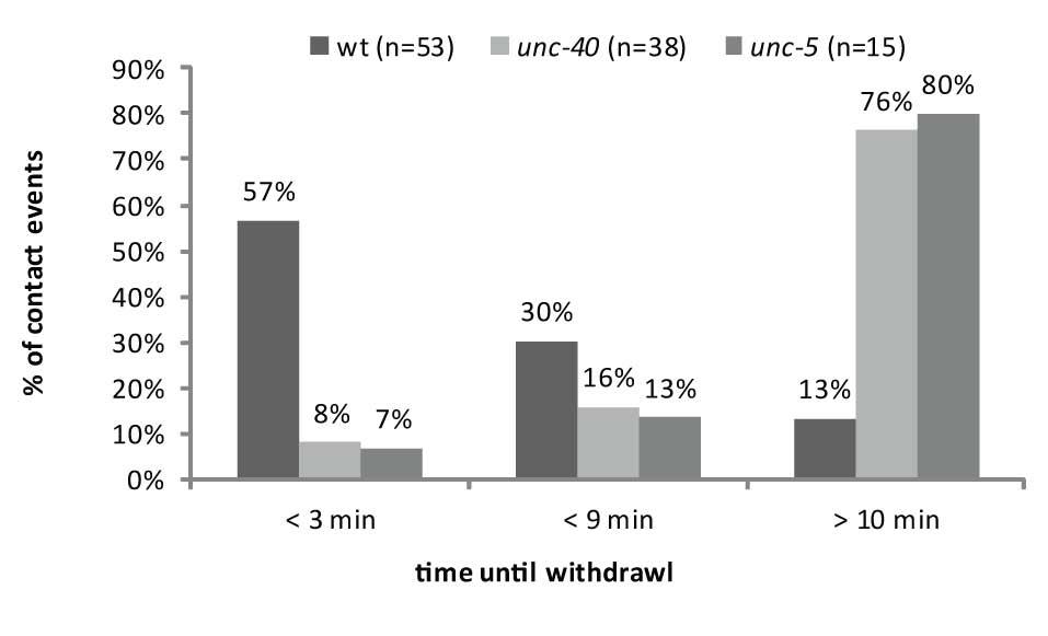
5 Supplemental figure 2. unc-40 and unc-5 mutants show defects in contactdependent self-avoidance. Quantification from movies of self-avoidance events in wild type (wt), unc-5 (e152) and unc-40 (e271) show that 3 O branches in unc-5 (e152) and unc-40 (e271) do not retract as quickly as in wild-type animals; a majority (>75%) of 3 O branches have failed to retract up to 10 minutes after initial contact in unc-40 and unc-5 mutants whereas only 13% of 3 O dendrites are still overlapping at this time point in wild type.
6
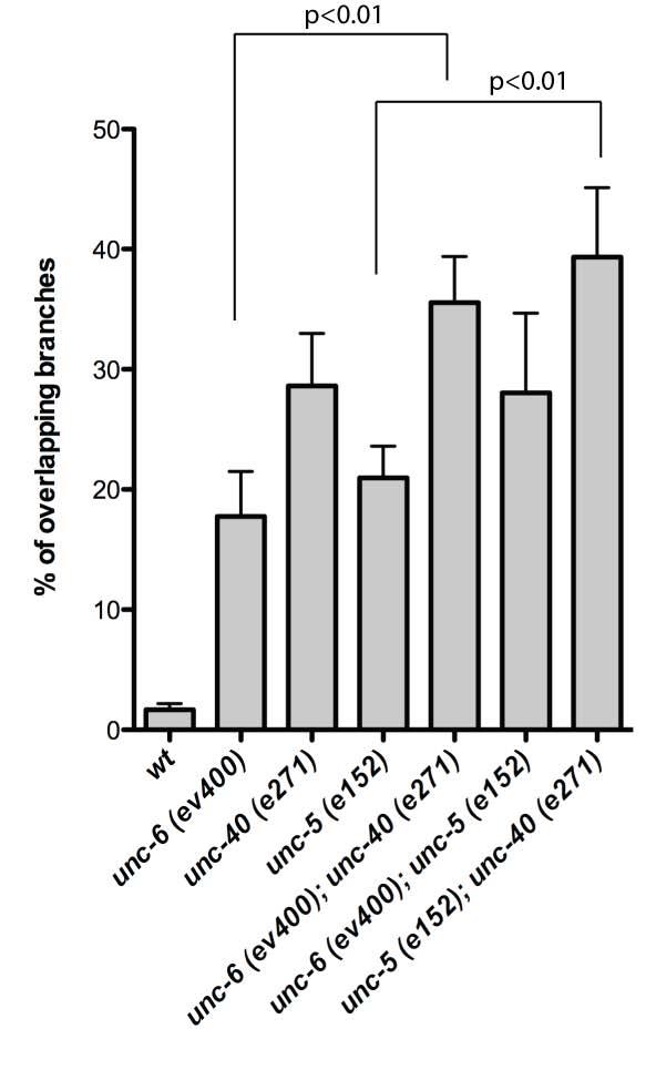
7 Supplemental Figure 3. Genetic interactions of unc-40, unc-5 and unc-6. Single mutants of unc-5 (e152), unc-40 (e271) and unc-6 (ev400) show comparable selfavoidance defects that are not statistically different from each other. The self-avoidance defect of the double mutant unc-5 (e152); unc-6 (ev400) is not significantly different from either unc-5 (e152) or unc-6 (ev400) single mutant which suggests that unc-5 and unc-6 function in a common pathway. unc-40 (e271); unc-5 (e152) double mutants do not show enhancement of the PVD self-avoidance defect vs unc-40 (e271) but do show a more severe self avoidance defect than unc-5 (e152) alone (p < 0.01, n= 20, Students t-test). unc-40 (e271); unc-6 (ev400) double mutants show enhancement of selfavoidance defects compared to unc-6 (ev400) but not to unc-40 (e271) (p=3e-3 vs unc- 6 (ev400)). These results suggest that unc-40 fulfills an additional unc-5/unc-6- independent role in self-avoidance.
8
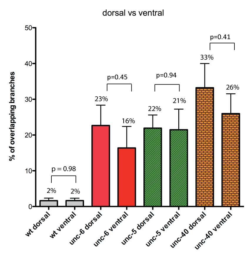
9 Supplemental Figure 4. UNC-6/Netrin signaling mutants do not show differences in dorsal vs. ventral 3 0 dendrite self-avoidance phenotypes. The fraction of overlapping 3 O branches in dorsal vs ventral regions was scored for unc-6(ev400), unc- 5(e152) and unc-40(e271). N = 20 animals
10
11 Supplemental Figure 5. UNC-6/Netrin is required for self-avoidance during the L3 larval stage. (a) Schematic of PVD development showing the elaboration of dendritic branches during larval development. (b) Experimental design for temperature shifts with the temperature sensitive mutant unc-6(rh46) to determine the temporal requirement for UNC-6 in PVD 3 0 dendritic branch self-avoidance. (c) Histogram showing fraction of overlapping 3 0 branches resulting from maintenance at either the permissive (15C) (15C control) or restrictive (25C) (25C control) temperatures and from upshift experiments (15C>25C) in which animals grown at permissive temperature are shifted to growth at the restrictive temperature. Note that the extent of overlapping 3 0 branches after shifting to restrictive temperature at the L2/L3 larval transition is not significantly different from the self-avoidance defect resulting from continuous exposure to 25 C whereas shifts to restrictive temperature at later developmental periods (i.e., L3/L4 transition, L4/adult transition) result in a significantly lower fraction of overlapping 3 0 dendritic branches that is not significantly different from the PVD self-avoidance defect from 15C control animals. These results indicate that UNC-6/Netrin function is required before the L3 larval stage for 3 0 branch self-avoidance but is not necessary in older animals.
12
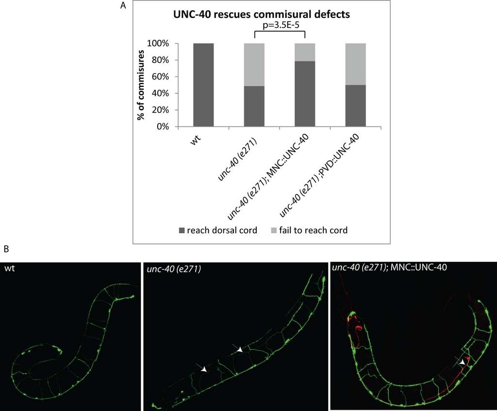
13 Supplemental Figure 6. Expression of UNC-40/DCC in ventral cord motor neurons rescues motor axon guidance defects. (a) Histogram showing that 100% of unc-25::gfp-labeled GABAergic motor neurons extend circumferential commissures (MNCs) to the dorsal cord whereas only ~45% of MNCs reach the dorsal nerve cord in unc-40 (e271) (n = 20). MNC guidance defects are largely rescued by expression of UNC-40 in ventral cord motor neurons with the unc-25 promoter (MNC::UNC-40). (b) Representative confocal images of wild type (wt), unc-40 (e271) and unc-40 (e271); MNC::UNC-40 adults. Arrows point to MNCs that fail to reach the dorsal nerve cord in unc-40(e271). Axon guidance defects are not rescued in the PDE neuron that is labeled by a co-injected marker (dat-1::mcherry) in which expression of UNC-40 is not restored (arrow in MNC::UNC-40)
14
15 Supplemental Figure 7. Expression of UNC-6::YFP in ventral motor neurons labels the ventral nerve cord but is not detected at the wild-type PVD neuron. (a,b) In a wild-type animal, YFP-labeled UNC-6 (UNC-6::YFP) is detected in the cell body of ventral cord motor neurons (double-headed arrowheads) where it is expressed (unc-6 promoter) and in the adjoining ventral nerve cord (arrow) but is not detectable in posterior lateral region in which the wild-type PVD neuron (arrowhead) and it dendritic arbor reside. (c) Schematic representation of UNC-6::YFP localization.
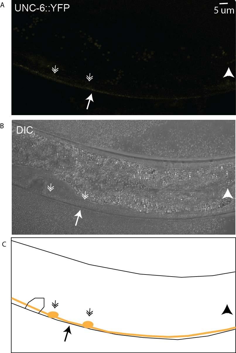
16
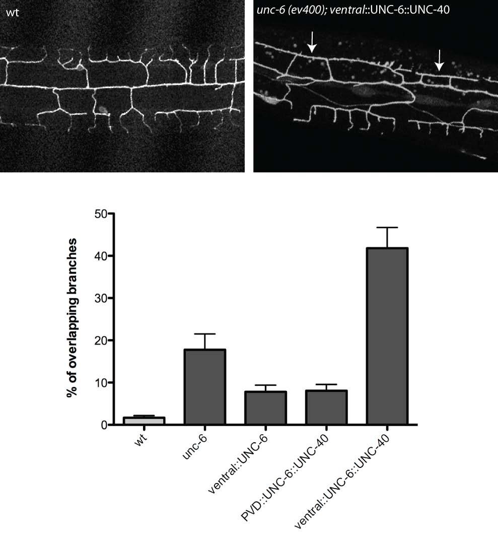
17 Supplemental figure 8. Expression of the UNC-6::UNC-40 chimeric protein in ventral neurons does not rescue the Unc-6 PVD self-avoidance defect. Expression of ventral::unc-6::unc-40 in unc-6 (ev400) does not restore self-avoidance (unc-6 vs ventral::unc-6::unc-40) whereas expression of a secreted form of UNC-6 in ventral neurons (ventral::unc-6) or membrane-tethered UNC-6 in PVD (PVD:UNC- 6::UNC-40) does rescue the Unc-6 self-avoidance defect. We note that expression of UNC-6::UNC-40 in ventral neurons enhances the PVD self avoidance defect of unc- 6(ev400); the mechanism of this effect is unclear. For histogram, genetic backgrounds are wild type (wt) (light grey box) or unc-6(ev400) (dark grey boxes).
18
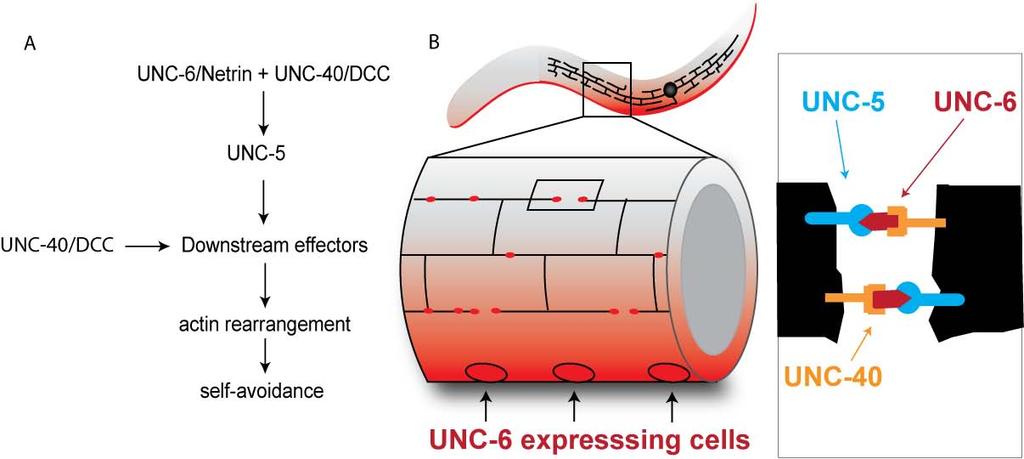
19 Supplemental figure 9. Model: UNC-40/DCC captures UNC-6/Netrin at the tips of growing dendrites to mediate UNC-5-dependent mutual repulsion. (a) UNC- 6/Netrin functions with UNC-40 and UNC-5 through downstream effectors to reorganize the actin cytoskeleton for self-avoidance. UNC-40 also signals through an UNC- 6/Netrin-independent pathway (b) Schematic showing distribution of UNC-6/Netrin expressed from ventral cells and focal UNC-6/Netrin localization to PVD dendritic branches. (c) Inset depicts the tips of adjacent sister dendrites where UNC-40/DCC captures UNC-6/Netrin for contact with UNC-5 and mutual repulsion.
20
21 Supplemental movie 1. Wildtype self-avoidance. Time-lapse confocal movie of PVD::GFP in wild-type background. 3 O dendrites contact but quickly retract (arrows). Note the intervening distance between 3 O dendrites at the end of the movie is comparable to distance visualized in mature PVD neurons. Arrows indicate location of contact-dependent self-avoidance. Supplemental movie 2. Self-avoidance defect in unc-40 (e271). Time-lapse confocal movie of PVD::GFP in unc-40 (e271). 3 O dendrites grow toward each other but upon contact fail to retract. Arrow indicates location of failed self-avoidance. Supplemental movie 3. Self-avoidance defect in unc-5 (e152). Imaging of unc-5 (e152) shows PVD dendrites fail to retract after contact. Arrow indicates location of failed self-avoidance.
Supporting Online Material for
 www.sciencemag.org/cgi/content/full/1171320/dc1 Supporting Online Material for A Frazzled/DCC-Dependent Transcriptional Switch Regulates Midline Axon Guidance Long Yang, David S. Garbe, Greg J. Bashaw*
SUPPLEMENTARY INFORMATION
 DOI: 10.1038/ncb3443 In the format provided by the authors and unedited. Supplementary Figure 1 TC and SC behaviour during ISV sprouting. (a) Predicted outcome of TC division and competitive Dll4-Notch-mediated
Nature Medicine doi: /nm.2860
 Supplemental Figure Legends Supplemental Figure 1: Hypomorphic expression of IFT88 results in olfactory signaling proteins no longer localizing to the ciliary layer. (a) ACIII localizes to the cilia and
Supplementary Table 1. List of primers used in this study
 Supplementary Table 1. List of primers used in this study Gene Forward primer Reverse primer Rat Met 5 -aggtcgcttcatgcaggt-3 5 -tccggagacacaggatgg-3 Rat Runx1 5 -cctccttgaaccactccact-3 5 -ctggatctgcctggcatc-3
Supplemental Information. Proprioceptive Opsin Functions. in Drosophila Larval Locomotion
 Neuron, Volume 98 Supplemental Information Proprioceptive Opsin Functions in Drosophila Larval Locomotion Damiano Zanini, Diego Giraldo, Ben Warren, Radoslaw Katana, Marta Andrés, Suneel Reddy, Stephanie
During Brain Development Final Destinations for Neurons and Glia Get Separated from Germinal Niches
 During Brain Development Final Destinations for Neurons and Glia Get Separated from Germinal Niches Two mayor forms of neuronal migration: Radial and Tangential Leber & Sanes, 95 How do young neurons actually
Investigating the role of EphAl ephrin-a signalling during trigeminal ganglion axon guidance
 Investigating the role of EphAl ephrin-a signalling during trigeminal ganglion axon guidance A thesis submitted for the degree of Doctor of Philosophy Molecular and Biomedical Science (Discipline of Genetics),
Supplementary Figure 1
 Supplementary Figure 1 Global TeNT expression effectively impairs synaptic transmission. Injection of 100 pg tent mrna leads to a reduction of vesicle mediated synaptic transmission in the spinal cord
Cell Migration II: CNS Cell Migration. Steven McLoon Department of Neuroscience University of Minnesota
 Cell Migration II: CNS Cell Migration Steven McLoon Department of Neuroscience University of Minnesota 1 Course News Coffee Hour Wednesday (Oct 18) 9:00-10:00am Surdyk s Café in Northrop Auditorium Stop
Zhu et al, page 1. Supplementary Figures
 Zhu et al, page 1 Supplementary Figures Supplementary Figure 1: Visual behavior and avoidance behavioral response in EPM trials. (a) Measures of visual behavior that performed the light avoidance behavior
T H E J O U R N A L O F C E L L B I O L O G Y
 Supplemental material Chen et al., http://www.jcb.org/cgi/content/full/jcb.201210119/dc1 T H E J O U R N A L O F C E L L B I O L O G Y Figure S1. Lack of fast reversibility of UVR8 dissociation. (A) HEK293T
CD3 coated cover slips indicating stimulatory contact site, F-actin polymerization and
 SUPPLEMENTAL FIGURES FIGURE S1. Detection of MCs. A, Schematic representation of T cells stimulated on anti- CD3 coated cover slips indicating stimulatory contact site, F-actin polymerization and microclusters.
SUPPLEMENTARY LEGENDS...
 TABLE OF CONTENTS SUPPLEMENTARY LEGENDS... 2 11 MOVIE S1... 2 FIGURE S1 LEGEND... 3 FIGURE S2 LEGEND... 4 FIGURE S3 LEGEND... 5 FIGURE S4 LEGEND... 6 FIGURE S5 LEGEND... 7 FIGURE S6 LEGEND... 8 FIGURE
ErbB4 migrazione I parte. 3- ErbB4- NRG1
 ErbB4 migrazione I parte 3- ErbB4- NRG1 1 In rodent brains postnatal neuronal migration is evident in three main areas: the cerebellum (CB), the hippocampus (Hipp) and the rostral migratory stream (RMS).
doi: /nature09554
 SUPPLEMENTARY INFORMATION doi:10.1038/nature09554 Supplementary Figure 1: Optical Tracing with New Photoactivatable GFP Variants Reveals Enhanced Labeling of Neuronal Processes We qualitatively compare
Supplementary Figure 1: GFAP positive nerves in patients with adenocarcinoma of
 SUPPLEMENTARY FIGURES AND MOVIE LEGENDS Supplementary Figure 1: GFAP positive nerves in patients with adenocarcinoma of the pancreas. (A) Images of nerves stained for GFAP (green), S100 (red) and DAPI
Supplementary Figure 1. Gene schematics of hyls-1, gasr-8 and k10g6.4, and TEM analysis of TFs in WT and hyls-1 cilia. (a) Gene structure of hyls-1,
 Gene structure of hyls-1,") Supplementary Figure 1. Gene schematics of hyls-1, gasr-8 and k10g6.4, and TEM analysis of TFs in WT and hyls-1 cilia. (a) Gene structure of hyls-1, gasr-8 and k10g6.4 based on WormBase (http://wormbase.org),
Supplementary Figure 1. Gene schematics of hyls-1, gasr-8 and k10g6.4, and TEM analysis of TFs in WT and hyls-1 cilia. (a) Gene structure of hyls-1, gasr-8 and k10g6.4 based on WormBase (http://wormbase.org),
Inner ear development Nervous system development
 Upcoming Sessions April 22: Nervous System Development Lecture April 24: Reviews of Axonal Pathfinding in Sensory Systems April 29: Inner Ear Development Lecture May 1: May 6: May 8: Auditory System Pathfinding
Development of the Nervous System. Leah Militello, class of 2018
 Development of the Nervous System Leah Militello, class of 2018 Learning Objectives 1. Describe the formation and fate of the neural tube and neural crest including timing and germ layer involved. 2. Describe
Structural basis for the role of inhibition in facilitating adult brain plasticity
 Structural basis for the role of inhibition in facilitating adult brain plasticity Jerry L. Chen, Walter C. Lin, Jae Won Cha, Peter T. So, Yoshiyuki Kubota & Elly Nedivi SUPPLEMENTARY FIGURES 1-6 a b M
SUPPLEMENTARY INFORMATION
 Supplementary Figure 1. Formation of the AA5x. a, Camera lucida drawing of embryo at 48 hours post fertilization (hpf, modified from Kimmel et al. Dev Dyn. 1995 203:253-310). b, Confocal microangiogram
Name: Answer Key. Question 1.
 2007 7.013 Problem Set 6 Due before 5 PM on FRIDAY, April 27, 2007. Turn answers in to the box outside of 68-120. PLEASE WRITE YOUR ANSWERS ON THIS PRINTOUT. Question 1. 1a. This is a diagram showing changes
Supplementary Materials for
 www.sciencesignaling.org/cgi/content/full/10/487/eaag2476/dc1 Supplementary Materials for Gene expression profiles of brain endothelial cells during embryonic development at bulk and single-cell levels
PHYSIOLOGICAL ADAPTATIONS FOR SURVIVAL
 PHYSIOLOGICAL ADAPTATIONS FOR SURVIVAL HOMEOSTASIS Homeostasis means staying similar or unchanging and refers to the constant internal environment or steady state of an organism. It also includes the processes
During Brain Development Final Destinations for Neurons and Glia Get Separated from Germinal Niches
 During Brain Development Final Destinations for Neurons and Glia Get Separated from Germinal Niches Progenitors are Contained within Unique Domains and Tangentially Fixed. EMBRYO ADULT Migratory Behavior
Nature Neuroscience: doi: /nn Supplementary Figure 1. MADM labeling of thalamic clones.
 Supplementary Figure 1 MADM labeling of thalamic clones. (a) Confocal images of an E12 Nestin-CreERT2;Ai9-tdTomato brain treated with TM at E10 and stained for BLBP (green), a radial glial progenitor-specific
Supplementary Figure S1 (a) (b)
 (b)") Supplementary Figure S1: IC87114 does not affect basal Ca 2+ level nor nicotineinduced Ca 2+ influx. (a) Bovine chromaffin cells were loaded with Fluo-4AM (1 μm) in buffer A containing 0.02% of pluronic
Supplementary Figure S1: IC87114 does not affect basal Ca 2+ level nor nicotineinduced Ca 2+ influx. (a) Bovine chromaffin cells were loaded with Fluo-4AM (1 μm) in buffer A containing 0.02% of pluronic
effect on the upregulation of these cell surface markers. The mean peak fluorescence intensity
 SUPPLEMENTARY FIGURE 1 Supplementary Figure 1 ASIC1 disruption or blockade does not effect in vitro and in vivo antigen-presenting cell activation. (a) Flow cytometric analysis of cell surface molecules
Nature Neuroscience: doi: /nn Supplementary Figure 1. Neuron class-specific arrangements of Khc::nod::lacZ label in dendrites.
 Supplementary Figure 1 Neuron class-specific arrangements of Khc::nod::lacZ label in dendrites. Staining with fluorescence antibodies to detect GFP (Green), β-galactosidase (magenta/white). (a, b) Class
Cell Migration II: CNS Cell Migration. Steven McLoon Department of Neuroscience University of Minnesota
 Cell Migration II: CNS Cell Migration Steven McLoon Department of Neuroscience University of Minnesota 1 Hey! The major concepts discussed relative to neural crest cell migration apply to cell migration
FGF22 signaling regulates synapse formation during postinjury remodeling of the spinal cord
 Manuscript EMBO-2014-90578 FGF22 signaling regulates synapse formation during postinjury remodeling of the spinal cord Anne Jacobi, Kristina Loy, Anja M Schmalz, Mikael Hellsten, Hisashi Umemori, Martin
Contralateral migration of oculomotor neurons is regulated by Slit/Robo signaling
 Bjorke et al. Neural Development (2016) 11:18 DOI 10.1186/s13064-016-0073-y RESEARCH ARTICLE Open Access Contralateral migration of oculomotor neurons is regulated by Slit/Robo signaling Brielle Bjorke
Concerted action of CB1 cannabinoid receptor and Deleted in Colorectal Cancer (DCC) in axon guidance
 in axon guidance") Anteneh Argaw et al. Supplemental Information and RGC axon guidance 1 Supplemental Information Concerted action of CB1 cannabinoid receptor and Deleted in Colorectal Cancer () in axon guidance Abbreviated
Anteneh Argaw et al. Supplemental Information and RGC axon guidance 1 Supplemental Information Concerted action of CB1 cannabinoid receptor and Deleted in Colorectal Cancer () in axon guidance Abbreviated
Supplementary Figure 1. EC-specific Deletion of Snail1 Does Not Affect EC Apoptosis. (a,b) Cryo-sections of WT (a) and Snail1 LOF (b) embryos at
 Cryo-sections of WT (a) and Snail1 LOF (b) embryos at") Supplementary Figure 1. EC-specific Deletion of Snail1 Does Not Affect EC Apoptosis. (a,b) Cryo-sections of WT (a) and Snail1 LOF (b) embryos at E10.5 were double-stained for TUNEL (red) and PECAM-1 (green).
Supplementary Figure 1. EC-specific Deletion of Snail1 Does Not Affect EC Apoptosis. (a,b) Cryo-sections of WT (a) and Snail1 LOF (b) embryos at E10.5 were double-stained for TUNEL (red) and PECAM-1 (green).
Proteins Regulate a Cell-Intrinsic Switch from Sonic Hedgehog-Mediated Commissural Axon Attraction to Repulsion after Midline Crossing
 Article 14-3-3 Proteins Regulate a Cell-Intrinsic Switch from Sonic Hedgehog-Mediated Commissural Axon Attraction to Repulsion after Midline Crossing Patricia T. Yam, 1,2,5,7 Christopher B. Kent, 2,5,7
SUPPLEMENTARY INFORMATION
 DOI: 10.1038/ncb2294 Figure S1 Localization and function of cell wall polysaccharides in root hair cells. (a) Spinning-disk confocal sections of seven day-old A. thaliana seedlings stained with 0.1% S4B
Early View Article: Online published version of an accepted article before publication in the final form.
 : Online published version of an accepted article before publication in the final form. Journal Name: Edorium Journal of Anatomy and Embryology Type of Article: Letter to Editors Title: The genetics in
Cells and reagents. Synaptopodin knockdown (1) and dynamin knockdown (2)
 and dynamin knockdown (2)") Supplemental Methods Cells and reagents. Synaptopodin knockdown (1) and dynamin knockdown (2) podocytes were cultured as described previously. Staurosporine, angiotensin II and actinomycin D were all obtained
Supplemental Methods Cells and reagents. Synaptopodin knockdown (1) and dynamin knockdown (2) podocytes were cultured as described previously. Staurosporine, angiotensin II and actinomycin D were all obtained
Bio 3411 Midterm Review:
 Bio 3411 Midterm Review: Structure/Development/Systems/ Plastics/Talents/Diseases/Genes Structure General Overview Wednesday October 26, 2011 1 2 THE BRAIN ATLAS 3 rd ed, p. 8! THE BRAIN ATLAS 3 rd ed,
Supplementary Figure 1. A microarray screen of organizers compared to non-organizer tissue reveals a putative organizer gene set.
 Supplementary Figure 1. A microarray screen of organizers compared to non-organizer tissue reveals a putative organizer gene set. (a, b) Venn diagrams of 31 enriched (a) and 17 depleted (b) genes significantly
fasting blood glucose [mg/dl]
![fasting blood glucose [mg/dl]](/thumbs/80/82129652.jpg "fasting blood glucose [mg/dl]") SUPPLEMENTL MTERIL Supplemental Figure I body weight [g] 5 5 5 fasting blood glucose [mg/dl] 5 5 5 C total cholesterol [mg/dl] 8 6 4 WT Has -/- Supplemental Figure I: Has-deficient mice exhibited no apparent
SUPPLEMENTL MTERIL Supplemental Figure I body weight [g] 5 5 5 fasting blood glucose [mg/dl] 5 5 5 C total cholesterol [mg/dl] 8 6 4 WT Has -/- Supplemental Figure I: Has-deficient mice exhibited no apparent
Supplementary Figure 1
 Supplementary Figure 1 Kif1a RNAi effect on basal progenitor differentiation Related to Figure 2. Representative confocal images of the VZ and SVZ of rat cortices transfected at E16 with scrambled or Kif1a
Dynamics of Compartmentalized HIV-1 Populations in the Central Nervous System Patrick R. Harrington
 Dynamics of Compartmentalized HIV-1 Populations in the Central Nervous System Patrick R. Harrington Postdoctoral Fellow Laboratory of Ronald Swanstrom Lineberger Comprehensive Cancer Center University
effects on organ development. a-f, Eye and wing discs with clones of ε j2b10 show no
 Supplementary Figure 1. Loss of function clones of 14-3-3 or 14-3-3 show no significant effects on organ development. a-f, Eye and wing discs with clones of 14-3-3ε j2b10 show no obvious defects in Elav
ErbB4 migrazione II parte
 ErbB4 migrazione II parte Control SVZ cells prefer to migrate on the NRG1 type III substrate the substrate preference of the neuroblasts migrating out of the SVZ explant was evaluated SVZ cells had a strong
A Cxcl12-Cxcr4 Chemokine Signaling Pathway Defines
 Supplemental Data A Cxcl12-Cxcr4 Chemokine Signaling Pathway Defines the Initial Trajectory of Mammalian Motor Axons Ivo Lieberam, Dritan Agalliu, Takashi Nagasawa, Johan Ericson, and Thomas M. Jessell
p75 NTR Mediates Ephrin-A Reverse Signaling Required for Axon Repulsion and Mapping
 Article p75 NTR Mediates Ephrin-A Reverse Signaling Required for Axon Repulsion and Mapping Yoo-Shick Lim, 1,3 Todd McLaughlin, 1,3 Tsung-Chang Sung, 2,3 Alicia Santiago, 1 Kuo-Fen Lee, 2 and Dennis D.M.
Nature Neuroscience: doi: /nn Supplementary Figure 1. Splenic atrophy and leucopenia caused by T3 SCI.
 Supplementary Figure 1 Splenic atrophy and leucopenia caused by T3 SCI. (a) Gross anatomy of representative spleens from control and T3 SCI mice at 28 days post-injury. (b and c) Hematoxylin and eosin
Supplementary Information
 1 Supplementary Information A role for primary cilia in glutamatergic synaptic integration of adult-orn neurons Natsuko Kumamoto 1,4,5, Yan Gu 1,4, Jia Wang 1,4, Stephen Janoschka 1,2, Ken-Ichi Takemaru
Supplemental Data. Beck et al. (2010). Plant Cell /tpc
. Plant Cell /tpc") Supplemental Figure 1. Phenotypic comparison of the rosette leaves of four-week-old mpk4 and Col-0 plants. A mpk4 vs Col-0 plants grown in soil. Note the extreme dwarfism of the mpk4 plants (white arrows)
Supplemental Figure 1. Phenotypic comparison of the rosette leaves of four-week-old mpk4 and Col-0 plants. A mpk4 vs Col-0 plants grown in soil. Note the extreme dwarfism of the mpk4 plants (white arrows)
Supplementary figure 1: LII/III GIN-cells show morphological characteristics of MC
 1 2 1 3 Supplementary figure 1: LII/III GIN-cells show morphological characteristics of MC 4 5 6 7 (a) Reconstructions of LII/III GIN-cells with somato-dendritic compartments in orange and axonal arborizations
Neurodevelopment II Structure Formation. Reading: BCP Chapter 23
 Neurodevelopment II Structure Formation Reading: BCP Chapter 23 Phases of Development Ovum + Sperm = Zygote Cell division (multiplication) Neurogenesis Induction of the neural plate Neural proliferation
Hierarchical assembly of presynaptic components in defined C. elegans synapses
 Hierarchical assembly of presynaptic components in defined C. elegans synapses Maulik R Patel 1,2, Emily K Lehrman 1, Vivian Y Poon 1,2, Justin G Crump 3, Mei Zhen 4, Cornelia I Bargmann 5 & Kang Shen
Supplementary Figure 1. Nature Neuroscience: doi: /nn.4547
 Supplementary Figure 1 Characterization of the Microfetti mouse model. (a) Gating strategy for 8-color flow analysis of peripheral Ly-6C + monocytes from Microfetti mice 5-7 days after TAM treatment. Living
Shh signaling guides spatial pathfinding of raphespinal tract axons by multidirectional repulsion
 ORIGINAL ARTICLE Cell Research (2012) 22:697-716. 2012 IBCB, SIBS, CAS All rights reserved 1001-0602/12 $ 32.00 www.nature.com/cr npg Shh signaling guides spatial pathfinding of raphespinal tract axons
Conserved properties of dentate gyrus neurogenesis across postnatal development revealed by single-cell RNA sequencing
 SUPPLEMENTARY INFORMATION Resource https://doi.org/10.1038/s41593-017-0056-2 In the format provided by the authors and unedited. Conserved properties of dentate gyrus neurogenesis across postnatal development
Lesson 33. Objectives: References: Chapter 16: Reading for Next Lesson: Chapter 16:
 Lesson 33 Lesson Outline: Nervous System Structure and Function Neuronal Tissue Supporting Cells Neurons Nerves Functional Classification of Neuronal Tissue Organization of the Nervous System Peripheral
Supplemental Information. Otic Mesenchyme Cells Regulate. Spiral Ganglion Axon Fasciculation. through a Pou3f4/EphA4 Signaling Pathway
 Neuron, Volume 73 Supplemental Information Otic Mesenchyme Cells Regulate Spiral Ganglion Axon Fasciculation through a Pou3f4/EphA4 Signaling Pathway Thomas M. Coate, Steven Raft, Xiumei Zhao, Aimee K.
SUPPLEMENTARY INFORMATION
 DOI: 10.1038/ncb2988 Supplementary Figure 1 Kif7 L130P encodes a stable protein that does not localize to cilia tips. (a) Immunoblot with KIF7 antibody in cell lysates of wild-type, Kif7 L130P and Kif7
Supplementary Figure 1.
 Supplementary Figure 1. Increased expression of cell cycle pathway genes in insulin + Glut2 low cells of STZ-induced diabetic islets. A) random blood glucose measuers of STZ and vehicle treated MIP-GFP
GFP/Iba1/GFAP. Brain. Liver. Kidney. Lung. Hoechst/Iba1/TLR9!
 Supplementary information a +KA Relative expression d! Tlr9 5!! 5! NSC Neuron Astrocyte Microglia! 5! Tlr7!!!! NSC Neuron Astrocyte! GFP/Sβ/! Iba/Hoechst Microglia e Hoechst/Iba/TLR9! GFP/Iba/GFAP f Brain
Axis Formation and Mesoderm Induction
 Developmental Biology Biology 4361 Axis Formation and Mesoderm Induction October 27, 2005 Amphibian anteroposterior specification polarized eggs animal/vegetal pigment yolk v. clear cytoplasm mitochondrial
Overview of the Nervous System (some basic concepts) Steven McLoon Department of Neuroscience University of Minnesota
 Steven McLoon Department of Neuroscience University of Minnesota") Overview of the Nervous System (some basic concepts) Steven McLoon Department of Neuroscience University of Minnesota 1 Coffee Hour Tuesday (Sept 11) 10:00-11:00am Friday (Sept 14) 8:30-9:30am Surdyk s
Overview of the Nervous System (some basic concepts) Steven McLoon Department of Neuroscience University of Minnesota 1 Coffee Hour Tuesday (Sept 11) 10:00-11:00am Friday (Sept 14) 8:30-9:30am Surdyk s
BIPN140 Lecture 13: Synapse Formation (Synaptogenesis)
") BIPN140 Lecture 13: Synapse Formation (Synaptogenesis) 1. Neuromuscular Junction (NMJ) Development 2. Synaptogenesis at Central Synapses Su (FA16) Ultrastructural Image of an NMJ Active Zone Basal Lamina
BIPN140 Lecture 13: Synapse Formation (Synaptogenesis) 1. Neuromuscular Junction (NMJ) Development 2. Synaptogenesis at Central Synapses Su (FA16) Ultrastructural Image of an NMJ Active Zone Basal Lamina
Bio 3411 Midterm Review:
 Midterm Review: Structure/Development/Systems/ Plastics/Talents/Diseases/Genes Structure General Overview Wednesday 1( 2( THE BRAIN ATLAS 3 rd ed, p. 8! THE BRAIN ATLAS 3 rd ed, p. 9! Mid-line (sagittal)
ns ns hp761(lf); daf-28(lf) daf-28(gf) Figure S1
; daf-28(lf) daf-28(gf) Figure S1") A ns ns B 100 100 80 80 60 60 40 40 20 20 0 0% 0 0% hp761(lf); daf-28(lf) daf-28(gf) Figure S1 A 100 80 15 o C 22 o C 25 o C % Dauers 60 40 20 0 0% 0% * 0% 0% 0% Class I alleles Class II alleles daf-2(lf;ts)
A ns ns B 100 100 80 80 60 60 40 40 20 20 0 0% 0 0% hp761(lf); daf-28(lf) daf-28(gf) Figure S1 A 100 80 15 o C 22 o C 25 o C % Dauers 60 40 20 0 0% 0% * 0% 0% 0% Class I alleles Class II alleles daf-2(lf;ts)
Supplementary Fig. 1 V-ATPase depletion induces unique and robust phenotype in Drosophila fat body cells.
 Supplementary Fig. 1 V-ATPase depletion induces unique and robust phenotype in Drosophila fat body cells. a. Schematic of the V-ATPase proton pump macro-complex structure. The V1 complex is composed of
Supplemental Data. Müller-Xing et al. (2014). Plant Cell /tpc
. Plant Cell /tpc") Supplemental Figure 1. Phenotypes of iclf (clf-28 swn-7 CLF pro :CLF-GR) plants. A, Late rescue of iclf plants by renewed DEX treatment; senescent inflorescence with elongated siliques (arrow; 90 DAG,
Supplemental Figure 1. Phenotypes of iclf (clf-28 swn-7 CLF pro :CLF-GR) plants. A, Late rescue of iclf plants by renewed DEX treatment; senescent inflorescence with elongated siliques (arrow; 90 DAG,
SUPPLEMENTARY FIGURE S1: nlp-22 is expressed in the RIA interneurons and is secreted. (a) An animal expressing both the RIA specific reporter
 An animal expressing both the RIA specific reporter") 1 SUPPLEMENTARY FIGURE S1: nlp-22 is expressed in the RIA interneurons and is secreted. (a) An animal expressing both the RIA specific reporter Pglr-3:mCherry (red) and Pnlp-22:gfp (green) shows co-localization
1 SUPPLEMENTARY FIGURE S1: nlp-22 is expressed in the RIA interneurons and is secreted. (a) An animal expressing both the RIA specific reporter Pglr-3:mCherry (red) and Pnlp-22:gfp (green) shows co-localization
Supplementary Information
 Supplementary Information Supplementary Figure 1: Luminal localization of CCM-3. (a) The CCM-3::GFP fusion protein localizes along the apical (luminal) surface of the pharynx (b) as well as the lumen of
A conserved neuronal DAF-16/FoxO plays an important role in conveying
 SREP-17-10421.R2 Supplementary Information (clean version) Scientific Reports A conserved neuronal DAF-16/FoxO plays an important role in conveying pheromone signals to elicit repulsion behavior in Caenorhabditis
Principles of Anatomy and Physiology
 Principles of Anatomy and Physiology 14 th Edition CHAPTER 12 Nervous Tissue Introduction The purpose of the chapter is to: 1. Understand how the nervous system helps to keep controlled conditions within
SUPPLEMENTARY FIGURE LEGENDS
 SUPPLEMENTARY FIGURE LEGENDS Supplemental FIG. 1. Localization of myosin Vb in cultured neurons varies with maturation stage. A and B, localization of myosin Vb in cultured hippocampal neurons. A, in DIV
glial cells missing and gcm2 Cell-autonomously Regulate Both Glial and Neuronal
 glial cells missing and gcm2 Cell-autonomously Regulate Both Glial and Neuronal Development in the Visual System of Drosophila Carole Chotard, Wendy Leung and Iris Salecker Supplemental Data Supplemental
Fig. S1. RT-PCR analyses of the expression and distribution of Xdscr6 transcripts during early development.
 Fig. S1. RT-PCR analyses of the expression and distribution of Xdscr6 transcripts during early development. (A) Temporal expression of Xdscr6 at various stages (numbers on the top) and its distribution
Name Group. 8. Growth 9. Insulin 10. Iodine 11. Manufactures 12. Nervous system 13. Neuron 14. Neurotransmitter
 THE NERVOUS AND ENDOCRINE SYSTEM Name Group 0. Translate all these words 1. Adrenaline 2. Brain 3. Cerebellum 4. Cerebrum 5. Dendrite 6. Dwarfism 7. Goiter 8. Growth 9. Insulin 10. Iodine 11. Manufactures
GAP-FREE NEURAL CIRCUITS CLASS #3: C. elegans touch-induced locomotion
 GAP-FREE NEURAL CIRCUITS CLASS #3: C. elegans touch-induced locomotion OUTLINE: C. elegans background o General background o Neural signaling: no action potentials Excitatory circuits: Touch-induced locomotion
Vesicular Trafficking of Semaphorin 3A is Activity- Dependent and Differs Between Axons and Dendrites
 Traffic 6; 7: 6 77 Blackwell Munksgaard Copyright # Blackwell Munksgaard 6 doi:./j.6-854.6.44.x Vesicular Trafficking of Semaphorin A is Activity- Dependent and Differs Between Axons and Dendrites Joris
Project report October 2012 March 2013 CRF fellow: Principal Investigator: Project title:
 Project report October 2012 March 2013 CRF fellow: Gennaro Napolitano Principal Investigator: Sergio Daniel Catz Project title: Small molecule regulators of vesicular trafficking to enhance lysosomal exocytosis
PHENOTYPIC DYNAMICS OF MICROGLIAL AND MONOCYTE-DERIVED CELLS IN GLIOBLASTOMA-BEARING MICE.
 SUPPLEMENTARY FIGURES, TABLES AND VIDEOS PHENOTYPIC DYNAMICS OF MICROGLIAL AND MONOCYTE-DERIVED CELLS IN GLIOBLASTOMA-BEARING MICE. Clément Ricard 1,2,3,4, Aurélie Tchoghandjian 2,4, Hervé Luche 5, Pierre
In Vivo Imaging of Virological Synapses
 In Vivo Imaging of Virological Synapses Xaver Sewald 1, David G. Gonzalez 2, Ann M. Haberman 2, and Walther Mothes 1 * 1 Department of Microbial Pathogenesis, Yale University School of Medicine, New Haven,
Embryonic MGE Cells as a Treatment for Epilepsy December 1, 2012
 Embryonic MGE Cells as a Treatment for Epilepsy December 1, 2012 Scott C. Baraban, PhD University of California, San Francisco American Epilepsy Society Annual Meeting Disclosure Name of Commercial Interest
Outline. Neuron Structure. Week 4 - Nervous System. The Nervous System: Neurons and Synapses
 Outline Week 4 - The Nervous System: Neurons and Synapses Neurons Neuron structures Types of neurons Electrical activity of neurons Depolarization, repolarization, hyperpolarization Synapses Release of
Supplementary Figures
 Supplementary Figures Supplementary Figure 1. nrg1 bns101/bns101 embryos develop a functional heart and survive to adulthood (a-b) Cartoon of Talen-induced nrg1 mutation with a 14-base-pair deletion in
Nature Neuroscience: doi: /nn Supplementary Figure 1. Large-scale calcium imaging in vivo.
 Supplementary Figure 1 Large-scale calcium imaging in vivo. (a) Schematic illustration of the in vivo camera imaging set-up for large-scale calcium imaging. (b) High-magnification two-photon image from
SUPPLEMENTARY FIG. S2. Representative counting fields used in quantification of the in vitro neural differentiation of pattern of dnscs.
 Supplementary Data SUPPLEMENTARY FIG. S1. Representative counting fields used in quantification of the in vitro neural differentiation of pattern of anpcs. A panel of lineage-specific markers were used
Capu and Spire Assemble a Cytoplasmic Actin Mesh
 Developmental Cell 13 Supplemental Data Capu and Spire Assemble a Cytoplasmic Actin Mesh that Maintains Microtubule Organization in the Drosophila Oocyte Katja Dahlgaard, Alexandre A.S.F. Raposo, Teresa
Cellular Neurobiology BIPN140
 Cellular Neurobiology BIPN140 1st Midterm Exam Ready for Pickup By the elevator on the 3 rd Floor of Pacific Hall (waiver) Exam Depot Window at the north entrance to Pacific Hall (no waiver) Mon-Fri, 10:00
Supplemental Materials Molecular Biology of the Cell
 Supplemental Materials Molecular Biology of the Cell Garcia-Alvarez et al. Supplementary Figure Legends Figure S1.Expression and RNAi-mediated silencing of STIM1 in hippocampal neurons (DIV, days in vitro).
Nature Neuroscience doi: /nn Supplementary Figure 1. Characterization of viral injections.
 Supplementary Figure 1 Characterization of viral injections. (a) Dorsal view of a mouse brain (dashed white outline) after receiving a large, unilateral thalamic injection (~100 nl); demonstrating that
Systems Neuroscience November 21, 2017 The autonomic nervous system
 Systems Neuroscience November 21, 2017 The autonomic nervous system Daniel C. Kiper kiper@ini.phys.ethz.ch http: www.ini.unizh.ch/~kiper/system_neurosci.html How is the organization of the autonomic nervous
Supplemental information contains 7 movies and 4 supplemental Figures
 1 2 3 4 5 6 7 8 9 10 11 12 13 14 15 16 17 18 19 20 21 22 23 24 25 26 27 Supplemental information contains 7 movies and 4 supplemental Figures Movies: Movie 1. Single virus tracking of A4-mCherry-WR MV
Supplementary Information A Hydrophobic Barrier Deep Within the Inner Pore of the TWIK-1 K2P Potassium Channel Aryal et al.
 Supplementary Information A Hydrophobic Barrier Deep Within the Inner Pore of the TWIK-1 K2P Potassium Channel Aryal et al. Supplementary Figure 1 TWIK-1 stability during MD simulations in a phospholipid
Supplemental Figure 1. Intracranial transduction of a modified ptomo lentiviral vector in the mouse
 Supplemental figure legends Supplemental Figure 1. Intracranial transduction of a modified ptomo lentiviral vector in the mouse hippocampus targets GFAP-positive but not NeuN-positive cells. (A) Stereotaxic
Nature Neuroscience: doi: /nn Supplementary Figure 1
 Supplementary Figure 1 Subcellular segregation of VGluT2-IR and TH-IR within the same VGluT2-TH axon (wild type rats). (a-e) Serial sections of a dual VGluT2-TH labeled axon. This axon (blue outline) has
ANSC/FSTC 607 Biochemistry and Physiology of Muscle as a Food INNERVATION AND DIFFERENTIATION OF MUSCLE
 ANSC/FSTC 607 Biochemistry and Physiology of Muscle as a Food INNERVATION AND DIFFERENTIATION OF MUSCLE I. Organization of the motor neuron and myofibers A. Motoneuron bifurcates into many branches (terminal
Regionalization of the nervous system. Paul Garrity 7.68J/9.013J February 25, 2004
 Regionalization of the nervous system Paul Garrity 7.68J/9.013J February 25, 2004 Patterning along: Rostral/Caudal (AP) axis Dorsal/Ventral (DV) axis Start with DV axial patterning in Spinal Cord Dorsal/Ventral
Polarity and Segmentation. Chapter Two
 Polarity and Segmentation Chapter Two Polarization Entire body plan is polarized One end is different than the other Head vs. Tail Anterior vs. Posterior Front vs. Back Ventral vs. Dorsal Majority of neural
SUPPLEMENTARY INFORMATION
 DOI: 1.138/ncb222 / b. WB anti- WB anti- ulin Mitotic index (%) 14 1 6 2 T (h) 32 48-1 1 2 3 4 6-1 4 16 22 28 3 33 e. 6 4 2 Time (min) 1-6- 11-1 > 1 % cells Figure S1 depletion leads to mitotic defects
Supplemental Data. Wang et al. (2013). Plant Cell /tpc
. Plant Cell /tpc") Supplemental Data. Wang et al. (2013). Plant Cell 10.1105/tpc.112.108993 Supplemental Figure 1. 3-MA Treatment Reduces the Growth of Seedlings. Two-week-old Nicotiana benthamiana seedlings germinated on
Supplemental Data. Wang et al. (2013). Plant Cell 10.1105/tpc.112.108993 Supplemental Figure 1. 3-MA Treatment Reduces the Growth of Seedlings. Two-week-old Nicotiana benthamiana seedlings germinated on