Figure S1: CtxA interacts with Notch and the endogenous Drosophila camp machinery. Figure S1, related to Figure 1.
|
|
|
- Sydney Higgins
- 5 years ago
- Views:
Transcription
1 Cell Host & Microbe, Volume 14 Supplemental Information Cholera Toxin Disrupts Barrier Function by Inhibiting Exocyst-Mediated Trafficking of Host Proteins to Intestinal Cell Junctions Annabel Guichard, Beatriz Cruz-Moreno, Berenice Aguilar, Nina M. van Sorge, Jennifer Kuang, Adrianne A. Kurkciyan, Zhipeng Wang, Saiyu Hang, Guillaume P. Pineton de Chambrun, Declan F. McCole, Paula Watnick, Victor Nizet, and Ethan Bier Inventory of Supplemental Information: Figure S1: CtxA interacts with Notch and the endogenous Drosophila camp machinery. Figure S1, related to Figure 1. Figure S2: The effects of CtxA in Drosophila are mediated by Rab11, PKA, and Epac. Figure S2, related to Figure 2. Figure S3: Additional effects of CT on the actin cytoskeleton. Figure S3, related to Figure 3. Figure S4: Additional effects of CT on tight junctions. Figure S4, related to Figure 4. Figure S5: Additional effects of CT on localization of Notch components and its mediation by both the PKA and Epac camp effectors. Figure S5, related to Figure 5. Figure S6: Sec15 staining is reduced in crypts with high CtxB levels. Figure S6, related to Figure 6. Figure S7: Additional effects of CtxA in the Drosophila gut. Figure S7, related to Figure 7.


2 Supplemental Figure 1 A B C N - /+ prdg4>ctxa! WT N - /+ D E F CtxA! N - /+; CtxA! L2G4>Dl! N/+! N/+; wingg4>ctxa! L2G4>Dl+CtxA!


3 Supplemental Figure 1, continued G H I WT J wingg4>ctxa wingg4>gsα60a wingg4>ctxa+gsα60a K L wingg4>gsα49b wingg4>ctxa+gsα49b M N O wingg4>gsα49b i P wingg4>ctxa+gsα49b i Q wingg4>gsα60a i R wingg4>ctxa+gsα60a i S wingg4>cta i T wingg4>ctxa+cta i wingg4>ac i wingg4>ctxa+ac i
4 Figure S1: CtxA interacts with Notch and the endogenous Drosophila camp machinery. A-F) CtxA interacts with Notch mutations. A) A wing from a Notch-/+ (N-/+) heterozygous fly with thickened veins (enlarged in the second frame from the left in panel C). B) A prd- GAL4>CtxA wing with margin notches (arrow) similar to those observed in N-/+ heterozygotes (e.g., panel A). C) Enlargements of a central wing region (boxed in panel A) showing the width of the L3 and L5 veins (marked with arrowheads) in wild-type (left panel), modestly thickened veins in N-/+ heterozygotes (second panel) and in wing-gal4>ctxa fly (third panel), and yet broader veins in a Notch heterozygote expressing CtxA (N-/+; wing-gal4>ctxa) (right panel). D) A wing from a fly carrying one copy of an activated Notch allele (N/+). E) Activated N almost fully suppresses the wing-gal4>ctxa phenotype (compare to panel B). F) Ectopic expression of Delta (Dl) in the L2 vein primordium using the L2-GAL4 driver results in truncation of the L2 vein (top panel, arrow), an effect that can be nearly reversed by coexpression of CtxA (bottom panel, arrow). G-T) The effects of CtxA are mediated by the endogenous Drosophila camp machinery. Co-expression of CtxA with the Gs subunits Gs 49B or Gs 60A results in a greatly enhanced CtxA-like phenotype while co-expression with RNAi constructs that knock-down levels of Gs 49B (Gs 49B i ), Gs 60A (Gs 60A i ), concertina (cta i ), or the adenylate cyclase (AC i ) encoded by the rutabaga gene suppress the effect of CtxA. Panels on the left are controls showing the effect of wild-type or RNAi expression constructs alone, which either have weak or no obvious effects, while those on the right show the effects of these various constructs in combination with CtxA. Supplemental Figure 1, related to Figure 1.


5 Sec15-GFP Delta DECad E C A wingg4>ctxa wingg4>ctxa wingg4>ctxa wingg4/+ H F D B wingg4>ctxa+rab11 wingg4>ctxa+rab11 wingg4>ctxa+rab11 wingg4>rab11 Supplemental Figure 2 G


6 Delta Sec15-GFP Rab11 Supplemental Figure 2, continued I wingg4>ctxa J wingg4>rap1 K wingg4>pka WT wingg4>rap1 wingg4>pka L M N O P Q R S T
7 Figure S2: The effects of CtxA in Drosophila are mediated by Rab11, PKA, and Epac. A-H) Rab11 over-expression rescues the effects of CtxA in Drosophila wing discs. Rab11was over-expressed in the wing disc using the wing-gal4 driver, which is expressed more strongly in dorsal versus ventral cells. Inserts in each panel are Z-sections (brackets indicate apical cell surface). Quantification of vesicle number and total Sec15-GFP- fluorescence (= total vesicle surface area) was measured in comparably located areas on the dorsal surface measuring 42.5 m X 27.5 m = 1174 m 2 = A). A) Expression of the UAS-Sec15-GFP construct alone reveals many large round vesicles (aver = 443 ± 60 vesicles per unit area = A) near the cell surface as well as a distribution of smaller vesicle sizes (error = St Dev.). B) Coexpression of Rab11 with Sec15 GFP results in a modest decrease in the number of Sec15 vesicles (aver. = 316 ± 32 vesicles) as in (A) but nearly all are uniformly large resulting in virtually equal total vesicle surface areas (aver = 53,225 ± 9676 pixels within vesicles delimited by the Image-J program in a unit area A = P/A) for Sec15-GFP alone compared to Sec15 + Rab11 (aver = ± 9092 P/A). This small difference is not statistically significant (p= 0.49). C) Expression of CtxA greatly reduces the number of Sec15-GFP vesicles (aver = 202 ± 51; p< compared to Sec15-GFP alone). The remaining vesicles are typically smaller resulting in a 71% reduction in total vesicle surface area (aver = 15,407 ± 4144 P/A) as compared to Sec15-GFP expression alone (p < ). The remaining Sec15-GFP vesicles are also frequently abnormally shaped and accumulate abnormally in basal locations (insert). For these quantitative measurements, all compared images were acquired on an SP2 Leica confocal microscope with the same settings: Objective 40X, optical zoom 6.3X, gain for cannel PMT1=468 (excitation 488). SP Mirror 1 (left)496, SP Mirror 1 (right) 556. Beam Exp 6. Logical Size:1024X1024. Scan Speed 400. Frame-Average 1, Line-Average 4. D) Co-expression of Rab11, CtxA, and Sec15-GFP rescues the effect of CtxA increasing the number of vesicles (331 ± 77, p< compared to Sec15+CtxA) and restoring total vesicle surface area (53,997± 11,185 P/A, p< compared to Sec15-GFP+CtxA) to nearly exactly that observed in discs expressing Sec15-GFP alone (compare with A). E,F) Reduction of Delta expression by CtxA on the dorsal surface of the wing disc (E, bracket) is rescued by coexpression with Rab11 (F, bracket). G,H) Reduction of DECad expression by CtxA (G) is also rescued by co-expression with Rab11 (H). These high magnification images are centered on regions from the dorsal surface of the wing disc. I-T) camp acts via both the PKA and Epac/Rap1 effectors in Drosophila. I) CtxA expressing wing. J) Expression of an activated form of the Epac target and effector Rap1 (Rap1) generates a wing phenotype similar of that
8 of CtxA. K) Expression of an activated form of PKA (PKA) generates a wing phenotype similar of that of CtxA. L-T) Expression of Rap1 (M,P,S) or PKA (N,Q,T) results in down regulation of Rab11 (L-N), Sec15-GFP (O-Q), and Delta (R-T) as observed for CtxA (see Fig. 2J-O). The wingg4 driver was used for all panels. Brackets in S,T denote dorsal surface of the wing disc where expression of wingg4 is strongest. Supplemental Figure 2, related to Figure 2.


9 Supplemental Figure 3 Untreated + CT B C D E F G H I J F-actin! A
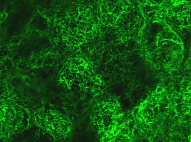
10 Figure S3: Additional effects of CT on the actin cytoskeleton. Recently confluent monolayers of CACO-2 cells, untreated or treated with CT (3X over 28 hours) were fixed in paraformaldehyde and stained with phalloidin for F-actin expression. A,B) Apical-most view of actin cap in untreated (A) and CT-treated (B) CACO-2 cells. Treated cells tend to have larger spaces between neighbors (paired arrowheads). C,D) Junctional (TJ/AJ) level view of untreated (C) and CT-treated (D) cells as shown in Fig. 4C,D. CT-treated cells have broader and less regular zones of junctional staining, with regions of high-level actin accumulation. E,F) Basal region of untreated (E) have a dense network of radial actin stress fibers that are virtually eliminated in CT-treated cells (F). G-J) In untreated cells (G,I), round cup-like structures comprised of 8-12 cells can be observed scattered throughout the monolayer. Cells at the edge of these cups are typically elongated and have an elevated inner lip labeling intensely for F-actin. These rim cells flatten abruptly into very thin pseudopodiallike processes that meet at the center of the cup (I). In CT-treated monolayers (H,J) these cup-like structures assume irregular shapes and the actin ring around the inner rim of the cup is lost. Whether these cup-like structures bear any relationship to crypts or other morphological specializations of the intact intestine is an interesting question to address in future experiments. Supplemental Figure 3, related to Figure 3.


11 Supplemental Figure 4 ZO-1 + β-tubulin A Untreated B + CT F-actin + ZO-1 Z-sect. C E G D F H


12 Supplemental Figure 4, continued I Untreated J + CT Z-sect. Occludin + ZO-1 K L M N ZO-1 + Rab11 sub-apical apical O P Z-sect. Q R
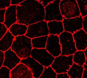
13 Figure S4: Additional effects of CT on tight junctions. A-D) CT treatment does not alter the distribution of tubulin. Recently confluent monolayers of CACO-2 cells, untreated or treated with CT (3X over 28 hours) were fixed in paraformaldehyde and double stained for -tubulin (green) and ZO-1 (red). A,B) Apical focal plane showing tubulin stain only. C,D) Z-section double stained for -tubulin (green) and ZO-1 (red). Brackets indicate apical zone of cells, and arrows in D indicate ectopic basal accumulation of ZO-1. Tubulin is similarly organized into an apical meshwork of fibers in both untreated and CT-treated cells. E-H) CT alters ZO-1 staining in fully confluent CACO-2 cells. Fully confluent monolayers of CACO-2 cells ( 3 weeks), untreated or treated with CT (3X over 28 h) as indicated, were fixed in paraformaldehyde and double stained for ZO-1 (red) and F-actin (green). Images represent mini-stack maximal projections spanning the full TJ and extending into the AJ level. E,G) Untreated cells have well developed junctions. F,H) CT-treated cells had occasional gaps in apical ZO-1 staining (F, arrows) and higher levels of cytoplasmic staining with abnormal accumulations (F,H, asterisks), and displayed a mis-alignment of apical-most and more basal staining contours (F,H, paired arrowheads), which were similar to albeit less pronounced than defects observed in newly confluent CT-treated cultures (Fig 4). I-L) CT alters occludin staining. Recently confluent monolayers of CACO-2 cells ( 1 week), untreated or treated with CT (3X over 28 hours) were fixed in methanol and stained for occludin (red) and ZO-1 (green) as indicated. Junctional expression of occludin and ZO-1 were coincidental in both untreated (I) and CT-treated (J) cells. In CT-treated cells both of these TJ proteins followed less regular contours than in untreated cells. In addition, cytoplasmic accumulations of occludin were observed in CT-treated cells (J,L, asterisks). Similar cytoplasmic accumulation of ZO-1 was observed in CT-treated cells fixed in paraformaldehyde (Fig. 4G,H), which apparently is not well preserved during methanol fixation. K,L) Z-sections of occludin staining. The apical-basal distribution of occludin, which is strongest at the TJ (K) is not appreciably altered by CT treatment (L). Brackets indicate the apical TJ level of the cell. M-R) Ctx alters Rab11 and ZO-1 co-localization in T84 cells. Recently confluent monolayers of T84 cells, untreated or treated with CT (3X over 28 hours), were fixed in paraformaldehyde and double stained for ZO-1 and Rab11. M,N) Apical TJ level view of untreated (M) and CT-treated (N) cells. O,P) Mid-level view of untreated cells in which a subset of large Rab11 positive vesicles co-label with ZO-1 (O, arrows) and CT-treated cells (P) in which Rab11/ZO-1 co-localization is abolished. Q,R) Z-section of untreated (Q) and CT-
14 treated cells (R) showing a large basal accumulation of ZO-1 (arrows in R). Supplemental Figure 4, related to Figure 4.


15 N3 Jg2 DLL4 A C E Supplemental Figure 5 Untreated + CT B D F


16 ZO-1 Supplemental Figure 5, continued Untreated + CT G H I CT+H89 CT+ESI-09 J 6Bnz 8Cpt K L
17 Figure S5: Additional effects of CT on localization of Notch components and its mediation by both the PKA and Epac camp effectors. A-F) CT disrupts localization of Notch components in CACO-2 cups. Recently confluent monolayers of CACO-2 cells, untreated or treated with CT (3X over 28 h) were fixed in paraformaldehyde and stained for expression of the Notch pathway components: A,B) Delta-like-4 (DLL4); C,D) Jagged-2 (Jg2); or E,F) Notch-3 (N3) in cup-like structures such as those shown in Supp. Fig. 4. The rims of the cups label intensely for DLL4, Jg2, and N3 (A,C,E) and also for F-actin (Supp. Fig 4G). In CT-treated cultures, strong expression of Notch components (B,D,F) and F-actin (Supp. Fig. 4H) is lost along the rims of the cups, which also loose their symmetric circular shape. G-L) CT disrupts junctions via both PKA and Epac in CACO-2 cells. G) ZO-1 staining at TJs in untreated CACO-2 cells outlines cells with regular straight borders between cells. H) In CT-treated cells (3X over 28 hours) the ZO-1 pattern becomes irregular and corrugated. The junctional effects of CT are greatly reduced by co-treating cells with the PKA inhibitor H89 (I) or the Epac inhibitor ESI-09 (J). Also, treatment of CACO-2 cells with camp analogs that specifically activate the PKA (6Bnz, K) or Epac (8Cpt, L) effectors cause CT-like defects. Similar alterations of Ecad staining were also observed in these cells (not shown). Supplemental Figure 5, related to Figure 5.


18 CtxB C Sec15 A Supplemental Figure 6 PBS + CT B D
19 Figure S6: Sec15 staining is reduced in crypts with high CtxB levels. Double labeling for Sec15 and the CtxB subunit in sections of PBS versus CT-treated ileal loops. A,C) In PBS treated loops nearly all crypts stain brightly for Sec15 (A, green). There is only low-level background signal in the CtxB channel (red). B,D) While nearly all villus cells in CT-treated loops stain strongly for CtxB (not shown), only a subset of crypts, which may be less accessible to the toxin, accumulate high levels of the label (D, red), and in such crypts, staining for Sec15 is most strongly reduced (B, green). Supplemental Figure 6, related to Figure 6.


20 α-cat + GFP A B NP1>Pyd-GFP D sc E ent sc F G ent sc sc J I NP1>Pyd+CtxA H GFP C Supplemental Figure 7 NP1>Pyd mg hg NP1/+ folds mv NP1>CtxA NP1>CtxA+Rab11 ]


21 K N bm N mv Np1/+ N zb ms L mv O >CtxA O O >CtxA bo >CtxA+Rab11 P Np1/+ O P AJ O N P bo bm O bm bm N N Supplemental Figure 7, continued mv >CtxA+Rab11 M P P P bo
22 Figure S7: Additional effects of CtxA in the Drosophila gut. A) Lethality caused by adult-specific expression of CtxA in the midgut can be rescued by coexpression with Rab11. Adult-specific NP1-GAL4-driven expression of UAS-transgenes was induced in flies carrying a ubiquitously expressed temperature sensitive GAL80 transgene (tub-gal80ts), which blocks the transactivating activity of GAL4 at permissive temperatures (<25 C), by raising the temperature to 31 C (non-permissive for GAL80ts) for the indicated time period. Once flies are shifted to the non-permissive temperature, high-level expression of NP1-GAL4-dependent UAS-genes is initiated in the midgut. Twenty flies of each genotype were placed in fresh vials at the start of the experiment, which were all done in triplicate and averaged (error bars = St. Dev.). Adult-specific expression of CtxA alone leads to rapid lethality (purple line) that can be reversed by co-expression with Rab11 (red line) (p<0.035 for the comparison of NP1/+; tubgal80ts versus NP1>CtxA; tubgal80ts and p< for the comparison NP1>CtxA; tubgal80ts versus NP1>CtxA+Rab11; tubgal80ts). B) CtxA acts via host camp machinery in the Drosophila gut. Weight-loss in flies caused by midgut-specific expression of CtxA ( = p< for comparison of NP1/+ versus NP1>CtxA) can be reduced by feeding flies the SK-channel blocker clotrimazole (Clot) ( = p< for comparison of NP1>Ctx versus NP1>Ctx + Clot), as has been previously observed in the context of ctxa-dependent infection with V.c. (Blow et al., 2005). Error bars = St. Dev. Likewise, co-expression of a Gs 60A RNAi CtxA construct reverses weight loss caused by CtxA ( = p< for comparison of NP1>Ctx versus NP1>CtxA+Gs 60A-RNAi). C-G) CtxA reduces levels of additional Drosophila junctional proteins in the gut. Junctional expression of a GFP-tagged form of Polycheatoid (Pyd), the Drosophila ortholog of ZO-1, and alpha-catenin ( -cat). C) Low magnification view of horizontal section of NP1>Pyd-GFP gut showing expression throughout the midgut: mg = midgut, hg = hindgut. D-G) High magnification surface view of a posterior region of midgut showing junctional expression of Pyd-GFP (green) in all mature enterocytes (ent) and -cat (red) in all cells of the midgut including smaller stem cells (sc) (note that NP1>Pyd does not label stem cells). Overlay of Pyd-GFP and -cat staining is shown in D,E and the -cat channel alone is shown in F,G. D,F) Pyd-GFP and -cat staining label the straight borders between wild-type enterocytes. E,G) CtxA expression in the midgut (NP1> Pyd-GFP+CtxA) results in a discontinuous (asterisk) and jagged (arrowheads) pattern of Pyd-GFP staining, which is similar to that of ZO- 1 expression in CT-treated CACO-2 cells (e.g., Fig. 4H,L compared to Fig. 4G,K). CtxA also
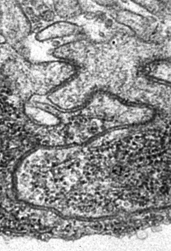
23 nearly eliminates junctional -cat staining in mature enterocytes, although ectopic vesicular cytoplasmic staining increases. Note that -cat staining in stem cells is much less reduced than in mature enterocytes consistent with the expression pattern of the NP1-GAL4 driver being restricted to mature enterocytes (the junctional staining, however, is lost in stem cells, which is presumably a consequence of the junctional defects in the neighboring enterocytes.) H-J) Light level micrographs of sections of Drosophila intestine analyzed by EM (in panels K-P below). Toluidine Blue stained thick sections of Drosophila midgut from NP1/+ (H), NP1>CtxA (I), or NP1>CtxA+Rab11-WT (J) flies that were processed for ultrastructural analysis (see below). CtxA causes thinning and flattening (loss of folds) of the intestinal epithelium, reduction in the height of microvilli, and the accumulation of large vesicular structures, which form both intracellular vesicles and greatly enlarged extracellular spaces (I). All of these CtxA phenotypes are significantly rescued by co-expression with Rab11 (J). Bars = thickness of the intestinal epithelium; brackets = thickness of microvilli (mv); arrows = folds in the intestinal epithelium; asterisk = large vacuolar structures present in significant numbers only in NP1>CtxA guts. K-P) Ultrastructural effects of CtxA in the Drosophila gut. K-M: Low magnification views of the Drosophila gut in NP1/+ (K), NP1>CtxA (L), or NP1>CtxA+Rab11- WT (M) flies spanning the apical-basal extent of the intestinal epithelium. Boxed areas = regions shown at higher magnification in panels N-N, O-O, P-P ; asterisk = large vacuolated intercellular space; mv = microvilli; bs = basal membrane; ms = circumferential muscle fiber; zb = Z-band. N-N ) High magnification views of the NP1/+ gut shown in panel K. N) Apical junctions are typically straight or bent with a single angle and closely apposed (i.e., with no large gaps between cells). AJ = adherens junction. N ) A mid-level view of closely aligned plasma membranes from adjacent cells that shows a series of small varicose gaps (arrows indicate points of membrane separation), which tend to be aligned as in a chain of small mountain lakes (see low magnification view in panel K). N ) A basal view of adjacent intestinal epithelial cells showing the basal opening (bo) between cells at the interface with the extracellular matrix comprising the basal membrane (bm). O-O ) High magnification view of the NP1>CtxA gut shown in panel L. O) Apical junction is convoluted and S-shaped and has a gap between cells (arrow: also see Fig. 7P) that is not typical of this region of the cell in NP1/+ controls. Note the cell junctions skirt a large vacuolated area (asterisk - see also panel L). O ) Paired cell membranes run along the edge of a large vacuolated area (see panel L) and then open into it (arrow). The vacuolated area is continuous with the extracellular space at the basal surface of the cells (see panels L and O ). O ) The basal connection between a
24 vacuolated varicosity and the basal membrane (arrow indicates final narrow open passage or basal opening = bo). P-P ) High magnification view of the NP1>CtxA+Rab11-WT gut shown in panel M. P) Apical junctions showing closely apposed membranes. P ) A typical example of small intercellular varicosities, which are also observed, albeit less frequently, in NP1/+ control guts. Arrows indicate border where paired cell membranes open into varicosity. P ) The basal connection between a series of narrow vacuolated extracellular spaces and the basal membrane (arrow indicates final basal opening = bo). Supplemental Figure 7, related to Figure 7.
Supplementary Figure S1: TIPF reporter validation in the wing disc.
 Supplementary Figure S1: TIPF reporter validation in the wing disc. a,b, Test of put RNAi. a, In wildtype discs the Dpp target gene Sal (red) is expressed in a broad stripe in the centre of the ventral
Supplemental Figure 1. Quantification of proliferation in thyroid of WT, Ctns -/- and grafted
 Supplemental Figure 1. Quantification of proliferation in thyroid of WT, Ctns -/- and grafted Ctns -/- mice. Cells immunolabeled for the proliferation marker (Ki-67) were counted in sections (n=3 WT, n=4
Supplementary Figure 1: Signaling centers contain few proliferating cells, express p21, and
 Supplementary Figure 1: Signaling centers contain few proliferating cells, express p21, and exclude YAP from the nucleus. (a) Schematic diagram of an E10.5 mouse embryo. (b,c) Sections at B and C in (a)
effects on organ development. a-f, Eye and wing discs with clones of ε j2b10 show no
 Supplementary Figure 1. Loss of function clones of 14-3-3 or 14-3-3 show no significant effects on organ development. a-f, Eye and wing discs with clones of 14-3-3ε j2b10 show no obvious defects in Elav
SUPPLEMENTARY INFORMATION
 b 350 300 250 200 150 100 50 0 E0 E10 E50 E0 E10 E50 E0 E10 E50 E0 E10 E50 Number of organoids per well 350 300 250 200 150 100 50 0 R0 R50 R100 R500 1st 2nd 3rd Noggin 100 ng/ml Noggin 10 ng/ml Noggin
Supplementary Figures
 J. Cell Sci. 128: doi:10.1242/jcs.173807: Supplementary Material Supplementary Figures Fig. S1 Fig. S1. Description and/or validation of reagents used. All panels show Drosophila tissues oriented with
Supplementary Figure 1 Madm is not required in GSCs and hub cells. (a,b) Act-Gal4-UAS-GFP (a), Act-Gal4-UAS- GFP.nls (b,c) is ubiquitously expressed
 Act-Gal4-UAS-GFP (a), Act-Gal4-UAS- GFP.nls (b,c) is ubiquitously expressed") Supplementary Figure 1 Madm is not required in GSCs and hub cells. (a,b) Act-Gal4-UAS-GFP (a), Act-Gal4-UAS- GFP.nls (b,c) is ubiquitously expressed in the testes. The testes were immunostained with GFP
Supplementary Figure 1 Madm is not required in GSCs and hub cells. (a,b) Act-Gal4-UAS-GFP (a), Act-Gal4-UAS- GFP.nls (b,c) is ubiquitously expressed in the testes. The testes were immunostained with GFP
Supplementary Information
 Supplementary Information Supplementary Figure 1: Luminal localization of CCM-3. (a) The CCM-3::GFP fusion protein localizes along the apical (luminal) surface of the pharynx (b) as well as the lumen of
marker. DAPI labels nuclei. Flies were 20 days old. Scale bar is 5 µm. Ctrl is
 Supplementary Figure 1. (a) Nos is detected in glial cells in both control and GFAP R79H transgenic flies (arrows), but not in deletion mutant Nos Δ15 animals. Repo is a glial cell marker. DAPI labels
J. Cell Sci. 129: doi: /jcs : Supplementary information
 Movie 1. AgLDL is contained in small sub-regions of the lysosomal synapse that are acidic. J774 cells were incubated with agldl dual labeled with a ph sensitive and a ph insensitive fluorophore for 1 hr.
The Fine Structure of the Epithelial Cells of the Mouse Prostate* II. Ventral Lobe Epithelium
 Published Online: 1 June, 1960 Supp Info: http://doi.org/10.1083/jcb.7.3.511 Downloaded from jcb.rupress.org on September 28, 2018 The Fine Structure of the Epithelial Cells of the Mouse Prostate* II.
African Trypanosomes
 African Trypanosomes Giemsa-stained blood smear of African trypanosomes viewed under the 100X objective lens. The block arrows denote trypomastigote forms of the African trypanosomes found within the blood
Bacterial translocation and pathogenesis in the digestive tract of larvae and fry: What are the indigenous bacteria and pathogens doing?
 Bacterial translocation and pathogenesis in the digestive tract of larvae and fry: What are the indigenous bacteria and pathogens doing? E. Ringø 1,2, R. Myklebust 3, T.M. Mayhew 4 and R.E. Olsen 2 1 Aquaculture
SUPPLEMENTARY INFORMATION
 DOI: 10.1038/ncb2419 Figure S1 NiGFP localization in Dl mutant dividing SOPs. a-c) time-lapse analysis of NiGFP (green) in Dl mutant SOPs (H2B-RFP, red; clones were identified by the loss of nlsgfp) showing
SUPPLEMENTARY INFORMATION
 doi: 10.1038/nature07173 SUPPLEMENTARY INFORMATION Supplementary Figure Legends: Supplementary Figure 1: Model of SSC and CPC divisions a, Somatic stem cells (SSC) reside adjacent to the hub (red), self-renew
04_polarity. The formation of synaptic vesicles
 Brefeldin prevents assembly of the coats required for budding Nocodazole disrupts microtubules Constitutive: coatomer-coated Selected: clathrin-coated The formation of synaptic vesicles Nerve cells (and
tom tom 24hpf tom tom 48hpf tom 60hpf tom tom 72hpf tom
 a 24hpf c 48hpf d e 60hpf f g 72hpf h i j k ISV ISV Figure 1. Vascular integrity defects and endothelial regression in mutant emryos. (a,c,e,g,i) Bright-field and (,d,f,h,j) corresponding fluorescent micrographs
Supplementary Figure 1. Electroporation of a stable form of β-catenin causes masses protruding into the IV ventricle. HH12 chicken embryos were
 Supplementary Figure 1. Electroporation of a stable form of β-catenin causes masses protruding into the IV ventricle. HH12 chicken embryos were electroporated with β- Catenin S33Y in PiggyBac expression
50mM D-Glucose. Percentage PI. L-Glucose
 Monica Dus, Minrong Ai, Greg S.B Suh. Taste-independent nutrient selection is mediated by a brain-specific Na+/solute cotransporter in Drosophila. Control + Phlorizin mm D-Glucose 1 2mM 1 L-Glucose Gr5a;Gr64a
Tissues. tissue = many cells w/ same structure and function. cell shape aids its function tissue shape aids its function
 Tissues tissue = many cells w/ same structure and function cell shape aids its function tissue shape aids its function Histology = study of tissues 4 types of tissues Epithelial coverings contact openings
Small intestine. Small intestine
 General features Tubular organ longest part; 5-6 m most of chemical digestion absorption of nutrients reabsorption of H2O occurs. Two structural features; maximize the lumenal surface area villi microvilli
Supplementary table and figures
 3D single molecule tracking with multifocal plane microscopy reveals rapid intercellular transferrin transport at epithelial cell barriers Sripad Ram, Dongyoung Kim, Raimund J. Ober and E. Sally Ward Supplementary
Supplementary Fig. 1 V-ATPase depletion induces unique and robust phenotype in Drosophila fat body cells.
 Supplementary Fig. 1 V-ATPase depletion induces unique and robust phenotype in Drosophila fat body cells. a. Schematic of the V-ATPase proton pump macro-complex structure. The V1 complex is composed of
Supporting Online Material for
 www.sciencemag.org/cgi/content/full/1171320/dc1 Supporting Online Material for A Frazzled/DCC-Dependent Transcriptional Switch Regulates Midline Axon Guidance Long Yang, David S. Garbe, Greg J. Bashaw*
Cystic Fibrosis. Na+ 2Cl - K+ Na+ Na+
 1 Cystic Fibrosis I. Overview of cystic fibrosis Among Caucasians, about one out of twenty people carry the gene for cystic fibrosis (CF), and one of 2,000 to 4,000 people is afflicted with the recessive
Nature Neuroscience: doi: /nn Supplementary Figure 1. Large-scale calcium imaging in vivo.
 Supplementary Figure 1 Large-scale calcium imaging in vivo. (a) Schematic illustration of the in vivo camera imaging set-up for large-scale calcium imaging. (b) High-magnification two-photon image from
Supplemental Information. Octopamine Neurons Mediate Flight-Induced Modulation of Visual Processing in Drosophila. Supplemental Inventory
 1 Current Biology, Volume 22 Supplemental Information Octopamine Neurons Mediate Flight-Induced Modulation of Visual Processing in Drosophila Marie P. Suver, Akira Mamiya, and Michael H. Dickinson Supplemental
Histology = the study of tissues. Tissue = a complex of cells that have a common function
 { EPITHELIAL TISSUE Histology = the study of tissues Tissue = a complex of cells that have a common function The Four Primary Tissue Types: Epithelium (epithelial tissue) covers body surfaces, lines body
Chapter 3. Expression of α5-megfp in Mouse Cortical Neurons. on the β subunit. Signal sequences in the M3-M4 loop of β nachrs bind protein factors to
 22 Chapter 3 Expression of α5-megfp in Mouse Cortical Neurons Subcellular localization of the neuronal nachr subtypes α4β2 and α4β4 depends on the β subunit. Signal sequences in the M3-M4 loop of β nachrs
SUPPLEMENTARY INFORMATION
 DOI: 0.038/ncb33 a b c 0 min 6 min 7 min (fixed) DIC -GFP, CenpF 3 µm Nocodazole Single optical plane -GFP, CenpF Max. intensity projection d µm -GFP, CenpF, -GFP CenpF 3-D rendering e f 0 min 4 min 0
Supplemental Information. Proprioceptive Opsin Functions. in Drosophila Larval Locomotion
 Neuron, Volume 98 Supplemental Information Proprioceptive Opsin Functions in Drosophila Larval Locomotion Damiano Zanini, Diego Giraldo, Ben Warren, Radoslaw Katana, Marta Andrés, Suneel Reddy, Stephanie
Supporting Information
 Supporting Information Fig. S1. Overexpression of Rpr causes progenitor cell death. (A) TUNEL assay of control intestines. No progenitor cell death could be observed, except that some ECs are undergoing
Supplemental information contains 7 movies and 4 supplemental Figures
 1 2 3 4 5 6 7 8 9 10 11 12 13 14 15 16 17 18 19 20 21 22 23 24 25 26 27 Supplemental information contains 7 movies and 4 supplemental Figures Movies: Movie 1. Single virus tracking of A4-mCherry-WR MV
Chapter 5 A Dose Dependent Screen for Modifiers of Kek5
 Chapter 5 A Dose Dependent Screen for Modifiers of Kek5 "#$ ABSTRACT Modifier screens in Drosophila have proven to be a powerful tool for uncovering gene interaction and elucidating molecular pathways.
SUPPLEMENTARY INFORMATION
 DOI: 10.1038/ncb3443 In the format provided by the authors and unedited. Supplementary Figure 1 TC and SC behaviour during ISV sprouting. (a) Predicted outcome of TC division and competitive Dll4-Notch-mediated
(a) Significant biological processes (upper panel) and disease biomarkers (lower panel)
 Significant biological processes (upper panel) and disease biomarkers (lower panel)") Supplementary Figure 1. Functional enrichment analyses of secretomic proteins. (a) Significant biological processes (upper panel) and disease biomarkers (lower panel) 2 involved by hrab37-mediated secretory
Supplementary Figure 1. Functional enrichment analyses of secretomic proteins. (a) Significant biological processes (upper panel) and disease biomarkers (lower panel) 2 involved by hrab37-mediated secretory
MII. Supplement Figure 1. CapZ β2. Merge. 250ng. 500ng DIC. Merge. Journal of Cell Science Supplementary Material. GFP-CapZ β2 DNA
 A GV GVBD MI DNA CapZ β2 CapZ β2 Merge B DIC GFP-CapZ β2 Merge CapZ β2-gfp 250ng 500ng Supplement Figure 1. MII A early MI late MI Control RNAi CapZαβ DNA Actin Tubulin B Phalloidin Intensity(A.U.) n=10
T H E J O U R N A L O F C E L L B I O L O G Y
 Supplemental material Wang and Page-McCaw, http://www.jcb.org/cgi/content/full/jcb.201403084/dc1 T H E J O U R N A L O F C E L L B I O L O G Y Figure S1. Extracellular anti-wg staining is specific. Note
Nature Biotechnology: doi: /nbt Supplementary Figure 1. Analysis of hair bundle morphology in Ush1c c.216g>a mice at P18 by SEM.
 Supplementary Figure 1 Analysis of hair bundle morphology in Ush1c c.216g>a mice at P18 by SEM. (a-c) Heterozygous c.216ga mice displayed normal hair bundle morphology at P18. (d-i) Disorganized hair bundles
Nature Neuroscience: doi: /nn Supplementary Figure 1
 Supplementary Figure 1 Expression of escargot (esg) and genetic approach for achieving IPC-specific knockdown. (a) esg MH766 -Gal4 UAS-cd8GFP (green) and esg-lacz B7-2-22 (red) show similar expression
Ahtiainen et al., http :// /cgi /content /full /jcb /DC1
 Supplemental material JCB Ahtiainen et al., http ://www.jcb.org /cgi /content /full /jcb.201512074 /DC1 THE JOURNAL OF CELL BIOLOGY Figure S1. Distinct distribution of different cell cycle phases in the
Supplementary Figure 1 Expression of Crb3 in mouse sciatic nerve: biochemical analysis (a) Schematic of Crb3 isoforms, ERLI and CLPI, indicating the
 Schematic of Crb3 isoforms, ERLI and CLPI, indicating the") Supplementary Figure 1 Expression of Crb3 in mouse sciatic nerve: biochemical analysis (a) Schematic of Crb3 isoforms, ERLI and CLPI, indicating the location of the transmembrane (TM), FRM binding (FB)
Supplementary Figure 1 Expression of Crb3 in mouse sciatic nerve: biochemical analysis (a) Schematic of Crb3 isoforms, ERLI and CLPI, indicating the location of the transmembrane (TM), FRM binding (FB)
20 2 Stomach Fig. 2.1 An illustration showing different patterns of the myenteric plexus peculiar to the regions in the guinea-pig stomach stained wit
 Stomach 2 The stomach is unique in that ICC have a different distribution in proximal and distal regions of the same organ. ICC-CM and ICC-LM are densely distributed throughout the thick circular and longitudinal
Supplementary Figure 1. EC-specific Deletion of Snail1 Does Not Affect EC Apoptosis. (a,b) Cryo-sections of WT (a) and Snail1 LOF (b) embryos at
 Cryo-sections of WT (a) and Snail1 LOF (b) embryos at") Supplementary Figure 1. EC-specific Deletion of Snail1 Does Not Affect EC Apoptosis. (a,b) Cryo-sections of WT (a) and Snail1 LOF (b) embryos at E10.5 were double-stained for TUNEL (red) and PECAM-1 (green).
Supplementary Figure 1. EC-specific Deletion of Snail1 Does Not Affect EC Apoptosis. (a,b) Cryo-sections of WT (a) and Snail1 LOF (b) embryos at E10.5 were double-stained for TUNEL (red) and PECAM-1 (green).
F-actin VWF Vinculin. F-actin. Vinculin VWF
 a F-actin VWF Vinculin b F-actin VWF Vinculin Supplementary Fig. 1. WPBs in HUVECs are located along stress fibers and at focal adhesions. (a) Immunofluorescence images of f-actin (cyan), VWF (yellow),
Supplemental Data. Wu et al. (2010). Plant Cell /tpc
. Plant Cell /tpc") Supplemental Figure 1. FIM5 is preferentially expressed in stamen and mature pollen. The expression data of FIM5 was extracted from Arabidopsis efp browser (http://www.bar.utoronto.ca/efp/development/),
Supplemental Figure 1. FIM5 is preferentially expressed in stamen and mature pollen. The expression data of FIM5 was extracted from Arabidopsis efp browser (http://www.bar.utoronto.ca/efp/development/),
Supplementary Figure 1. Gene schematics of hyls-1, gasr-8 and k10g6.4, and TEM analysis of TFs in WT and hyls-1 cilia. (a) Gene structure of hyls-1,
 Gene structure of hyls-1,") Supplementary Figure 1. Gene schematics of hyls-1, gasr-8 and k10g6.4, and TEM analysis of TFs in WT and hyls-1 cilia. (a) Gene structure of hyls-1, gasr-8 and k10g6.4 based on WormBase (http://wormbase.org),
Supplementary Figure 1. Gene schematics of hyls-1, gasr-8 and k10g6.4, and TEM analysis of TFs in WT and hyls-1 cilia. (a) Gene structure of hyls-1, gasr-8 and k10g6.4 based on WormBase (http://wormbase.org),
SUPPLEMENTARY INFORMATION
 DOI: 10.1038/ncb2294 Figure S1 Localization and function of cell wall polysaccharides in root hair cells. (a) Spinning-disk confocal sections of seven day-old A. thaliana seedlings stained with 0.1% S4B
Tanimoto et al., http ://www.jcb.org /cgi /content /full /jcb /DC1
 Supplemental material JCB Tanimoto et al., http ://www.jcb.org /cgi /content /full /jcb.201510064 /DC1 THE JOURNAL OF CELL BIOLOGY Figure S1. Method for aster 3D tracking, extended characterization of
Supplementary Figure 1 hlrrk2 promotes CAP dependent protein translation.
 ` Supplementary Figure 1 hlrrk2 promotes CAP dependent protein translation. (a) Overexpression of hlrrk2 in HeLa cells enhances total protein synthesis in [35S] methionine/cysteine incorporation assays.
a 0,8 Figure S1 8 h 12 h y = 0,036x + 0,2115 y = 0,0366x + 0,206 Labeling index Labeling index ctrl shrna Time (h) Time (h) ctrl shrna S G2 M G1
 Time (h) ctrl shrna S G2 M G1") (GFP+ BrdU+)/GFP+ Labeling index Labeling index Figure S a, b, y =,x +, y =,x +,,,,,,,, Time (h) - - Time (h) c d S G M G h M G S G M G S G h Time of BrdU injection after electroporation (h) M G S G M
(GFP+ BrdU+)/GFP+ Labeling index Labeling index Figure S a, b, y =,x +, y =,x +,,,,,,,, Time (h) - - Time (h) c d S G M G h M G S G M G S G h Time of BrdU injection after electroporation (h) M G S G M
SUPPLEMENTARY INFORMATION
 Supplementary Figure 1. Ras V12 expression in the entire eye-antennal disc does not cause invasive tumours. a, Eye-antennal discs expressing Ras V12 in all cells (marked with GFP, green) overgrow moderately
Nature Medicine doi: /nm.2860
 Supplemental Figure Legends Supplemental Figure 1: Hypomorphic expression of IFT88 results in olfactory signaling proteins no longer localizing to the ciliary layer. (a) ACIII localizes to the cilia and
CD4 and CD8 T cells show a similar accumulation in the tumor stroma.
 Fig S1 CD4 Fibronectin EpCM CD8 CD4 and CD8 T cells show a similar accumulation in the tumor stroma. Fluorescently-labeled CD4 (CMFD, green) and CD8 (Hoechst, yellow) T cells were added to a human lung
Supplementary Figure 1. Mother centrioles can reduplicate while in the close association
 C1-GFP distance (nm) C1-GFP distance (nm) a arrested HeLa cell expressing C1-GFP and Plk1TD-RFP -3 s 1 2 3 4 5 6 7 8 9 11 12 13 14 16 17 18 19 2 21 22 23 24 26 27 28 29 3 b 9 8 7 6 5 4 3 2 arrested HeLa
T H E J O U R N A L O F C E L L B I O L O G Y
 T H E J O U R N A L O F C E L L B I O L O G Y Supplemental material Lu et al., http://www.jcb.org/cgi/content/full/jcb.201012063/dc1 Figure S1. Kinetics of nuclear envelope assembly, recruitment of Nup133
SUPPLEMENTARY INFORMATION
 doi: 10.1038/nature06994 A phosphatase cascade by which rewarding stimuli control nucleosomal response A. Stipanovich*, E. Valjent*, M. Matamales*, A. Nishi, J.H. Ahn, M. Maroteaux, J. Bertran-Gonzalez,
Supplementary Figure 1. Procedures to independently control fly hunger and thirst states.
 Supplementary Figure 1 Procedures to independently control fly hunger and thirst states. (a) Protocol to produce exclusively hungry or thirsty flies for 6 h water memory retrieval. (b) Consumption assays
Rescue of mutant rhodopsin traffic by metformin-induced AMPK activation accelerates photoreceptor degeneration Athanasiou et al
 Supplementary Material Rescue of mutant rhodopsin traffic by metformin-induced AMPK activation accelerates photoreceptor degeneration Athanasiou et al Supplementary Figure 1. AICAR improves P23H rod opsin
Supplementary Figure 1
 Supplementary Figure 1 Kif1a RNAi effect on basal progenitor differentiation Related to Figure 2. Representative confocal images of the VZ and SVZ of rat cortices transfected at E16 with scrambled or Kif1a
Influenza virus exploits tunneling nanotubes for cell-to-cell spread
 Supplementary Information Influenza virus exploits tunneling nanotubes for cell-to-cell spread Amrita Kumar 1, Jin Hyang Kim 1, Priya Ranjan 1, Maureen G. Metcalfe 2, Weiping Cao 1, Margarita Mishina 1,
Supplementary Figure 1: Uncropped western blots for Figure 1B. Uncropped blots shown in Figure 1B, showing that NOTCH intracellular domain (NICD) is
 is") Supplementary Figure 1: Uncropped western blots for Figure 1B. Uncropped blots shown in Figure 1B, showing that NOTCH intracellular domain (NICD) is increased with exposure of HUVEC to 1h FSS, and that
Supplementary Figure 1: Uncropped western blots for Figure 1B. Uncropped blots shown in Figure 1B, showing that NOTCH intracellular domain (NICD) is increased with exposure of HUVEC to 1h FSS, and that
Supplemental Information. Memory-Relevant Mushroom Body. Output Synapses Are Cholinergic
 Neuron, Volume 89 Supplemental Information Memory-Relevant Mushroom Body Output Synapses Are Cholinergic Oliver Barnstedt, David Owald, Johannes Felsenberg, Ruth Brain, John-Paul Moszynski, Clifford B.
El Azzouzi et al., http ://www.jcb.org /cgi /content /full /jcb /DC1
 Supplemental material JCB El Azzouzi et al., http ://www.jcb.org /cgi /content /full /jcb.201510043 /DC1 THE JOURNAL OF CELL BIOLOGY Figure S1. Acquisition of -phluorin correlates negatively with podosome
Supplemental Figure 1
 mrn/ mirn expression 3.5 3.5.5.5 +Jagged mir-5+jagged Supplemental Figure HS mir-5 Z H HS mir-5 Z H HS mir-5 Z H HM MF PT HM JG HS Percentage of 4-44 + cells (%) mrn/mirn 8 6 4.5.5.5 mir-5 sh-jg +Jagged
Supplemental Information. Autophagy in Oncogenic K-Ras. Promotes Basal Extrusion. of Epithelial Cells by Degrading S1P. Current Biology, Volume 24
 Current Biology, Volume 24 Supplemental Information Autophagy in Oncogenic K-Ras Promotes Basal Extrusion of Epithelial Cells by Degrading S1P Gloria Slattum, Yapeng Gu, Roger Sabbadini, and Jody Rosenblatt
Supplemental Information. Helicobacter pylori Employs a Unique Basolateral. Type IV Secretion Mechanism for CagA Delivery
 Cell Host & Microbe, Volume 22 Supplemental Information Helicobacter pylori Employs a Unique Basolateral Type IV Secretion Mechanism for CagA Delivery Nicole Tegtmeyer, Silja Wessler, Vittorio Necchi,
SUPPLEMENTARY INFORMATION
 Supplementary Figure 1. Formation of the AA5x. a, Camera lucida drawing of embryo at 48 hours post fertilization (hpf, modified from Kimmel et al. Dev Dyn. 1995 203:253-310). b, Confocal microangiogram
Supplementary Figure 1. Flies form water-reward memory only in the thirsty state
 1 2 3 4 5 6 7 Supplementary Figure 1. Flies form water-reward memory only in the thirsty state Thirsty but not sated wild-type flies form robust 3 min memory. For the thirsty group, the flies were water-deprived
Supplementary Figure 1. Rab27a-KD inhibits speed and persistence of HEp3 cells migrating in the chick CAM. (a) Western blot analysis of Rab27a
 Western blot analysis of Rab27a") Supplementary Figure 1. Rab27a-KD inhibits speed and persistence of HEp3 cells migrating in the chick CAM. (a) Western blot analysis of Rab27a expression in GFP-expressing HEp3 cells. (b) Representative
Supplementary Figure 1. Rab27a-KD inhibits speed and persistence of HEp3 cells migrating in the chick CAM. (a) Western blot analysis of Rab27a expression in GFP-expressing HEp3 cells. (b) Representative
Head of College Scholars List Scheme. Summer Studentship Report Form
 Head of College Scholars List Scheme Summer Studentship 2019 Report Form This report should be completed by the student with his/her project supervisor. It should summarise the work undertaken during the
SUPPLEMENTARY INFORMATION
 doi:10.1038/nature10188 Supplementary Figure 1. Embryonic epicardial genes are down-regulated from midgestation stages and barely detectable post-natally. Real time qrt-pcr revealed a significant down-regulation
Lack of cadherins Celsr2 and Celsr3 impairs ependymal ciliogenesis, leading to fatal
 Lack of cadherins Celsr2 and Celsr3 impairs ependymal ciliogenesis, leading to fatal hydrocephalus Fadel TISSIR, Yibo QU, Mireille MONTCOUQUIOL, Libing ZHOU, Kouji KOMATSU, Dongbo SHI, Toshihiko FUJIMORI,
Supplemental Figure 1: Lrig1-Apple expression in small intestine. Lrig1-Apple is observed at the crypt base and in insterstial cells of Cajal, but is
 Supplemental Figure 1: Lrig1-Apple expression in small intestine. Lrig1-Apple is observed at the crypt base and in insterstial cells of Cajal, but is not co-expressed in DCLK1-positive tuft cells. Scale
Supplementary Materials for
 www.sciencetranslationalmedicine.org/cgi/content/full/4/117/117ra8/dc1 Supplementary Materials for Notch4 Normalization Reduces Blood Vessel Size in Arteriovenous Malformations Patrick A. Murphy, Tyson
Supplementary Figure S1
 Supplementary Figure S1 Supplementary Figure S1. PARP localization patterns using GFP-PARP and PARP-specific antibody libraries GFP-PARP localization in non-fixed (A) and formaldehyde fixed (B) GFP-PARPx
Transgenic Expression of the Helicobacter pylori Virulence Factor CagA Promotes Apoptosis or Tumorigenesis through JNK Activation in Drosophila
 Transgenic Expression of the Helicobacter pylori Virulence Factor CagA Promotes Apoptosis or Tumorigenesis through JNK Activation in Drosophila Anica M. Wandler, Karen Guillemin* Institute of Molecular
Mass Histology Service
 Mass Histology Service A complete anatomical pathology laboratory www.masshistology.com Telephone: (877) 286-6004 Report on Pathology A Time Course Study of the Local Effects of Intramuscular XXXXXXX Injection
Rearrangements of the Actin Cytoskeleton and E-Cadherin Based Adherens Junctions Caused by Neoplasic Transformation Change Cell Cell Interactions
 Rearrangements of the Actin Cytoskeleton and E-Cadherin Based Adherens Junctions Caused by Neoplasic Transformation Change Cell Cell Interactions Dmitry V. Ayollo., Irina Y. Zhitnyak., Jury M. Vasiliev,
klp-18 (RNAi) Control. supplementary information. starting strain: AV335 [emb-27(g48); GFP::histone; GFP::tubulin] bleach
![klp-18 (RNAi) Control. supplementary information. starting strain: AV335 [emb-27(g48); GFP::histone; GFP::tubulin] bleach](/thumbs/91/104639484.jpg "klp-18 (RNAi) Control. supplementary information. starting strain: AV335 [emb-27(g48); GFP::histone; GFP::tubulin] bleach") DOI: 10.1038/ncb1891 A. starting strain: AV335 [emb-27(g48); GFP::histone; GFP::tubulin] bleach embryos let hatch overnight transfer to RNAi plates; incubate 5 days at 15 C RNAi food L1 worms adult worms
DOI: 10.1038/ncb1891 A. starting strain: AV335 [emb-27(g48); GFP::histone; GFP::tubulin] bleach embryos let hatch overnight transfer to RNAi plates; incubate 5 days at 15 C RNAi food L1 worms adult worms
MULTIPLE CHOICE. Choose the one alternative that best completes the statement or answers the question.
 Exam Name MULTIPLE CHOICE. Choose the one alternative that best completes the statement or answers the question. 1) All of the following are synthesized along various sites of the endoplasmic reticulum
HISTOLOGICAL PARAMETERS
 HISTOLOGICAL PARAMETERS ADDITIONAL FILE FOR THE MANUSCRIPT: Peritoneal Negative Pressure Therapy Prevents Multiple Organ Injury in a Chronic Porcine Sepsis and Ischemia/Reperfusion model Brian D. Kubiak
The Alimentary Canal of the Aphid Prociphilus Tesselata Fitch
 The Ohio State University Knowledge Bank kb.osu.edu Ohio Journal of Science (Ohio Academy of Science) Ohio Journal of Science: Volume 38, Issue 3 (May, 1938) 1938-05 The Alimentary Canal of the Aphid Prociphilus
SUPPLEMENTARY INFORMATION
 DOI: 10.1038/ncb2988 Supplementary Figure 1 Kif7 L130P encodes a stable protein that does not localize to cilia tips. (a) Immunoblot with KIF7 antibody in cell lysates of wild-type, Kif7 L130P and Kif7
T H E J O U R N A L O F C E L L B I O L O G Y
 Supplemental material Chen et al., http://www.jcb.org/cgi/content/full/jcb.201210119/dc1 T H E J O U R N A L O F C E L L B I O L O G Y Figure S1. Lack of fast reversibility of UVR8 dissociation. (A) HEK293T
Nature Methods: doi: /nmeth.4257
 Supplementary Figure 1 Screen for polypeptides that affect cellular actin filaments. (a) Table summarizing results from all polypeptides tested. Source shows organism, gene, and amino acid numbers used.
AP VP DLP H&E. p-akt DLP
 A B AP VP DLP H&E AP AP VP DLP p-akt wild-type prostate PTEN-null prostate Supplementary Fig. 1. Targeted deletion of PTEN in prostate epithelium resulted in HG-PIN in all three lobes. (A) The anatomy
glial cells missing and gcm2 Cell-autonomously Regulate Both Glial and Neuronal
 glial cells missing and gcm2 Cell-autonomously Regulate Both Glial and Neuronal Development in the Visual System of Drosophila Carole Chotard, Wendy Leung and Iris Salecker Supplemental Data Supplemental
CELL BIOLOGY - CLUTCH CH CELL JUNCTIONS AND TISSUES.
 !! www.clutchprep.com CONCEPT: CELL-CELL ADHESION Cells must be able to bind and interact with nearby cells in order to have functional and strong tissues Cells can in two main ways - Homophilic interactions
Nature Neuroscience: doi: /nn Supplementary Figure 1
 Supplementary Figure 1 Relative expression of K IR2.1 transcript to enos was reduced 29-fold in capillaries from knockout animals. Relative expression of K IR2.1 transcript to enos was reduced 29-fold
Karen L.P. McNally, Amy S. Fabritius, Marina L. Ellefson, Jonathan R. Flynn, Jennifer A. Milan, and Francis J. McNally
 Developmental Cell, Volume 22 Supplemental Information Kinesin-1 Prevents Capture of the Oocyte Meiotic Spindle by the Sperm Aster Karen L.P. McNally, Amy S. Fabritius, Marina L. Ellefson, Jonathan R.
Nature Neuroscience: doi: /nn Supplementary Figure 1
 Supplementary Figure 1 Subcellular segregation of VGluT2-IR and TH-IR within the same VGluT2-TH axon (wild type rats). (a-e) Serial sections of a dual VGluT2-TH labeled axon. This axon (blue outline) has
Supplemental Information. Otic Mesenchyme Cells Regulate. Spiral Ganglion Axon Fasciculation. through a Pou3f4/EphA4 Signaling Pathway
 Neuron, Volume 73 Supplemental Information Otic Mesenchyme Cells Regulate Spiral Ganglion Axon Fasciculation through a Pou3f4/EphA4 Signaling Pathway Thomas M. Coate, Steven Raft, Xiumei Zhao, Aimee K.
Apical and Basolateral Endocytic Pathways of MDCK Cells Meet in Acidic Common Endosomes Distinct from a Nearly-Neutral Apical Recycling Endosome
 Traffic 2000 1: 480 493 Munksgaard International Publishers Apical and Basolateral Endocytic Pathways of MDCK Cells Meet in Acidic Common Endosomes Distinct from a Nearly-Neutral Apical Recycling Endosome
T H E J O U R N A L O F C E L L B I O L O G Y
 Supplemental material Brooks and Wallingford, http://www.jcb.org/cgi/content/full/jcb.201204072/dc1 T H E J O U R N A L O F C E L L B I O L O G Y Figure S1. Quantification of ciliary compartments in control
Cell Overview. Hanan Jafar BDS.MSc.PhD
 Cell Overview Hanan Jafar BDS.MSc.PhD THE CELL is made of: 1- Nucleus 2- Cell Membrane 3- Cytoplasm THE CELL Formed of: 1. Nuclear envelope 2. Chromatin 3. Nucleolus 4. Nucleoplasm (nuclear matrix) NUCLEUS
Department of Cell and Developmental Biology, Vanderbilt University, Nashville, TN
 UNC-6/Netrin mediates dendritic self-avoidance Cody J. Smith 1, Joseph D. Watson 1,4,5, Miri K. VanHoven 2, Daniel A. Colón-Ramos 3 and David M. Miller III 1,4,6 1 Department of Cell and Developmental
Tissues. Tissues - Overview. Bio211 Laboratory 2. Epithelial and Connective Tissues
 Bio211 Laboratory 2 Epithelial and Connective Tissues 1 Tissues Tissues to be examined under the microscope Epithelial Tissue (p. 79 Lab Manual) [TODAY] Connective Tissue (p. 93 Lab Manual) [TODAY] Muscle/Nervous
Type of file: PDF Title of file for HTML: Supplementary Information Description: Supplementary Figures
 Type of file: PDF Title of file for HTML: Supplementary Information Description: Supplementary Figures Type of file: MOV Title of file for HTML: Supplementary Movie 1 Description: NLRP3 is moving along
Supplementary Figure 1. The CagA-dependent wound healing or transwell migration of gastric cancer cell. AGS cells transfected with vector control or
 Supplementary Figure 1. The CagA-dependent wound healing or transwell migration of gastric cancer cell. AGS cells transfected with vector control or 3xflag-CagA expression vector were wounded using a pipette